. 2024 Mar;25(3):512-524.
doi: 10.1038/s41590-024-01755-7. Epub 2024 Feb 14.

IL-23 stabilizes an effector Treg cell program in the tumor microenvironment

Affiliations

IL-23 stabilizes an effector Treg cell program in the tumor microenvironment

Tobias Wertheimer et al. Nat Immunol. 2024 Mar.

Abstract

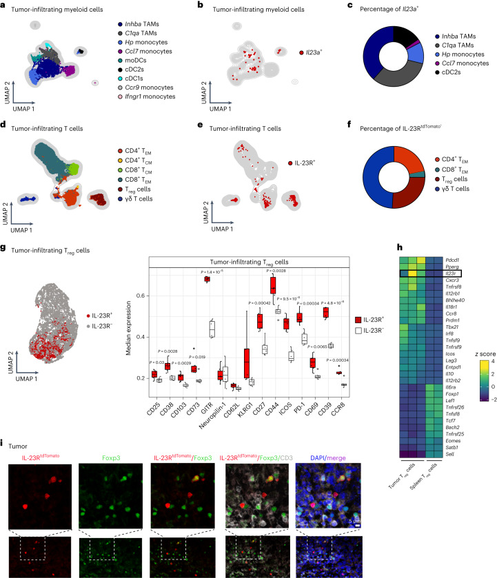
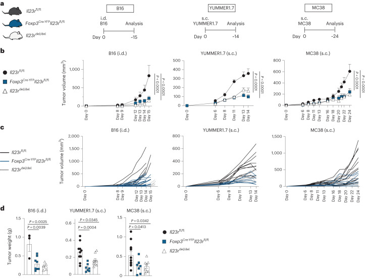
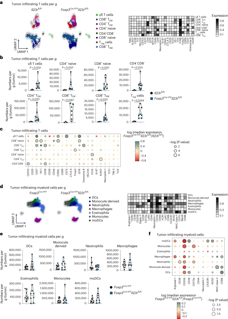
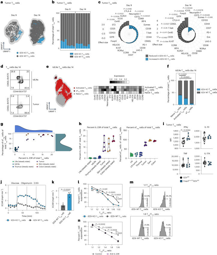
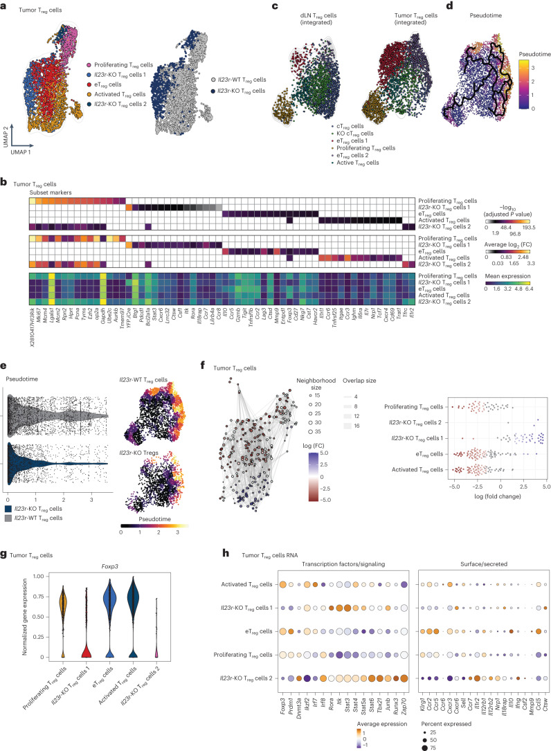
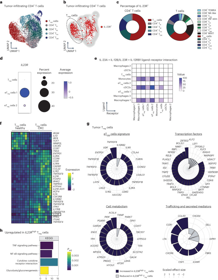
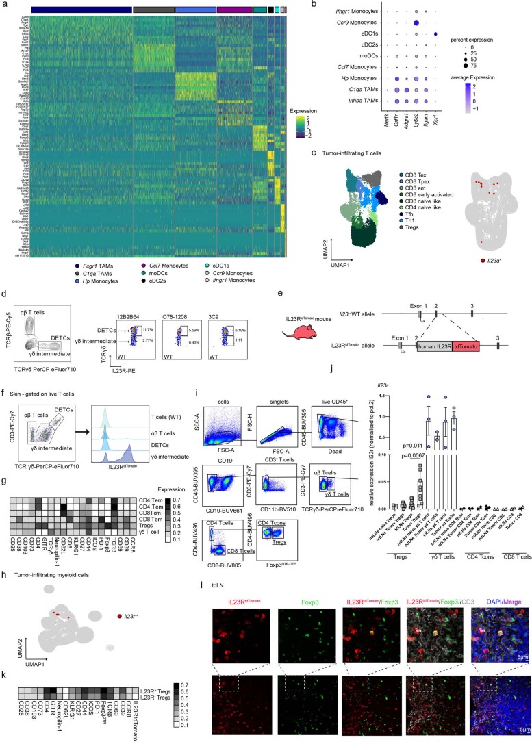
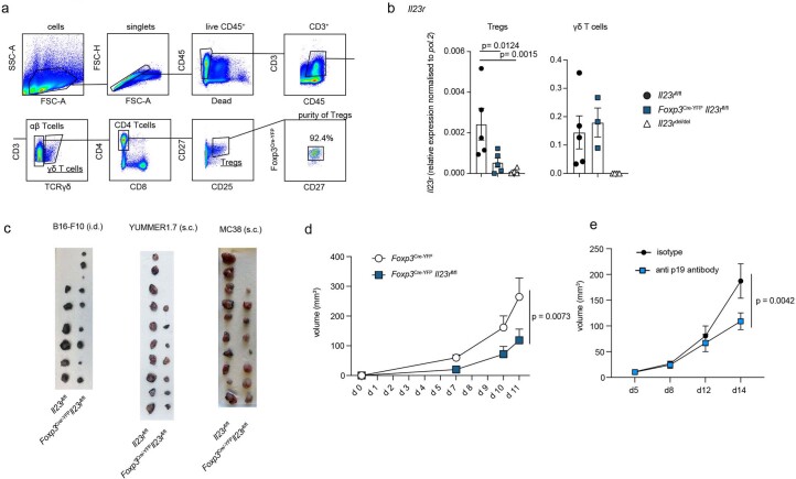
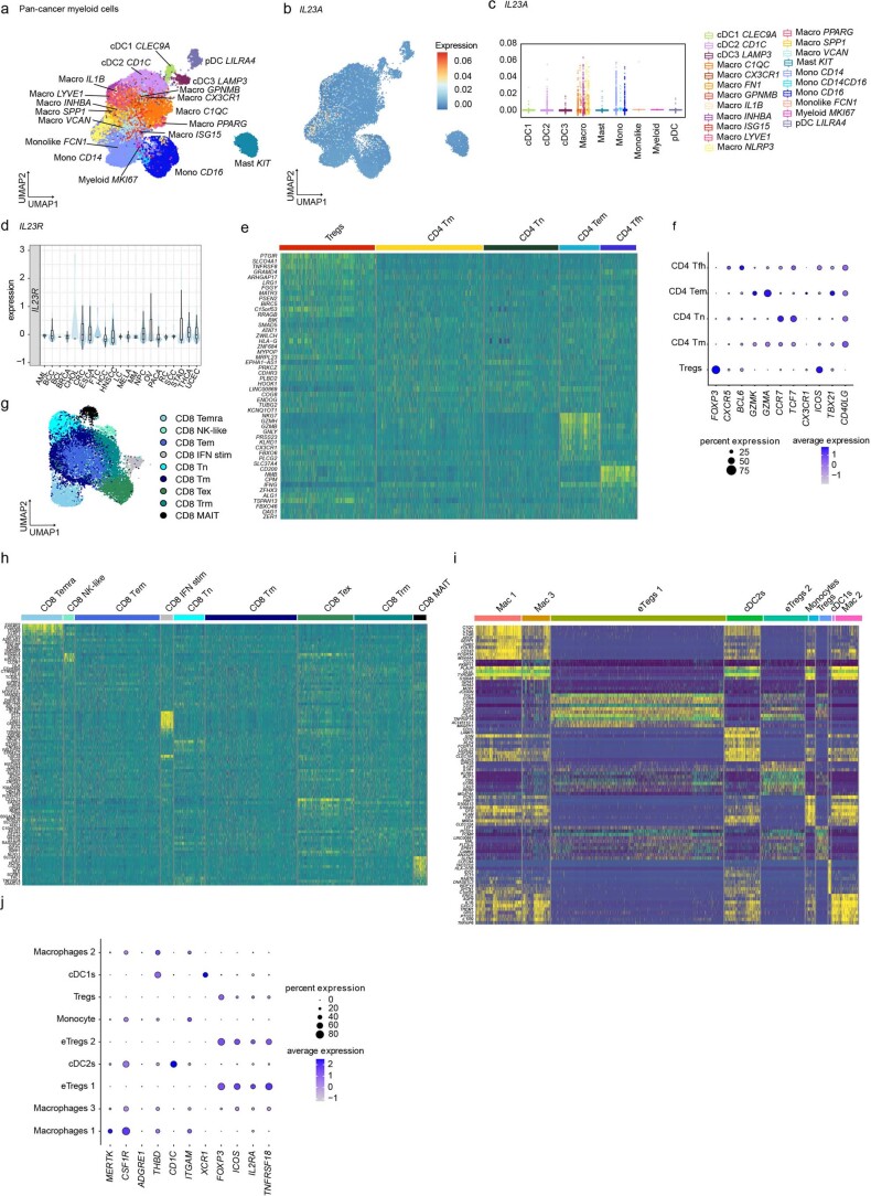
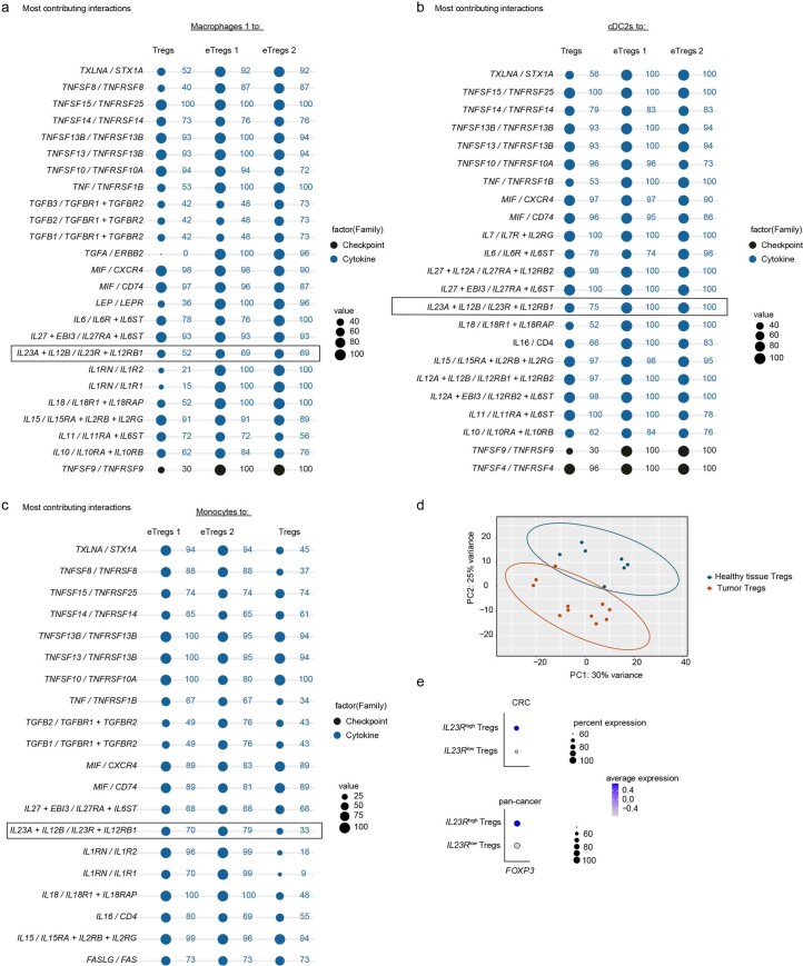
Interleukin-23 (IL-23) is a proinflammatory cytokine mainly produced by myeloid cells that promotes tumor growth in various preclinical cancer models and correlates with adverse outcomes. However, as to how IL-23 fuels tumor growth is unclear. Here, we found tumor-associated macrophages to be the main source of IL-23 in mouse and human tumor microenvironments. Among IL-23-sensing cells, we identified a subset of tumor-infiltrating regulatory T (Treg) cells that display a highly suppressive phenotype across mouse and human tumors. The use of three preclinical models of solid cancer in combination with genetic ablation of Il23r in Treg cells revealed that they are responsible for the tumor-promoting effect of IL-23. Mechanistically, we found that IL-23 sensing represents a crucial signal driving the maintenance and stabilization of effector Treg cells involving the transcription factor Foxp3. Our data support that targeting the IL-23/IL-23R axis in cancer may represent a means of eliciting antitumor immunity.


Conflict of interest statement

The authors declare no competing interests.

Figures

Fig. 1
Fig. 1. IL-23R marks a highly suppressive Treg cell subset in the mouse TME.
ac, Analysis of a myeloid cell scRNA-seq dataset from mouse B16 tumors (GSE188548; WT tumor). a, UMAP depicting tumor-infiltrating myeloid cell clusters. b, UMAP displaying Il23a+ myeloid cells. c, Pie chart displaying the frequencies of myeloid cell subsets among total Il23a+ myeloid cells. dg, Foxp3DTR-GFPIL-23RtdTomato mice were inoculated intradermally (i.d.) with B16 tumors. TILs were analyzed by flow cytometry on day 14. Data are shown from one representative experiment out of two independent experiments with n = 5–6 biologically independent animals. d, UMAP with overlaid FlowSOM clustering (gated on CD45+TCRβ+TCRγδ+ cells). e, UMAP displaying IL-23RtdTomato+ T cells. f, Pie chart depicting the frequencies of T cell subsets among total IL-23RtdTomato+ T cells. g, UMAP with overlaid FlowSOM clustering displaying IL-23RtdTomato+Foxp3+ and IL-23RtdTomato–Foxp3+ Treg cell clusters (left). Box plots showing median expression of surface markers on IL-23R+ and IL-23R Treg cells are shown on the right. Box plots display the median and interquartile range (IQR; 25–75%), with whiskers representing the upper and lower quartiles ± IQR. Statistical significance was calculated using two-tailed t-tests. h, Analysis of a bulk next-generation sequencing dataset of Treg cells sorted from B16 tumors or spleens (Magnuson et al.). A heat map depicting selected genes among the top 50 DEGs is shown. Expression of Il23r is highlighted. i, Immunofluorescence stainings of tumors from i.d. inoculated B16 tumor-bearing IL-23RtdTomato mice showing Foxp3 (green), IL-23RTdtomato (red), CD3 (white), DAPI (blue) and merged signals (purple). Scale bar: 5 μm. Images shown (n = 4) are representative of two independent experiments; moDCs, monocyte-derived DCs; TCM, central memory T cells. Source data
Fig. 2
Fig. 2. Treg cells mediate the tumor-promoting functions of IL-23.
ad, Il23rfl/fl, Foxp3Cre-YFPIl23rfl/fl and Il23rdel/del mice were inoculated i.d. with B16 tumor cells, inoculated subcutaneously (s.c.) with YUMMER1.7 tumor cells or inoculated s.c. with MC38 tumor cells, and tumors were analyzed around days 15, 14 and 24 after inoculation. The data show the results of three independent experiments (B16: n = 3 Il23rfl/fl mice, n = 5 Il23rdel/del mice, n = 8 Foxp3Cre-YFPIl23rfl/fl mice; MC38: n = 12 Il23rfl/fl mice, n = 7 Foxp3Cre-YFPIl23rfl/fl mice, n = 7 Il23rdel/del mice; YUMMER1.7: n = 10 Il23rfl/fl mice, n = 7 Foxp3Cre-YFPIl23rfl/fl mice, n = 8 Il23rdel/del mice). a, Schematic illustration of the experimental approach. b, Tumor volume kinetics of the experimental groups measured by caliper gauge. Data are shown as mean ± s.e.m. Statistical significance was determined by two-way analysis of variance (ANOVA) with a Sidak’s post hoc test. c, Tumor volume kinetics of individual mice measured by caliper gauge. d, Bar graph displaying the final tumor weight. Data are displayed as mean ± s.e.m. Statistical significance was determined using two-tailed t-tests. Source data
Fig. 3
Fig. 3. IL-23R signaling in Treg cells suppresses antitumor immunity.
af, Il23rfl/fl and Foxp3Cre-YFPIl23rfl/fl mice (ac) or Foxp3Cre-YFP and Foxp3Cre-YFPIl23rfl/fl mice (df) were inoculated i.d. with B16 tumor cells, and TILs (gated on CD45+TCRβ+TCRγδ+ cells; ac) or myeloid cells (gated on CD45+CD90.2CD19NK1.1 cells; df) were analyzed by flow cytometry on day 14 after inoculation. Data are shown from one representative experiment out of two independent experiments with n = 3–7. a,d, UMAP with overlaid FlowSOM clustering (left) and heat map depicting relative marker expression among identified cell clusters (right). b,e, Violin plots depicting cell numbers of identified cell clusters per gram (tumor). Data are displayed as mean ± s.e.m. Statistical significance was determined using a two-tailed Mann–Whitney U-test. c,f, Dot plot displaying median marker expression in identified cell clusters comparing Foxp3Cre-YFPIl23rfl/fl and Il23rfl/fl (control group) mice (c) or Foxp3Cre-YFPIl23rfl/fl and Foxp3Cre-YFP (control group) mice (f). Statistical significance was determined using two-tailed t-tests. Color represents log (median expression Foxp3Cre-YFPIl23rfl/fl/median expression control group); that is, red indicates that median expression is decreased in Foxp3Cre-YFPIl23rfl/fl mice compared to in the control group, and green indicates that median expression is increased in Foxp3Cre-YFPIl23rfl/fl mice compared to in the control group. Circle size represents log (P value). Statistically significant changes (P < 0.05) are highlighted with black lines around the circles; TRM, resident memory T cells. Source data
Fig. 4
Fig. 4. IL-23R signaling confers a selective advantage on eTreg cells.
af, Foxp3Cre-YFP/+Il23rfl/fl female mice were inoculated i.d. with B16 cells and Treg cells from tumors, and tdLNs were analyzed by flow cytometry on day 9 or 14 after injection. Data are shown from one representative experiment out of two independent experiments with n = 5–6. a, UMAP and FlowSOM clustering displaying Treg cell subsets in tumors. b, Frequency plots of Il23r-KO and WT Treg cells out of total Treg cells in tumors. c, Spiral plot displaying the effect size of differential marker expression between Il23r-KO and WT Treg cells in tumors. d, Representative contour plots depicting Treg cells on day 14 after tumor inoculation. e, UMAP and FlowSOM clustering displaying Treg cells in tdLNs on day 14 after tumor inoculation (left). A heat map depicting relative marker expression is shown on the right. f, Frequency plots of subsets of Il23r-KO and WT Treg cells in tdLNs on day 14 after tumor injection. Statistical significance was determined by two-way ANOVA with a Sidak’s post hoc test. g,h, Scatter plot (g) and violin plots (h) displaying frequencies of IL-23R+ Treg and eTreg cells among total Treg cells. Data are pooled from one to two experiments with n = 3–5. i, Il23rfl/fl and Foxp3Cre-YFPIl23rfl/fl mice were inoculated i.d. with MC38 tumor cells. Treg cells were analyzed by flow cytometry on day 14 after inoculation. Violin plots display the median expression (median fluorescence intensity (MFI)) of cytokines. The data shown are from one experiment with n = 6–12. j, Extracellular acidification rate (ECAR) measurement of Treg cells from spleens and LNs of Foxp3Cre-YFPIl23rfl/fl or Foxp3Cre-YFPmice under the specified conditions after stimulation with anti-CD3/anti-CD28, IL-2 and IL-23 for 72 h. Data are representative of the results of two independent experiments with n = 4. k, Quantification of glycolysis in Il23r-KO and WT Treg cells. ln, Ex vivo suppression of CellTrace Violet-labeled CD4+ conventional T (Tcon) cell proliferation by Il23r-KO and WT Treg cells (l and m) or WT Treg cells ± anti-IL-23R (n). Data shown are from two independent experiments with n = 5. Statistical significance was assessed by two-way ANOVA with a Sidak’s post hoc test. Data in f, i, k, l and n are displayed as mean ± s.e.m. Statistical significance in c and ik was determined using two-tailed t-tests; 2-DG, 2-deoxyglucose; NS, not significant. Source data
Fig. 5
Fig. 5. IL-23 sensing by Treg cells initiates an eTreg cell program in the murine TME.
ah, Foxp3Cre-YFP/+ (heterozygous) Il23rfl/fl female mice were inoculated i.d. with B16 tumor cells, and a combined transcriptome (scRNA-seq) and protein expression analysis of sorted CD4+ T cells was performed on day 13 after inoculation. Data are shown from one experiment with n = 6. a, UMAP displaying identified tumor-infiltrating Treg cell clusters (transcriptome; left) and UMAP highlighting Il23-KO and WT Treg cells (right). b, Heat maps showing adjusted P value (top), average log2 (fold change) (log2 (FC); middle) and mean expression (bottom) of subset markers in the identified Treg cell clusters assessed by Wilcoxon rank-sum test and Benjamini–Hochberg correction. The complete list is available in Supplementary Table 1. c, UMAP displaying identified clusters of integrated tumor-infiltrating and tdLN-derived Treg cells. d, UMAP of tdLN and tumor Treg cells with overlayed pseudotime and principal graph lines calculated with Monocle 3. e, Violin plot (left) and UMAP (right) comparing the distribution of WT and Il23r-KO Treg cells along pseudotime (corresponding to d). f, Neighborhood graph of DA testing results (left). Coloring indicates log (fold change) of differentially abundant neighborhoods between WT and Il23r-KO Treg cells. White neighborhoods are not differentially abundant (false discovery rate of 10%). Dot size corresponds to the number of cells per neighborhood, and edges indicate the number of overlapping cells between neighborhoods. The index cell position in UMAP space (a) determines ordering of the neighborhood nodes. The Beeswarm plot (right) indicates the distribution of differentially abundant neighborhoods across clustering-based Treg cell subsets. g, Violin plots depicting the normalized Foxp3 RNA abundance among identified Treg cell subsets. h, Dot plot displaying selected DEGs between identified Treg cell subsets encoding transcription factors/signaling proteins (left) and surface or secreted proteins (right). The complete list is available in Supplementary Table 2; cTreg, central Treg cells.
Fig. 6
Fig. 6. IL-23R signaling induces an eTreg cell program in the human TME.
ac,g,h, Analyses of a human pan-cancer single-cell sequencing dataset. a, UMAP displaying pan-cancer CD4+ T cells. b, UMAP highlighting IL23R+CD4+ T cells. c, Pie chart depicting the frequencies of T cell subsets among total IL23R+CD4+ T cells (left) and among total IL23R+ T cells (right). d,e, Analysis of an scRNA-seq dataset of human colorectal carcinomas (Liu et al.). d, Dot plot displaying IL23R expression across Treg cell subsets. e, Interaction heat map based on inferred ligand–receptor score between myeloid and Treg cell subsets of the IL-23A + IL-12B/IL-23R + IL-12RB1 axis computed with ICELLNET. The intensity of communication score is depicted as color intensity value. f, Heat map depicting the median expression of selected genes among the top 50 DEGs between Treg cells isolated from healthy colon biopsies and from tumor tissue of individuals with colorectal cancer from a bulk next-generation sequencing dataset (Magnuson et al.). Expression of IL23R is highlighted. g, Spiral plots displaying the scaled (positive values between 1 and 2; negative values between −1 and −2) effect size of selected DEGs between IL23Rhigh (IL23R expression > 0) and IL23Rlow (IL23R expression = 0) Treg cells. The complete list of DEGs is available in Supplementary Table 3. h, Selected significantly enriched pathways from KEGG pathway analysis using G:Profiler comparing IL23Rhigh and IL23Rlow Treg cells. Significance was calculated by g:GOSt using a Fisher’s one-tailed test. No downregulated pathways were detected. The complete list is available in Supplementary Table 4; TN, naive T cells; TEX, exhausted T cells; TM, memory T cells; TFH, follicular helper T cells; MAIT, mucosal-associated invariant T cells; Padj, adjusted P value. Source data
Extended Data Fig. 1
Extended Data Fig. 1. IL23R marks a highly suppressive Treg cell subset in the murine TME.
(a, b, h) Myeloid cell-scRNAseq data from murine B16 tumors (Mujal et al. 2022, GSE188548). (a) Heatmap depicting the expression of cell-type-defining genes. (b) Dotplot showing the expression of selected linage markers. (c) scRNAseq data of murine pan-tumor T cells (Andreatta et al. 2022, E-MTAB-9274). UMAP displaying T cell subsets (left) or Il23a+ T cells (right). (d) Representative FACS plot (left) of steady state murine skin CD45+cells (left) (pregated on live, CD45+cells). Representative FACS plots (right, pregated on live, CD45+, TCRγδ+) with gates of positive signal of respective IL23R-PE antibody clones of dendritic epidermal T cells (DETCs) (upper gate) and γδintermediate T cells (lower gate). Displayed data are from one experiment with n = 4. (e) Schematic illustration of the IL23RtdTomatoallele. (f) Representative FACS plot (left) of murine skin T cells and histograms (right) depicting IL23RtdTomato expression among T cell subsets from IL23RtdTomato mice and total T cells (‘T cells’) from WT mice. Data shown from one representative experiment out of two independent experiments with n = 3. (g, k) T cells from i.d. inoculated B16 tumors in Foxp3DTR-GFP IL23RtdTomato mice were analyzed by flow cytometry on day 14. Data shown from one representative experiment out of two independent experiments with n = 5-6. (g) Heatmap displaying marker expression among tumor-infiltrating T cell subsets. (h) UMAP displaying Il23r+ myeloid cells. (i) Gating strategy used to FACS-sort γδ, CD8+, CD4+ T cells and Foxp3+ Treg cells from Foxp3DTR-GFP mice. (j) Bar graph depicting relative Il23r mRNA expression level (qPCR) in FACS sorted T cells from steady state LNs or tdLNs, ndLNs and tumors from i.d. inoculated B16 tumor-bearing Foxp3DTR-GFP mice. Pooled data from three independent experiments. Biologically independent samples: n = 2: ndLNs: γδ T cells, naïve CD4 Tcon; ndLNs: naïve CD8; tdLNs: CD8, CD4 Tcon; Tumor: CD8. n = 3: ndLNs: naïve Treg cell, naïve γδ T cells, γδ T cells; Tumor: γδ T cells, CD4 Tcon. n = 8: ndLNs Tumor Treg cell. n = 10: tdLNs: Treg cell; Tumor: Treg cell. Data are displayed as mean +/- SEM. (k) Heatmap displaying relative marker expression among IL23R+ and IL23RTreg cellclusters. (l) Immunofluorescence-stainings of tdLNs from i.d. inoculated B16-tumor bearing IL23RtdTomato mice. Foxp3 (green), IL23RtdTomato (red), CD3 (white) DAPI (blue), merged (purple). Images (n = 4) are representatives from 2 independent experiments. Source data
Extended Data Fig. 2
Extended Data Fig. 2. Treg cells mediate the tumor-promoting functions of IL-23.
(a) Gating strategy to FACS sort γδ T cells and Treg cells from LNs of Foxp3Cre-YFPIl23rfl/fl and Il23rfl/fl mice for qPCR and example plot showing purity of sorted Treg cells. (b) Bar graphs depicting relative mRNA expression level of Il23r normalized to pol.2 in FACS sorted γδ T cells and Treg cells from steady state LNs of Foxp3Cre-YFPIl23rfl/fl, Il23rfl/fl and Il23rdel/del mice. Data shown from one representative experiment out of two independent experiments with n = 5 (Foxp3Cre-YFPIl23rfl/fl and IL23Rfl/fl mice) or n = 3 (Il23rdel/del) biologically independent samples. Data are displayed as mean +/- SEM. Statistical significance was determined using t-tests. (c) Il23rfl/fl and Foxp3Cre-YFPIl23rfl/fl mice were inoculated i.d. with B16 tumor cells, inoculated s.c. with YUMMER1.7 tumor cells or inoculated s.c. with MC38 tumor cells. Tumors were analyzed on day 15, 14 or 24 post-inoculation. Pictures depict tumors after harvest. Data from 3 independent experiments with n = 6-10. (d) Kinetics of tumor volume measured by caliper gauge in B16 tumor cell-inoculated i.d. Foxp3Cre-YFP and Foxp3Cre-YFPIl23rfl/fl mice. Mean +/- SEM is displayed. Statistical significance was determined using 2-way Anovas. n = 4 biologically independent animals. (e) Kinetics of tumor volume measured by caliper gauge in MC38 tumor cell-inoculated s.c. C57Bl/6 mice i.p. injected either with anti-p19 blocking or isotype antibodies. n = 6 (isotype) and n = 7 (anti p19 antibody) biologically independent animals. Mean +/− SEM is displayed. Statistical significance was determined using 2-way Anovas. Source data
Extended Data Fig. 3
Extended Data Fig. 3. IL23R signaling in Treg cells suppresses anti-tumor immunity.
(a-f) Il23rfl/fl and Foxp3Cre-YFPIl23rfl/fl mice were inoculated s.c. with MC38 tumor cells (a-c) or inoculated s.c. with YUMMER1.7 tumor cells (d-f) and tumor-infiltrating T cells were analyzed by flow cytometry on day 24 (a-c) or 14 (d-f) post-inoculation. TILs from MC38 tumors were re-stimulated with PMA/Ionomycin prior flow cytometry analysis. Data display 2 independent experiments with n = 6-10. (a, d) UMAP with overlaid FlowSOM clustering (left) (gated on CD45+ TCRβ+ and TCRγδ+ cells) and heatmap depicting relative marker expression among identified cell clusters (right). (b, e) Violin plots depicting cell numbers of identified T cell clusters per gram tumor. Data are displayed as mean +/- SEM. Statistical significance was determined using two-tailed Mann-Whitney U-tests. (c, f) Dotplots displaying median marker expression in identified T cell clusters comparing Foxp3Cre-YFP Il23Rfl/fl and Il23Rfl/fl mice. Color represents log(median expression Foxp3Cre-YFP Il23Rfl/fl / median expression Il23Rfl/fl); that is red means that median expression is decreased in Foxp3Cre-YFP Il23Rfl/fl in comparison to Il23Rfl/fl mice; green means that median expression is increased in Foxp3Cre-YFP Il23Rfl/fl mice in comparison to Il23Rfl/fl mice. Circle size represent log(p value). Statistically significant changes (p < 0.05) are highlighted with black lines around the circles. Statistical significance was determined using t-tests. (g) C57Bl/6 mice were s.c. inoculated with MC38 tumor cells and tumor-infiltraring T cells were analyzed by flow cytometry on day 14 post-inoculation. Dotplot displaying median marker expression of CD4+T cell and Treg cell clusters comparing anti-p19 and isotype antibody treated mice. Color represents log(median expression anti-p19/median expression isotype). Circle size represent log(p value). Statistically significant changes (p < 0.05) are highlighted with black lines around the circles. Data display one out of 2 independent experiments with n = 4-7. Statistical significance was determined using t-tests. Source data
Extended Data Fig. 4
Extended Data Fig. 4. IL23R signaling confers a selective advantage on eTreg cells.
(a) Schematic illustration of Tregs in Foxp3Cre-YFP/+ Il23rfl/fl female mice. (b,c) FACS-gating strategy (b) and bar graphs (c) displaying Il23r mRNA expression levels as assessed by qPCR in YFP+ and YFP Treg cells from LNs of Foxp3Cre-YFP/+Il23rfl/fl female mice. Data depict one experiment with n = 2 (YFP-) or n = 3 (YFP+). (d-h) Foxp3Cre-YFP/+Il23rfl/fl female (d,e,f,h) and Foxp3Cre-YFP/+ non-floxed (g) mice were inoculated i.d. with B16 cells or left untreated. Treg cells in steady state LNs or tumor and tdLNs on day 9 or 14 post-inoculation were analyzed by flow cytometry. Data shown from one out of two independent experiments with n = 5-6. (d) Heatmap depicting marker expression among Treg cell clusters (tumor). (e, f) Frequency plots of Il23r KO and WT of total Treg cells in tdLNs (e) or steady state LNs (f). (g) Contour plots showing YFP+ Treg cells on day 14. (h) Spiral plot displaying differential marker expression between Il23r KO and WT Treg cells (tdLNs). (i) Bar graph displaying Foxp3-expression in YFP-/YFP+Treg cells (tdLNs) of Foxp3Cre-YFP/+non-floxed mice on day 14 post-inoculation. Data from 1 experiment with n = 3. (j) Contour plots displaying IL23R expression as assessed by flow cytometry in human Tregs from steady state PBMCs (gated on CD45+ CD3+ CD4+CD25+CD27+ FOXP3+ cells) or 2 days anti-CD3/CD28 + IL2 stimulated FACS sorted (CD45+CD3+CD4+CD27+CD25+CD127 from steady state PBMCs) Tregs (gated on CD45+ CD3+ CD4+CD25+CD27+ FOXP3+ cells). Data shown from one experiment with n = 2. (k) Il23rfl/fl and Foxp3Cre-YFPIl23rfl/fl mice were inoculated i.d. with B16 cells. Treg cells in the tdLNs were analyzed by flow cytometry on day 14 post-inoculation. Violin plots displaying median expression (MFI) of cytokines. Data from one experiment with n = 6-12. (l,m,n) Murine Treg cells were ex vivo stimulated with IFN-γ + IL-6, + /- IL-23 for 5 days and analysed by flow cytometry. Boxplots showing the MFI of Foxp3 (l) and total cell numbers (m). (l,m) n = 5.(n) Histograms (left) and boxplots showing normalized MFIs of pSTAT3/pSTAT5 (right). Combined result from two independent experiments with n = 8. (o) Murine Treg cells were ex vivo stimulated with anti-CD3/CD28 + IL-2 for 5 days and +/-IL-23 for 30 min. Histograms (left) and boxplots showing normalized MFIs (flow cytometry). Representative result from two independent experiments with n = 5 (control) or n = 6 (IL-23). (c,i,k,l,n,o) Data are displayed as mean +/- SEM. (c,h,i,k,l,n,o) Statistical significance was determined using, two-tailed t-test (c,h), t-tests (i,k,l) or the two-tailed Mann-Whitney U-test (n,o). Source data
Extended Data Fig. 5
Extended Data Fig. 5. IL-23 sensing by Treg cells initiates an eTreg cell program in the murine TME.
(a-i) Foxp3Cre-YFP/+ (heterozygous) Il23rfl/fl female mice were inoculated i.d. with B16 tumor cells and combined transcriptome (scRNAseq) and protein expression analysis of sorted CD4+T cells was performed on day 13 post-inoculation. Data shown from one experiment with n = 6. (a) Schematic illustration of the experimental workflow. (b) UMAP displaying clustered (based on transcriptome expression) and manually annotated cell subsets passing quality control. (c) Dotplot depicting the 10 most variable features in all identified cell subsets. (d) Dotplot displaying the 10 most variable features across identified Treg cell subsets. (e) Bar chart displaying the frequencies of Treg cell subsets of total tumor Treg cells. (f) Dotplot of the most variable features of Treg cell subsets of integrated tumor and tumor-draining lymph node Treg cells. (g) UMAPs highlighting the distribution of Il23r KO and WT Treg cells across identified clusters in tumors and tumor-draining lymph nodes. (h) Bar chart of percentages of Il23r KO and WT Treg cells across Treg cell clusters. (i) Cellular state plot displaying enrichment of module scores of identified Treg cell subsets compared to gene modules of activated vs. eTreg cells (x-axis) and KO Treg cells (y-axis).
Extended Data Fig. 6
Extended Data Fig. 6. IL-23 sensing by Treg cells initiates an eTreg cell program in the murine TME.
(a-c) Foxp3Cre-YFP/+ (heterozygous) Il23rfl/fl female mice were inoculated i.d. with B16 tumor cells and combined transcriptome (scRNAseq) and protein expression analysis of sorted CD4+T cells was performed on day 13 post-inoculation. (a) Log fold change of DA neighborhoods marked in red, neighborhoods with spatial FDR > 0.1 marked in grey (top). Heatmap depicting average gene expression of most variable markers across neighborhoods (bottom). (b) Dotplot depicting differentially expressed protein markers between the identified Treg cell clusters. (c) Expression density of selected variable features overlayed on UMAP displaying tumor Treg cells.
Extended Data Fig. 7
Extended Data Fig. 7. IL23R signaling induces an eTreg cell program in the human TME.
(ac) Analyses of human myeloid pan-cancer single-cell sequencing data set from (Cheng et al. 2021) from n = 210 individual patients across 15 human cancer types. (a) UMAP displaying pan-cancer myeloid cells. (b) UMAP depicting pan-cancer myeloid cells with overlaid IL23A expression. (c) Box plot displaying average IL23A expression in different pan-cancer myeloid cell clusters (grouped by cell type). Boxplots display the median and interquartile range (IQR; 25-75%) with whiskers representing the upper and lower quartile +/- IQR. (d) Box plot displaying IL23R expression among Treg cells from patients with different cancer types. Boxplots display the median and interquartile range (IQR; 25-75%) with whiskers representing the upper and lower quartile +/- IQR. (e-i) Analyses of human pan-cancer single-cell sequencing data set from Zheng et al. (e, f, g, h) and analyses of scRNAseq dataset of human colorectal carcinomas (Liu et al.) (i, j). (g) UMAP depicting pan-cancer CD8+ T cells. (e, h, i) Heatmap depicting the expression of cell-type defining genes. (f, j) Dotplots showing the expression of selected cell lineage markers.
Extended Data Fig. 8
Extended Data Fig. 8. IL-23 signaling induces an eTreg cell program in the human TME.
(ac) Analyses of scRNAseq dataset of human colorectal carcinomas (Liu et al.). Dotplots displaying the top 25 statistically significant outgoing interactions from the macrophages 1 (a), cDC2s (b) or monocytes (c) cell clusters to Treg cell clusters computed with the ICELLNET framework. (d) PCA depicting data from bulk NGS data set from (Magnuson et al.). Comparison between Treg cells isolated from healthy colon biopsies and tumor tissue from colorectal cancer patients. (e) Dotplots displaying FOXP3 gene expression in IL23Rhigh (IL23R expression > 0) and IL23Rlow(IL23R expression = 0) Treg cells from the human pan cancer T cell atlas (Zheng et al. 2021) and colorectal carcinoma (Liu et al.) scRNA seq data sets.

References

    1. Sakaguchi S, et al. Regulatory T cells and human disease. Annu. Rev. Immunol. 2020;38:541–566. doi: 10.1146/annurev-immunol-042718-041717. - DOI - PubMed
    1. Rosenblum MD, Way SS, Abbas AK. Regulatory T cell memory. Nat. Rev. Immunol. 2016;16:90–101. doi: 10.1038/nri.2015.1. - DOI - PMC - PubMed
    1. Tanaka A, Sakaguchi S. Regulatory T cells in cancer immunotherapy. Cell Res. 2017;27:109–118. doi: 10.1038/cr.2016.151. - DOI - PMC - PubMed
    1. Teh PP, Vasanthakumar A, Kallies A. Development and function of effector regulatory T cells. Prog. Mol. Biol. Transl. Sci. 2015;136:155–174. doi: 10.1016/bs.pmbts.2015.08.005. - DOI - PubMed
    1. Labani-Motlagh A, Ashja-Mahdavi M, Loskog A. The tumor microenvironment: a milieu hindering and obstructing antitumor immune responses. Front. Immunol. 2020;11:940. doi: 10.3389/fimmu.2020.00940. - DOI - PMC - PubMed

LinkOut - more resources