

Adipose-Derived Stem Cells' Secretome Attenuates Lesion Size and Parasite Loading in Leishmaniasis Caused by Leishmania Major in Mice
- PMID: 38356483
- PMCID: PMC10862109
- DOI: 10.30476/IJMS.2023.96413.2795
Adipose-Derived Stem Cells' Secretome Attenuates Lesion Size and Parasite Loading in Leishmaniasis Caused by Leishmania Major in Mice
Abstract
Background: Stem cell-derived secretome (SE) released into the extracellular space contributes to tissue repair. The present study aimed to investigate the impact of isolated secretome (SE) from adipose-derived mesenchymal stem cells (ASCs) on Leishmania major (L. major) lesions in BALB/c mice.
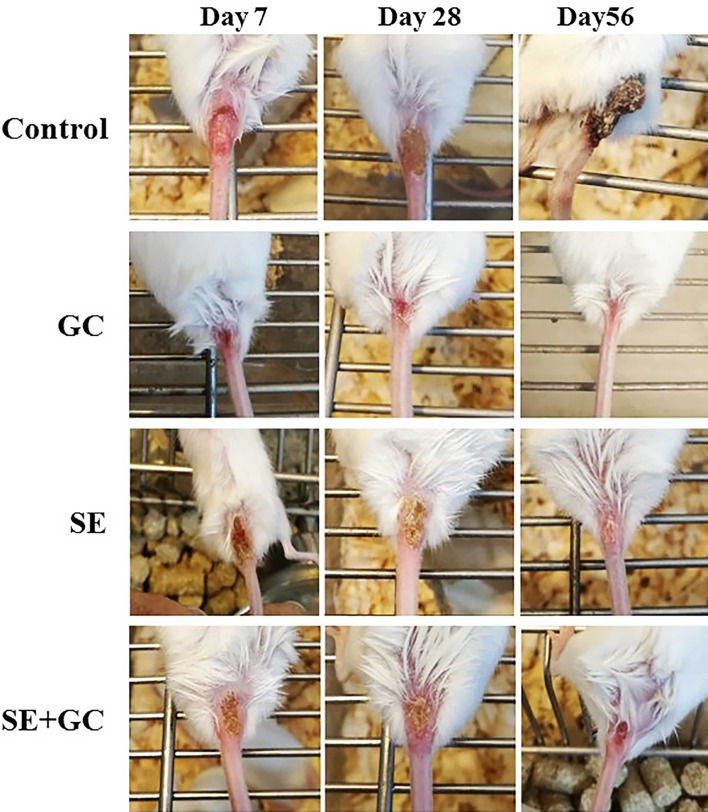
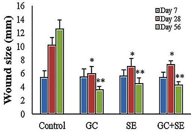
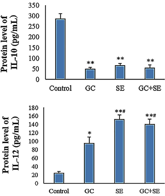
Methods: This experimental study was conducted at Ahvaz University of Medical Sciences (Ahvaz, Iran) in 2021. Forty female BALB/c mice were infected with stationary phase promastigotes through intradermal injection in the bottom of their tail and randomly divided into four groups (n=10 per group). The mice were given SE (20 mg/mL), either alone or in combination with Glucantime (GC, 20 mg/mL/Kg), meglumine antimoniate (20 mg/mL/Kg) for the GC group, and phosphate-buffered saline (PBS) for the control group. After eight weeks, the lesion size, histopathology, the levels of Interleukin 10 (IL-10), and Interleukin 12 (IL-12) were assessed. For the comparison of values between groups, the parametric one-way ANOVA was used to assess statistical significance.
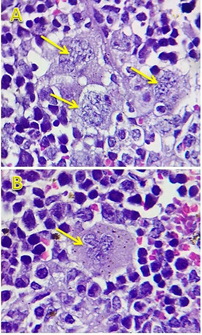
Results: At the end of the experiment, the mice that received SE had smaller lesions (4.56±0.83 mm versus 3.62±0.59 mm, P=0.092), lower levels of IL-10 (66.5±9.7 pg/mL versus 285.4±25.2 pg/mL, P<0.001), and higher levels of IL-12 (152.2±14.2 pg/mL versus 24.2±4.4 pg/mL, P<0.001) than the control. Histopathology findings revealed that mice treated with SE had a lower parasite burden in lesions and spleen than the control group.
Conclusion: The current study demonstrated that ADSC-derived SE could protect mice infected with L. major against leishmaniasis.
Keywords: Adipose-derived mesenchymal stem cells; Cytokines; Leishmania; Mice; Secretome; Wound.
Copyright: © Iranian Journal of Medical Sciences.
Conflict of interest statement
None declared.
Figures
References
-
- Hajjaran H, Saberi R, Borjian A, Fakhar M, Hosseini SA, Ghodrati S, et al. The Geographical Distribution of Human Cutaneous and Visceral Leishmania Species Identified by Molecular Methods in Iran: A Systematic Review With Meta-Analysis. Front Public Health. 2021;9:661674. doi: 10.3389/fpubh.2021.661674. [ PMC Free Article ] - DOI - PMC - PubMed
-
- Akhoundi M, Hajjaran H, Baghaei A, Mohebali M. Geographical distribution of leishmania species of human cutaneous leishmaniasis in fars province, southern iran. Iran J Parasitol. 2013;8:85–91. [ PMC Free Article ] - PMC - PubMed
MeSH terms
Substances
LinkOut - more resources
Full Text Sources
Miscellaneous