

Novel advanced imaging techniques for cerebral oedema
- PMID: 38356883
- PMCID: PMC10865379
- DOI: 10.3389/fneur.2024.1321424
Novel advanced imaging techniques for cerebral oedema
Abstract
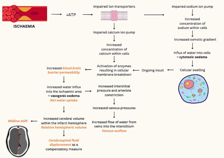
Cerebral oedema following acute ischemic infarction has been correlated with poor functional outcomes and is the driving mechanism of malignant infarction. Measurements of midline shift and qualitative assessment for herniation are currently the main CT indicators for cerebral oedema but have limited sensitivity for small cortical infarcts and are typically a delayed sign. In contrast, diffusion-weighted (DWI) or T2-weighted magnetic resonance imaging (MRI) are highly sensitive but are significantly less accessible. Due to the need for early quantification of cerebral oedema, several novel imaging biomarkers have been proposed. Based on neuroanatomical shift secondary to space-occupying oedema, measures such as relative hemispheric volume and cerebrospinal fluid displacement are correlated with poor outcomes. In contrast, other imaging biometrics, such as net water uptake, T2 relaxometry and blood brain barrier permeability, reflect intrinsic tissue changes from the influx of fluid into the ischemic region. This review aims to discuss quantification of cerebral oedema using current and developing advanced imaging techniques, and their role in predicting clinical outcomes.
Keywords: edema; imaging; infarction; malignant infarct; net water uptake; stroke.
Copyright © 2024 Pham and Ng.
Conflict of interest statement
The authors declare that the research was conducted in the absence of any commercial or financial relationships that could be construed as a potential conflict of interest.
Figures
Similar articles
-
[Follow-up monitoring with magnetic resonance tomography after decompressive trephining in experimental "malignant" hemispheric infarct].Zentralbl Neurochir. 1998;59(3):157-65. Zentralbl Neurochir. 1998. PMID: 9816666 German.
-
[Early assessment of severe hypoxic-ischemic encephalopathy in neonates by diffusion-weighted magnetic resonance imaging techniques and its significance].Zhonghua Er Ke Za Zhi. 2007 Nov;45(11):843-7. Zhonghua Er Ke Za Zhi. 2007. PMID: 18282417 Chinese.
-
Probability of cortical infarction predicted by flumazenil binding and diffusion-weighted imaging signal intensity: a comparative positron emission tomography/magnetic resonance imaging study in early ischemic stroke.Stroke. 2004 Aug;35(8):1892-8. doi: 10.1161/01.STR.0000134746.93535.9b. Epub 2004 Jun 24. Stroke. 2004. PMID: 15218157
-
Malignant MCA Infarction: Pathophysiology and Imaging for Early Diagnosis and Management Decisions.Cerebrovasc Dis. 2016;41(1-2):1-7. doi: 10.1159/000441627. Epub 2015 Nov 19. Cerebrovasc Dis. 2016. PMID: 26581023 Review.
-
Imaging biomarkers guided anti-angiogenic therapy for malignant gliomas.Neuroimage Clin. 2018 Jul 5;20:51-60. doi: 10.1016/j.nicl.2018.07.001. eCollection 2018. Neuroimage Clin. 2018. PMID: 30069427 Free PMC article. Review.
Cited by
-
A CT-based machine learning model for using clinical-radiomics to predict malignant cerebral edema after stroke: a two-center study.Front Neurosci. 2024 Oct 3;18:1443486. doi: 10.3389/fnins.2024.1443486. eCollection 2024. Front Neurosci. 2024. PMID: 39420983 Free PMC article.
-
Diabetic ketoacidosis and cerebral edema: a rare case of infarct-like MRI findings in a pediatric patient.Ann Med Surg (Lond). 2025 May 12;87(6):3963-3968. doi: 10.1097/MS9.0000000000003332. eCollection 2025 Jun. Ann Med Surg (Lond). 2025. PMID: 40486604 Free PMC article.
References
-
- Minnerup J, Broocks G, Kalkoffen J, Langner S, Knauth M, Psychogios MN, et al. . Computed tomography-based quantification of lesion water uptake identifies patients within 4.5 hours of stroke onset: a multicenter observational study. Ann Neurol. (2016) 80:924–34. doi: 10.1002/ana.24818, PMID: - DOI - PubMed
Publication types
LinkOut - more resources
Full Text Sources