. 2024 Jan 31:15:1298477.
doi: 10.3389/fneur.2024.1298477. eCollection 2024.

Accurate diagnosis and treatment of sacral meningeal cysts without spinal nerve root fibres: identifying leakage orificium using high-resolution spherical arbitrary-dimensional reconstructing magnetic resonance imaging

Affiliations

Accurate diagnosis and treatment of sacral meningeal cysts without spinal nerve root fibres: identifying leakage orificium using high-resolution spherical arbitrary-dimensional reconstructing magnetic resonance imaging

Chenlong Yang et al. Front Neurol. .

Abstract

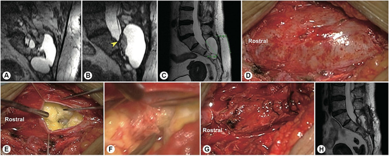
Objective: This study aimed to develop an arbitrary-dimensional nerve root reconstruction magnetic resonance imaging (ANRR-MRI) technique for identifying the leakage orificium of sacral meningeal cysts (SMCs) without spinal nerve root fibres (SNRFs).

Methods: This prospective study enrolled 40 consecutive patients with SMCs without SNRFs between March 2021 and March 2022. Magnetic resonance neural reconstruction sequences were performed for preoperative evaluation. The cyst and the cyst-dura intersection planes were initially identified based on the original thin-slice axial T2-weighted images. Sagittal and coronal images were then reconstructed by setting each intersecting plane as the centre. Then, three-dimensional reconstruction was performed, focusing on the suspected leakage point of the cyst. Based on the identified leakage location and size of the SMC, individual surgical plans were formulated.

Results: This cohort included 30 females and 10 males, with an average age of 42.6 ± 12.2 years (range, 17-66 years). The leakage orificium was located at the rostral pole of the cyst in 23 patients, at the body region of the cyst in 12 patients, and at the caudal pole in 5 patients. The maximum diameter of the cysts ranged from 2 cm to 11 cm (average, 5.2 ± 1.9 cm). The leakage orificium was clearly identified in all patients and was ligated microscopically through a 4 cm minimally invasive incision. Postoperative imaging showed that the cysts had disappeared.

Conclusion: ANRR-MRI is an accurate and efficient approach for identifying leakage orificium, facilitating the precise diagnosis and surgical treatment of SMCs without SNRFs.

Keywords: leakage orificium; magnetic resonance imaging; sacral cyst; sectional reconstruction; spinal nerve root.


Conflict of interest statement

The authors declare that the research was conducted in the absence of any commercial or financial relationships that could be construed as a potential conflict of interest. The reviewer HT declared a shared affiliation with the author JS to the handling editor at the time of review.

Figures

Figure 1
Figure 1
A precise incision was made for a sacral meningeal cyst without spinal nerve root fibres. (A) Spinal T2-weighted MR image demonstrating a sacral meningeal cyst without spinal nerve root fibres inside, and a precise incision was made as indicated by the green line. (B) The precise incision on the skin was only 4 cm in length. (C) After the cyst neck was ligated, the posterior wall of the sacral canal was reset using bone fixators.
Figure 2
Figure 2
A representative patient with a filum terminale cyst. (A) Preoperatively, reconstructed spinal MR image showing a sacral cyst at the S2-4 level with a thickened filum terminale and wide leakage neck (the arrowhead indicates the cystic leakage orificium, and the arrow indicates the filum terminale). (B) A 4 cm precise minimally invasive incision was made (yellow line segment). (C) Intraoperatively, the filum terminale was found within the cyst cavity. (D) The filum terminale was cut off after electric coagulation. (E) The leakage orificium was ligated. (F) Follow-up MRI showed that the cyst had disappeared and that there was no recurrence. (G) The 4 cm long skin incision healed well.
Figure 3
Figure 3
A representative patient with a fistula-type cyst. (A,B) Preoperatively, reconstructed MR image showing a fistula between the dural sac and the cyst (yellow arrowhead). (C) A 5 cm precise minimally invasive incision was designed (yellow line segment). (D) Intraoperatively, a fistula-type cyst was found, and there was no nerve root. (E) The cyst wall was dissected and turned over. (F) The cyst neck was ligated. (G) Follow-up MRI showed that the cyst had disappeared and that there was no recurrence.
Figure 4
Figure 4
A representative case of an arachnoid hernia-type cyst and a representative case of a spinal dura mater leakage cyst. (A) Preoperatively, reconstructed MR image revealing an arachnoidal structure within the cyst (the yellow arrowhead indicates the cystic leakage orificium). (B) A 4 cm precise and minimally invasive incision was made (yellow line segment). (C–E) Intraoperatively, an arachnoidal diverticulum was found as the origin of the cerebrospinal fluid, and this cystic leakage orificium was ligated. (F) Follow-up MRI showed that the cyst had disappeared and that there was no recurrence. (G) Preoperatively, reconstruction MRI showed that the narrow end of the dural sac was squeezed to the left by a giant cyst without nerve roots (the yellow arrowhead indicates the cystic leakage orificium). (H) A 4 cm precise minimally invasive incision was made (yellow line segment). (I) Intraoperatively, we found that the lateral wall of the dural sac was weak, leading to leakage. (J) The cyst wall was dissected and turned over. (K) Follow-up MRI showed that the cyst had disappeared and that there was no recurrence.
Figure 5
Figure 5
A representative patient with a massive sacral meningeal cyst. A 56 years-old woman with a massive sacral cyst was treated with glue injection and fat filling at the local hospital. (A) After referral to our institute, reconstructed MR image showed a sacral meningeal cyst and an extrasacral cyst, which were separated by the filled fat mass. (B) Sagittal spinal T2-weighted MR image showing the distorted dura sac end, dorsal fat mass, and residual cyst. (C) The precision of the incision is indicated by the green line. (D) Intraoperatively, a pseudocyst was found under the muscle layer. (E) After the pseudocyst was opened, cerebrospinal fluid was found to leak from the centre of the tightly packed fat. (F) After the filled fat was separated, the hardened biological glue was exposed. (G) After the neck of the cyst leakage site was sutured, the end of the dural sac was reinforced by an artificial dural membrane. (H) Two months after surgery, repeated MRI showed no cyst recurrence.
Figure 6
Figure 6
A representative patient with both sacroanterior and sacral cysts. A 22 years-old woman presenting with abdominal pain was found to have a pelvic mass. (A) Laparoscopic exploration revealed that the pelvic lesion was connected to the sacral canal. (B) Coronal MR image showing wide neck leakage between the end of the dural sac and the sacral canal. (C) Sagittal MR image revealing a giant sacroanterior cyst connected to a sacral cyst without nerve root fibres inside. (D) Sagittal CT image showing a sacroanterior bone defect. (E) A precise incision was made. (F) Intraoperatively, abundant cerebrospinal fluid leaked from the orificium, suggesting high intracystic pressure. (G) No spinal nerve root fibres were identified in the leakage orificium. (H) The leakage orificium was ligated. (I) On MRI, 1 week after the operation, the sacroanterior cyst had shrunk. (J) Three months after surgery, the sacroanterior cyst had almost completely disappeared on MRI.
Figure 7
Figure 7
The other representative patient had both sacroanterior and sacral cysts. A 45 years-old female presenting with abdominal pain was found to have a pelvic mass on CT at the local hospital. The intrapelvic part of the cyst was resected under laparoscopy. (A) After referral to our institute, coronal reconstructed MR image showed a narrow-neck leakage orificium (yellow arrowhead) between the end of the dural sac and the sacral canal cyst, and the filum terminale was thickened. (B) Sagittal reconstructed MR image showing the leakage orificium (yellow arrowhead) at the end of the dural sac connected to the sacral cyst and the residual sac. (C) A 4 cm precise incision was made (yellow line segment). (D) After the leakage orificium was ligated and the tethered spinal cord was released, a follow-up MRI showed that the cyst had completely vanished.

Similar articles

Cited by

References

    1. Ma Q, Wu C, Zhang J, Yin X, Yang C, Si Y, et al. . Arachnoidal diverticula of sacral extradural meningeal cyst: a novel definition and case series. World Neurosurg. (2022) 163:e106–12. doi: 10.1016/j.wneu.2022.03.052 - DOI - PubMed
    1. Sun J, Wang Z, Li Z, Wu H, Yen R, Zheng M, et al. . Reconstruction of nerve root sheaths for sacral extradural spinal meningeal cysts with spinal nerve root fibers. Sci China Life Sci. (2013) 56:1007–13. doi: 10.1007/s11427-013-4536-7 - DOI - PubMed
    1. Sun JJ, Wang ZY, Teo M, Li ZD, Wu HB, Yen RY, et al. . Comparative outcomes of the two types of sacral extradural spinal meningeal cysts using different operation methods: a prospective clinical study. PLoS One. (2013) 8:e83964. doi: 10.1371/journal.pone.0083964 - DOI - PMC - PubMed
    1. Sun JJ, Wang ZY, Liu B, Li ZD, Wu HB, Yen RY, et al. . Neck transfixion for sacral extradural spinal meningeal cysts without spinal nerve root fibers. Eur Spine J. (2016) 25:1945–52. doi: 10.1007/s00586-014-3471-z, PMID: - DOI - PubMed
    1. Maselli F, Palladino M, Barbari V, Storari L, Rossettini G, Testa M. The diagnostic value of red flags in thoracolumbar pain: a systematic review. Disabil Rehabil. (2022) 44:1190–206. doi: 10.1080/09638288.2020.1804626, PMID: - DOI - PubMed

LinkOut - more resources