. 2024;55(1-3):187-205.
doi: 10.1007/s00723-023-01611-1. Epub 2023 Sep 24.

Spectroscopically Orthogonal Labelling to Disentangle Site-Specific Nitroxide Label Distributions

Affiliations

Spectroscopically Orthogonal Labelling to Disentangle Site-Specific Nitroxide Label Distributions

Valentina Vitali et al. Appl Magn Reson. 2024.

Abstract

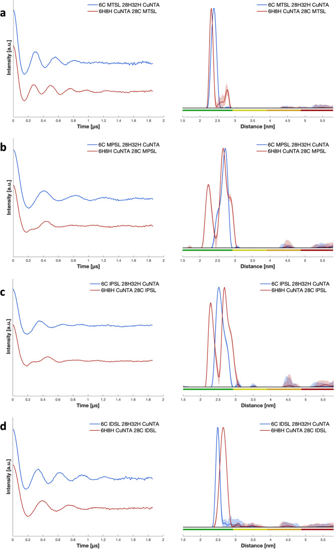
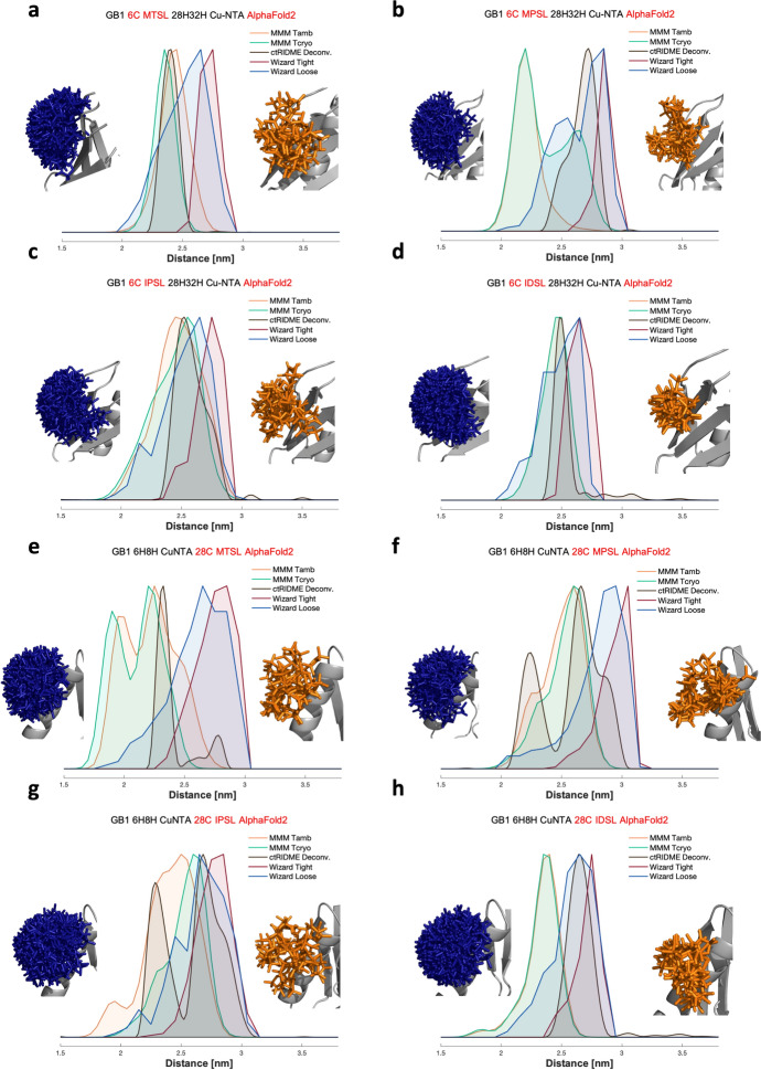
Biomolecular applications of pulse dipolar electron paramagnetic resonance spectroscopy (PDS) are becoming increasingly valuable in structural biology. Site-directed spin labelling of proteins is routinely performed using nitroxides, with paramagnetic metal ions and other organic radicals gaining popularity as alternative spin centres. Spectroscopically orthogonal spin labelling using different types of labels potentially increases the information content available from a single sample. When analysing experimental distance distributions between two nitroxide spin labels, the site-specific rotamer information has been projected into the distance and is not readily available, and the contributions of individual labelling sites to the width of the distance distribution are not obvious from the PDS data. Here, we exploit the exquisite precision of labelling double-histidine (dHis) motifs with CuII chelate complexes. The contribution of this label to the distance distribution widths in model protein GB1 has been shown to be negligible. By combining a dHis CuII labelling site with cysteine-specific nitroxide labelling, we gather insights on the label rotamers at two distinct sites, comparing their contributions to distance distributions based on different in silico modelling approaches and structural models. From this study, it seems advisable to consider discrepancies between different in silico modelling approaches when selecting labelling sites for PDS studies.

Supplementary information: The online version contains supplementary material available at 10.1007/s00723-023-01611-1.


Conflict of interest statement

Conflict of InterestAll authors declare that they have no conflict of interest.

Figures

Fig. 1
Fig. 1
a Structures of the four free nitroxide labels used for conjugation of the cysteine residues, respectively, in position 28 (α-helix) and 6 (β-sheet) of the GB1 protein and the CuNTA label directly coordinating the dHis motif (dHis-CuNTA). b The two GB1 constructs I6C/K28H/Q32H and I6H/N8H/K28C employed in this study
Fig. 2
Fig. 2
Constant time RIDME traces after background correction with DeerAnalysis2021, for both GB1 constructs I6C/K28H/Q32H (nitroxide on the β-sheet, in blue) and the I6H/N8H/K28C (nitroxide on the α-helix, in red) for the four nitroxide labels, MTSL, MPSL, IPSL, and IDSL, and their respective distance distributions. Colour bars represent reliability ranges (green: shape reliable; yellow: mean and width reliable; orange: mean reliable; red: no quantification possible). a MTSL, b MPSL, c IPSL, and d IDSL
Fig. 3
Fig. 3
Modelled distance distributions for both GB1 constructs (I6C/K28H/Q32H and I6H/N8H/K28C), both labelled with MTSL, MPSL, IPSL, and IDSL, based on the AlphaFold2 structure, superimposed with their respective experimental distance distributions (derived from ctRIDME deconvoluted data, in black). The in silico approaches compared are MMM at ambient (orange) and cryogenic (green) temperature and MtsslWizard with Tight (red) and Loose (blue) settings. Next to the distance distributions label rotamers modelled using MtsslWizard Loose (blue) and MMM at ambient temperature (orange) are given. a 6C MTSL, b 6C MPSL, c 6C IPSL, d 6C IDSL, e 28C MTSL, f 28C MPSL, g 28C IPSL, and h 28C IDSL

Similar articles

Cited by

References

    1. Verhalen B, Dastvan R, Thangapandian S, Peskova Y, Koteiche HA, Nakamoto RK, Tajkhorshid E, McHaourab HS. Energy transduction and alternating access of the mammalian ABC transporter P-glycoprotein. Nature. 2017;543:738–741. doi: 10.1038/nature21414. - DOI - PMC - PubMed
    1. Edwards DT, Huber T, Hussain S, Stone KM, Kinnebrew M, Kaminker I, Matalon E, Sherwin MS, Goldfarb D, Han S. Determining the oligomeric structure of proteorhodopsin by Gd3+-based pulsed dipolar spectroscopy of multiple distances. Structure. 2014;22:1677–1686. doi: 10.1016/j.str.2014.09.008. - DOI - PubMed
    1. Kasanmascheff M, Lee W, Nick TU, Stubbe J, Bennati M. Radical transfer in E. coli ribonucleotide reductase: a NH2Y731/R411A-α mutant unmasks a new conformation of the pathway residue 731. Chem. Sci. 2016;7:2170–2178. doi: 10.1039/c5sc03460d. - DOI - PMC - PubMed
    1. Schiemann O, Piton N, Mu Y, Stock G, Engels JW, Prisner TF. A PELDOR-based nanometer distance ruler for oligonucleotides. J. Am. Chem. Soc. 2004;126:5722–5729. doi: 10.1021/ja0393877. - DOI - PubMed
    1. Babaylova ES, Malygin AA, Lomzov AA, Pyshnyi DV, Yulikov M, Jeschke G, Krumkacheva OA, Fedin MV, Karpova GG, Bagryanskaya EG. Complementary-addressed site-directed spin labeling of long natural RNAs. Nucl. Acids Res. 2016;44:7935–7943. doi: 10.1093/nar/gkw516. - DOI - PMC - PubMed

LinkOut - more resources