

doi: 10.1016/j.vgie.2023.09.017.
eCollection 2024 Feb.
Light at the end of the tunnel: bridging the gap from the duodenum to the jejunum
Affiliations
- PMID: 38357030
- PMCID: PMC10861807
- DOI: 10.1016/j.vgie.2023.09.017
Item in Clipboard
Light at the end of the tunnel: bridging the gap from the duodenum to the jejunum
VideoGIE.
.
Abstract
Video 1Video describing the case, procedure, and outcomes.
© 2024 American Society for Gastrointestinal Endoscopy. Published by Elsevier Inc.
Conflict of interest statement
The authors did not disclose any financial relationships.
Figures
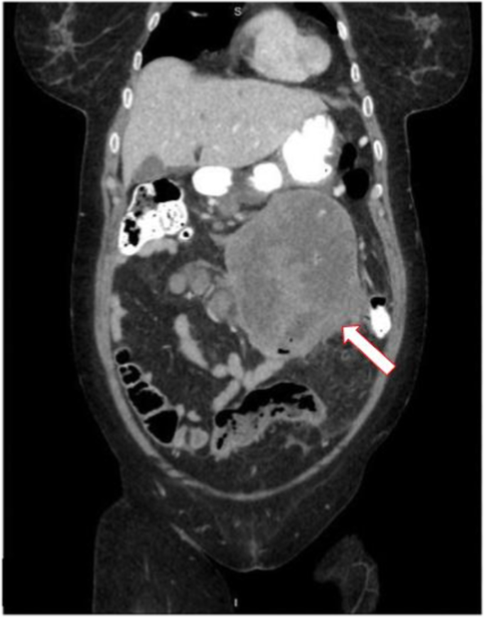
Coronal CT image of a large abdominal mass involving loops of bowel and jejunum. The arrow points to the mass.
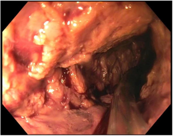
Endoscopic view of the mass-like lesion in the distal duodenum with no normal mucosa present.
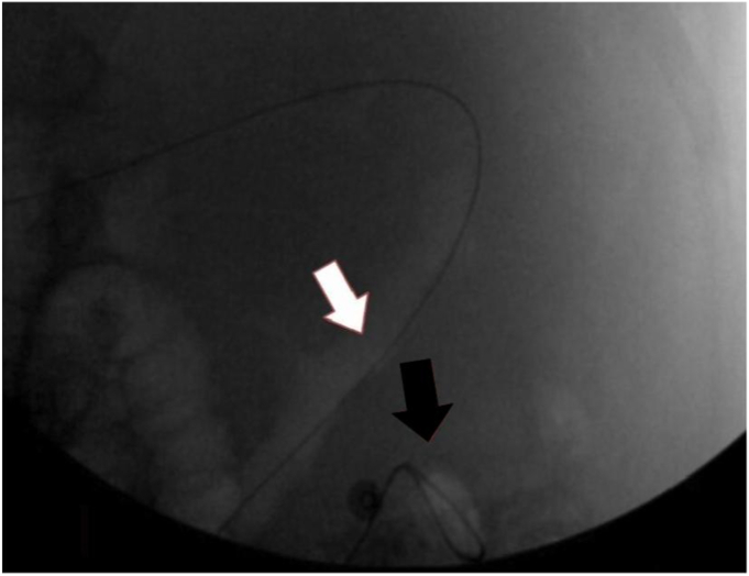
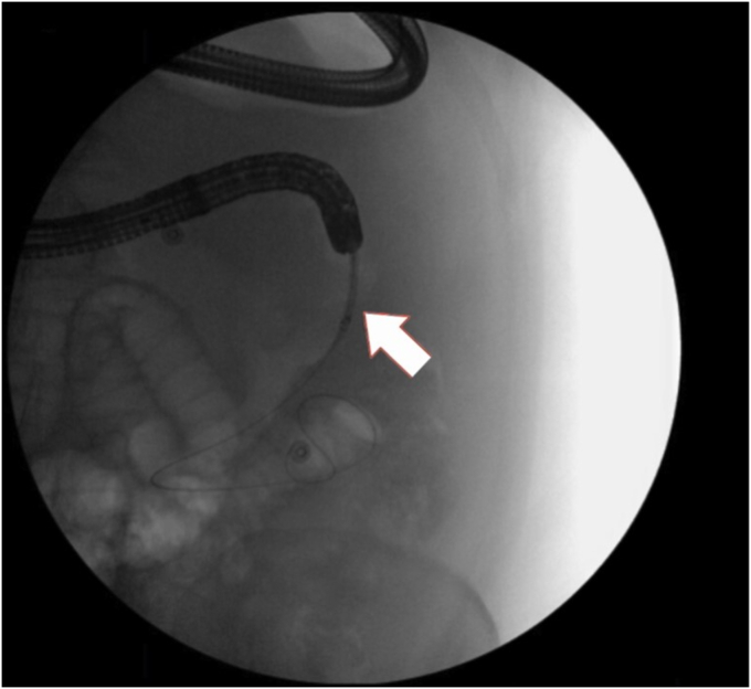
Fluoroscopic image of the guidewire passing through the stricture. The white arrow indicates the beginning of the stricture. The black arrow shows the guidewire passing through the stricture.
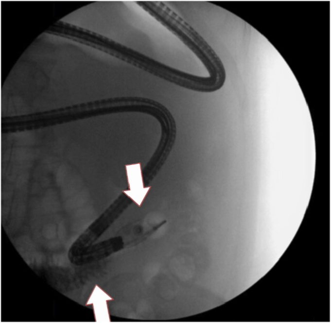
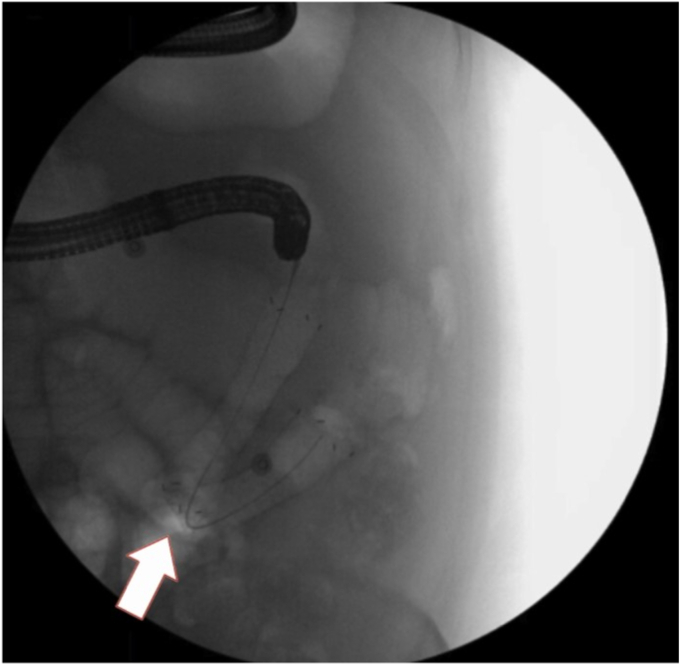
Fluoroscopic image of contrast being delivered by a 20-mm stone-extraction balloon showing luminal patency. The top arrow indicates the tip of the balloon where contrast was introduced into the lumen. The bottom arrow indicates contrast past the area of stricture.
Fluoroscopic image of the first esophageal stent being advanced. The arrow points to the stent.
Fluoroscopic image of the U-shaped bend in the first esophageal stent placement. The arrow indicates the bend in the stent.
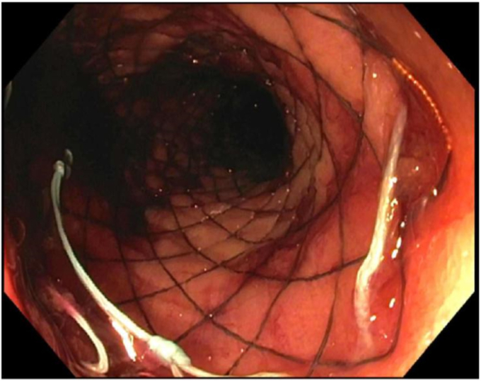
Endoscopic view of the third esophageal stent after placement.
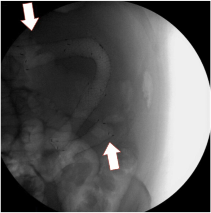
Fluoroscopic images of all 3 esophageal stents bridging the entire stricture. The top arrow indicates the beginning of the first esophageal stent. The bottom arrow indicates the end of the third esophageal stent.
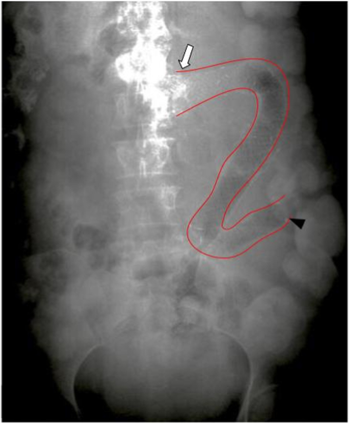
Abdominal radiograph showing interval placement of stents from the distal duodenum to the proximal jejunum. The entirety of the stenting is outlined by red lines. The white arrow indicates the beginning of the stenting. The black arrowhead indicates the end of the stenting.
References
-
- Soetikno R.M., Carr-Locke D.L. Expandable metal stents for gastric-outlet, duodenal, and small-intestinal obstruction. Gastrointest Endosc Clinics North America. 1999;9:447–458. - PubMed
-
- Yates M.R., 3rd, Morgan D.E., Baron T.H. Palliation of malignant gastric and small intestinal strictures with self-expandable metal stents. Endoscopy. 1998;30:266–272. - PubMed
-
- Rejchrt S., Kopacova M., Brozik J., et al. Biodegradable stents for the treatment of benign stenoses of the small and large intestines. Endoscopy. 2011;43:911–917. - PubMed
-
- Hirai F., Beppu T., Sou S., et al. Endoscopic balloon dilatation using double-balloon endoscopy is a useful and safe treatment for small intestinal strictures in Crohn’s disease. Digestive Endosc. 2010;22:200–204. - PubMed
LinkOut - more resources
Full Text Sources
Miscellaneous
