Review
. 2024 Feb;19(1):100883.
doi: 10.1016/j.ajps.2023.100883. Epub 2023 Dec 29.

Recent advances in zwitterionic nanoscale drug delivery systems to overcome biological barriers

Affiliations
Review

Recent advances in zwitterionic nanoscale drug delivery systems to overcome biological barriers

Xumei Ouyang et al. Asian J Pharm Sci. 2024 Feb.

Abstract

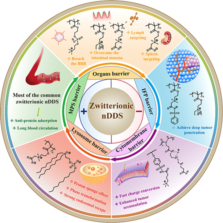
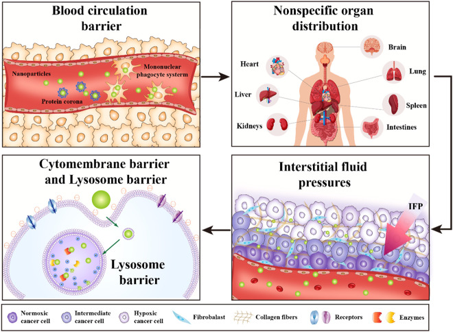
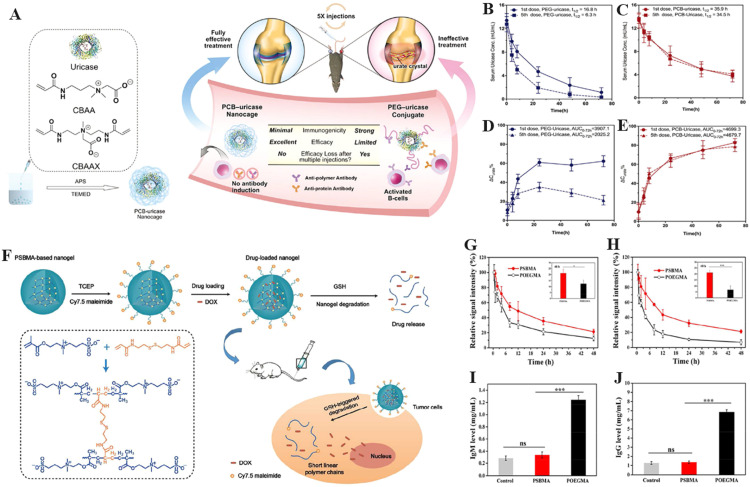
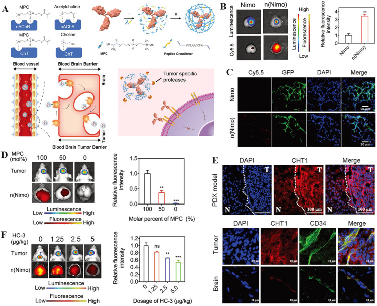
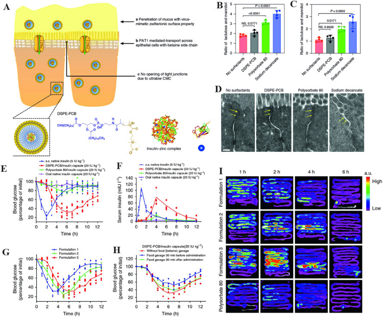
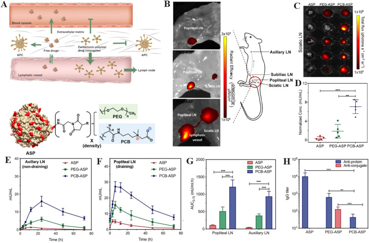
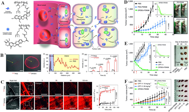
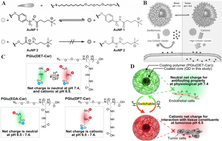
Nanoscale drug delivery systems (nDDS) have been employed widely in enhancing the therapeutic efficacy of drugs against diseases with reduced side effects. Although several nDDS have been successfully approved for clinical use up to now, biological barriers between the administration site and the target site hinder the wider clinical adoption of nDDS in disease treatment. Polyethylene glycol (PEG)-modification (or PEGylation) has been regarded as the gold standard for stabilising nDDS in complex biological environment. However, the accelerated blood clearance (ABC) of PEGylated nDDS after repeated injections becomes great challenges for their clinical applications. Zwitterionic polymer, a novel family of anti-fouling materials, have evolved as an alternative to PEG due to their super-hydrophilicity and biocompatibility. Zwitterionic nDDS could avoid the generation of ABC phenomenon and exhibit longer blood circulation time than the PEGylated analogues. More impressively, zwitterionic nDDS have recently been shown to overcome multiple biological barriers such as nonspecific organ distribution, pressure gradients, impermeable cell membranes and lysosomal degradation without the need of any complex chemical modifications. The realization of overcoming multiple biological barriers by zwitterionic nDDS may simplify the current overly complex design of nDDS, which could facilitate their better clinical translation. Herein, we summarise the recent progress of zwitterionic nDDS at overcoming various biological barriers and analyse their underlying mechanisms. Finally, prospects and challenges are introduced to guide the rational design of zwitterionic nDDS for disease treatment.

Keywords: Biological barrier; Disease treatment; Nano drug delivery system; Targeting delivery; Zwitterionic polymer.


Conflict of interest statement

The authors declare no conflicts of interest.

Figures

Image, graphical abstract
Graphical abstract
Fig 1
Fig. 1
The scheme of zwitterionic nDDS to overcome multiple biological barriers including MPS barrier, organs barrier, IFP barrier, cytomembrane barrier and lysosome barrier.
Fig 2
Fig. 2
The scheme of multiple biological barriers in drug delivery process.
Fig 3
Fig. 3
Zwitterionic PCB and PSB based nDDS to overcome the blood circulation barriers. (A) Encapsulation of uricase with PCB nDDS. The PK of PEGylated uricase (B) and uricase encapsulated by PCB nDDS (C) after the first and fifth injections. The urate-eliminating ability of PEGylated uricase (D) and uricase encapsulated by PCB nDDS (E) after the first and fifth injections (Reproduced with permission from . Copyright 2018 WILEY-VCH Verlag GmbH & Co. KGaA). (F) A schematic of DOX-loaded biodegradable zwitterionic PSB nanogels. PK profiles of PEGylated nanogels and PSB nanogels after the first (G) and second (H) injection in BALB/c mice (1 mg/ml). (I) Blood levels of immune globulin M (IgM) 5 d after the initial nanogel injection (n=5). (J) Blood levels of immune globulin G (IgG) 5 d post the second nanogel injection (n=5). The ns means that there is no significance (*P < 0.05, **P < 0.01, ***P < 0.001). (Reproduced with permission from . Copyright 2018 American Chemical Society).
Fig 4
Fig. 4
Zwitterionic PTMAO and polymer membrane-based nDDS to overcome the blood circulation barriers. (A) The PTMAO with excellent anti-fouling properties was originally identified in saltwater fish. Following the first (B) and third (C) intravenous injection, mice sera were used to determine the PK profiles of each sample. The urate level in mice sera was assessed following the first (D) and third (E) intravenous injections (Reproduced with permission from . Copyright 2019 The Authors). (F) A schematic depicting of the fabrication of zwitterionic polymer membranes-coated Fe3O4 and their biological properties. (G) The mass proportion of zwitterionic polymer membranes to zwitterionic polymer brushes coated on Fe3O4. (H) The protein adsorption of nanoparticles following a 30-min incubation in mouse blood. (I) The PK of nanoparticles upon intravenous administration. (J) The blood circulation half-life of nanoparticles in the bloodstream (Reproduced with permission from . Copyright 2019 Elsevier Ltd.) (*P < 0.05, **P < 0.01 and ***P < 0.001)
Fig 5
Fig. 5
The illustration of zwitterionic nDDS to achieve brain targeting. (A) Schematic of mAb encapsulation with PMPC nanocapsules for glioma treatment. (B) Fluorescence and bioluminescence images taken 10 d after injecting Cy5.5-labeled n(Nimo) into brain tissues dissected from patients with gliomas. The histogram summarizes the relative fluorescence intensity of tumor-bearing brain tissue. (C) The colocalization of n(Nimo) with the tumor tissue. (D) Photographs of brain ex vivo fluorescence and bioluminescence obtained 4 h after a single injection of Cy5.5-labeled n(Nimo) (5 mg/kg) in orthotopic U87-EGFRwt glioma xenograft mice. (E) Images of human tumor tissue xenograft (PDX) in mouse brain taken with confocal microscopy (separated by a dashed line, T indicates tumor and N indicates normal tissue). (F) Orthotopic U87-EGFRwt glioma xenograft mice bioluminescence and ex vivo fluorescence images of the dissected brain 4 h after a single injection of Cy5.5-labeled n(Nimo) (5 mg/kg) (Reproduced with permission from . Copyright 2019 WILEY-VCH Verlag GmbH & Co. KGaA, Weinheim). (*P < 0.05, **P < 0.01 and ***P < 0.001)
Fig 6
Fig. 6
The illustration of zwitterionic nDDS to achieve intestinal targeting. (A) The scheme of oral administration of insulin using DSPE-PCB micelles. Diabetic mice (B) and healthy mice (C) underwent intestinal permeability tests by evaluating the ratio of lactulose and mannitol concentration in urine collected 1 h after co-administering lactulose, mannitol and surfactants through the ileum. (D) Images captured by TEM of representative epithelial tissues 1 h after injection into ileum with various surfactants. Arrows indicate tight junctions. Scale bar indicates 0.2 µm. (E) Oral gavage of a DSPE-PCB/insulin capsule produced hypoglycemic effects in diabetic rats. (F) Oral gavage of a DSPE-PCB/insulin capsule into diabetic rats and the resulting serum insulin concentration (bioavailability). (G) Oral gavage testing of the hypoglycemic effectiveness of different DSPE-PCB/insulin capsule formulations in diabetic rats. Formulations 1, 2 and 3 were fed with insulin/ZnCl2 at ratios of 50:1, 20:1 and 2.5:1 (weight ratio) during encapsulation, respectively. (H) The impact of food on the DSPE-PCB/insulin capsule's ability to lower blood sugar levels. (I) Oral insulin absorption locations and kinetics. After fasting for 12 h, healthy rats received either DSPE-PCB/Cy7-insulin enteric capsules (formulations 1-3) or polysorbate 80/Cy7-insulin enteric capsules (20 IU/kg) via oral gavage. The ns indicates no statistical significance(*P < 0.05, **P < 0.01, ***P < 0.001). (Reproduced with permission from . Copyright 2020, The Authors.)
Fig 7
Fig. 7
The illustration of zwitterionic nDDS to achieve lymphatic and spleen targeting. (A) Intravascular distribution of zwitterionic and PEGylated nDDS is depicted schematically. NIR fluorescence pictures of (B) skin-removed ventral left hind leg muscle tissue and (C) the sciatic LNs at 180 min after subcutaneous foot injection of native L-ASP, PEG-ASP, and PCB-ASP conjugates. (D) The relative abundance of ASP in sciatic LNs was measured against the total flux of fluorescent signal. Mice were given native ASP, PEG-ASP, and PCB-ASP conjugates through subcutaneous injection once weekly for three weeks. Non-draining axillary LNs (E and draining popliteal LNs (F) from mice were obtained at various time intervals after the third injection for lymph PK investigation. (G) The bioavailability of each sample after third dosage in axillary and popliteal LNs. (H) Antibody titers were discovered in the popliteal LNs 72 h following the third injection. (**P<0.01; ***P<0.001). (Reproduced with permission from . Copyright 2020 American Chemical Society).
Fig 8
Fig. 8
The illustration of zwitterionic nDDS to overcome IFP barriers. (A) Intracellular processes following the combination of the anticancer drug SN38 with OPDEA-based conjugates or block copolymers. (B) Penetration of Cy5.5OPDEA and Cy5.5PEG (shown in red) in HepG2 MTSs. (i) Representative images of MTS slices after 4 h incubation. Scale bars = 200 μm. (ii) The intensity of fluorescence is plotted from the MTS surface to the center along arbitrary lines (yellow arrow). (iii) Cy5.5 integrated fluorescence intensity of MTSs at different times. (C) Fluorescence in vivo image of Cy5.5OPDEA or Cy5.5PEG (red) extravasation and distribution in a subcutaneous 4T1 tumour in mice following injection of the polymer via tail vein. Representative images and zoomed photographs of the area chosen at random (dotted outline) is shown graphically (left) along with the average total fluorescence intensity of the selected area was plotted (right). Scale bars indicate 500 µm. (D) Subcutaneous HepG2 xenograft tumours are effectively inhibited by OPDEA-PSN38 micelles. (E) OPDEA-PSN38 micelles exhibited stronger antitumor effect than CPT-11 against big subcutaneous HepG2 tumors. (F) Antitumor efficacy of OPDEA-PSN38 micelles in a PDX model of liver cancer. (*P < 0.05, **P < 0.01, ***P < 0.001). (Reproduced with permission from . Copyright 2021, The Author(s), under exclusive licence to Springer Nature Limited.)
Fig 9
Fig. 9
The illustration of zwitterionic nDDS to overcome cell membrane barriers. (A) Chemical structure of the zwitterionic acylsulfonamide monolayer-protected gold nanoparticles. (B) The strategy for pH-responsive zwitterionic acylsulfonamide delivery system into tumors. (Reproduced with permission from . Copyright 2015 WILEY-VCH Verlag GmbH & Co. KGaA, Weinheim.) (C) Illustration of the charge-conversion mechanism of PGlu(DET-Car) in different pH. (D) Illustration of tumor cellular uptake of PGlu(DET-Car) controlled by the pH value (Reproduced with permission from . Copyright 2018Wiley-VCHVerlagGmbH&Co. KGaA,Weinheim).
Fig 10
Fig. 10
The illustration of zwitterionic nDDS to overcome endosome/lysosome barriers. (A) iPhos lipids had one ionizable amine, one phosphate group, and three hydrophobic alkyl tails. Protonation of the tertiary amine in acidic endosomes/lysosomes resulted in a zwitterionic head group that could easily insert into membranes. (B) The tiny ion pair head and numerous hydrophobic tails of iPhos lipids formed a cone shape that allowed for hexagonal transformation as the lipids were combined and inserted into the membranes of endosomes and lysosomes. (C) Analysis of endosomal mimic and endosomal mimic-iPhos 9A1P9 mixtures by 31P NMR spectroscopy. Membrane hexagonal HII transformation was triggered by iPhos lipid mixing. (D) Endosomal escape scheme for lipid 17A and lipid 10A1P10. (E) Lipid 17A and 10A1P10 haemolysis at pH 5.5. (Reproduced with permission from . Copyright 2021, The Author(s), under exclusive licence to Springer Nature Limited.)

Similar articles

Cited by

References

    1. Tu Z, Zhong Y, Hu H, Shao D, Haag R, Schirner M, et al. Design of therapeutic biomaterials to control inflammation. Nat Rev Mater. 2022;7(7):557–574. - PMC - PubMed
    1. Kong XY, Cheng R, Wang J, Fang Y, Hwang KC. Nanomedicines inhibiting tumor metastasis and recurrence and their clinical applications. Nano Today. 2021;36
    1. Liu D, Yang F, Xiong F, Gu N. The smart drug delivery system and its clinical potential. Theranostics. 2016;6(9):1306–1323. - PMC - PubMed
    1. Hu QY, Sun WJ, Wang C, Gu Z. Recent advances of cocktail chemotherapy by combination drug delivery systems. Adv Drug Deliv Rev. 2016;98:19–34. - PMC - PubMed
    1. Bae YH, Park K. Advanced drug delivery 2020 and beyond: perspectives on the future. Adv Drug Deliv Rev. 2020;158:4–16. - PMC - PubMed

LinkOut - more resources