. 2024 Feb 1;19(2):359-371.
doi: 10.1016/j.jtumed.2024.01.002. eCollection 2024 Apr.

The synthetic cannabinoid 5-fluoro ABICA upregulates angiogenic markers and stimulates tube formation in human brain microvascular endothelial cells

Affiliations

The synthetic cannabinoid 5-fluoro ABICA upregulates angiogenic markers and stimulates tube formation in human brain microvascular endothelial cells

Laith Al-Eitan et al. J Taibah Univ Med Sci. .

Abstract

Objective: Synthetic cannabinoids (SCs), a class of psychoactive compounds emulating the effects of natural cannabis, have prompted addiction and psychosis concerns. However, recent research has suggested potential pharmacological applications, particularly in brain angiogenesis-an essential physiological process for growth, repair, and tissue maintenance, in which new blood vasculature is formed from existing vasculature. This study explored the in vitro ability of the SC 5-fluoro ABICA to enhance new blood formation processes in human brain microvascular endothelial cells (HBMECs).

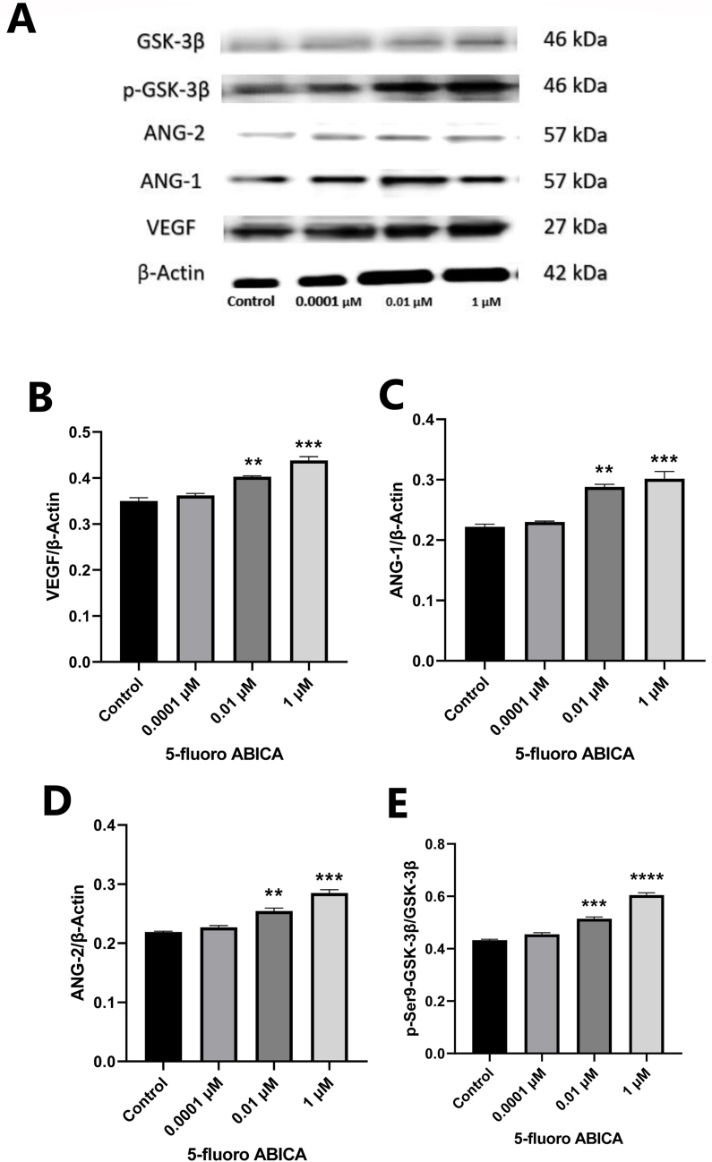
Methods: HBMECs were treated with various concentrations of 5-fluoro ABICA (1 μM, 0.1 μM, 0.01 μM, 0.001 μM, and 0.0001 μM). A comprehensive analysis was conducted, including MTT assays indicating cell viability, wound healing assays indicating migration ability, and tube formation assays indicating the angiogenesis potential of endothelial cells. Additionally, mRNA expression and protein levels of specific pro-angiogenic factors were measured, and the phosphorylation levels of glycogen synthase kinase-3β were detected in treated HBMECs through ELISA, real-time PCR, and western blotting.

Results: Treatment with 5-fluoro ABICA effectively stimulated proliferation, migration, and tube formation in HBMECs in a dose-dependent manner; markedly increased the expression of pro-angiogenic factors; and upregulated levels of phosphorylated-GSK-3β.

Conclusion: Our findings demonstrate that 5-fluoro ABICA stimulates angiogenesis in endothelial cells, thus potentially offering therapeutic options for diseases associated with angiogenesis. However, further research is needed to fully understand the molecular mechanism of 5-fluoro ABICA in angiogenesis, including ethical considerations regarding its use in medical research.

أهداف البحث: ارتبطت الكانابينويدات الاصطناعية، وهي فئة من المركبات ذات التأثير النفساني التي تحاكي تأثيرات الكانابينويدات الطبيعية، بمخاوف الإدمان والذهان. ومع ذلك، تشير الأبحاث الحديثة إلى تطبيقات دوائية محتملة، خاصة في تكوين الأوعية الدموية في الدماغ، وهي عملية فسيولوجية أساسية للنمو والإصلاح وصيانة الأنسجة من خلال تكوين أوعية دموية جديدة من الأوعية الدموية الموجودة. تستكشف هذه الدراسة القدرة المختبرية للكانابينويد الاصطناعي 5-فلورو أبيكا، لتعزيز عملية تكوين الدم الجديدة في الخلايا البطانية للأوعية الدموية الدقيقة في الدماغ البشري.

طرق البحث: تم إعطاء 5-فلورو أبيكا إلى الخلايا البطانية للأوعية الدموية الدقيقة في الدماغ البشري بتركيزات مختلفة (1، 0.1، 0.01، 0.001، 0.0001 مايكرو مولر). تم إجراء تحليل شامل، بما في ذلك فحص ام تي تي لبقاء الخلية، وفحص التئام الجروح لقدرة الهجرة، وفحص تكوين الأنبوب لتقييم إمكانية تكوين الأوعية الدموية للخلايا البطانية. بالإضافة إلى ذلك، تضمن التحليل قياس تعبير الرنا المرسال، ومستويات البروتين لعوامل معينة مؤيدة لتولد الأوعية، واكتشاف مستويات الفسفرة من سينثيز الجليكوجين كيناز-3بيتا في الخلايا البطانية للأوعية الدموية الدقيقة في الدماغ البشري المعالجة باستخدام تحليل الإليزا، و تحليل تفاعل البوليميراز المتسلسل في الوقت الحقيقي، وتقنيات الويسترن بلوت.

النتائج: 5-فلورو أبيكا يحفز بشكل فعال انتشار وهجرة وتكوين أنبوب الخلايا البطانية للأوعية الدموية الدقيقة في الدماغ البشري بطريقة تعتمد على الجرعة. كما أنه زاد بشكل ملحوظ من مستويات التعبير عن العوامل المؤيدة للتولد الوعائي، إلى جانب تنظيم مستويات الفسفرة من سينثيز الجليكوجين كيناز-3بيتا.

الاستنتاجات: بشكل عام، توفر النتائج التي توصلنا إليها أدلة دامغة على أن 5-فلورو أبيكا له تأثيرات تحفيزية على عملية تكوين الأوعية الدموية في الخلايا البطانية، مما يوفر خيارات علاجية في علاج الأمراض المرتبطة بتكوين الأوعية الدموية. ومع ذلك، هناك حاجة إلى مزيد من البحث لفهم الآلية الجزيئية لـ 5-فلورو أبيكا بشكل كامل في تكوين الأوعية، بما في ذلك الاعتبارات الأخلاقية لاستخدامه في البحوث الطبية.

Keywords: 5-Fluoro ABICA; Angiogenesis; Human brain endothelial cells; Synthetic cannabinoids.


Figures

Figure 1
Figure 1
Structure of 5-fluoro ABICA.
Figure 2
Figure 2
Effects of 5-fluoro ABICA on cell metabolic activity in HBMECs. For assessment of cell viability, HBMECs (5 × 103) were initially seeded in a 96-well plate and incubated 24 h. Subsequently, the cells were exposed to five concentrations of 5-fluoro ABICA (ranging from 0.0001 μM to 1 μM) for 24 h. After treatment, the culture medium containing 5-fluoro ABICA was removed and replaced with MTT (5 mg/ml), and the cells were incubated 4 h at 37 °C under 5 % CO2. The cells were then treated with DMSO and agitated for 10 min, and the resulting absorbance was quantified at 570 nm with an ELISA reader. The data are presented as SD ± mean (n = 3). Significantly greater cell viability was observed with concentrations ranging from 0.01 μM to 1 μM than the control. (∗∗) indicates p < 0.01 (∗) indicates p < 0.05.
Figure 3
Figure 3
Treatment with 5-fluoro ABICA enhances t HBMEC migration rate. The effects of 5-fluoro ABICA on cell migration in HBMECs were evaluated with the following protocol. HBMECs were cultured in a 12-well plate for 24 h. After the desired confluence was reached, a cell monolayer was gently scratched with a 1000 μl pipette tip to induce a wound. (A) Microscopic images were captured to document migration levels at the starting point (time zero) and 24 h after treatment with various concentrations of 5-fluoro ABICA at 0.0001 μM–0.1 μM. (B) Quantitative analysis of the migration data shown in A, revealing that doses of 5-fluoro ABICA ranging from 1 μM, and 0.1 μM elicited greater rates of migration than the control. The data were measured and assessed in three separate experiments, each conducted in duplicate, and are depicted as SD ± mean (n = 3). (∗∗∗∗) indicates p < 0.0001.
Figure 4
Figure 4
Treatment with 5-fluoro ABICA enhances HBMEC functionality. Angiogenic potential was assessed with tube formation assays. BME-coated plates were initially seeded with 2 × 104 HBMECs maintained in serum-free medium and treated with varying concentrations of 5-fluoro ABICA (0.001 μM, 0.1 μM, and 1 μM) or control for 24 h. (A) After 24 h of treatment, microscopic visual images of tubular structures produced by HBMECs were captured. (B) Quantity of tube-like structures, (C) number of loops, (D) number of branch points, and (E) total tube length. The data are presented as SD ± mean (n = 3). (∗∗∗∗) indicates a significance level of p < 0.0001.
Figure 5
Figure 5
Treatment with 5-fluoro ABICA enhances angiogenesis-associated gene expression in HBMECs: RT-PCR analysis. (A) RT-PCR quantification of VEGF mRNA expression in HBMECs treated with different concentrations of 5-fluoro ABICA. (B) Effects of 5-fluoro ABICA on the mRNA expression levels of ANG-1 and (C) ANG-2 in comparison to the control. Gene expression levels were calculated for each sample and are presented in the graph as units. The data are presented as SD ± mean (n = 3). (∗∗∗∗) indicates p < 0.0001, (∗∗∗) indicates p < 0.001.
Figure 6
Figure 6
ELISA quantification of gene expression in HBMECs treated with different concentrations of 5-fluoro ABICA. ELISA was conducted on HBMECs treated with 5-fluoro ABICA at different concentrations, to quantify the levels of secreted proangiogenic factors. (A–C) Significant increase in the release of VEGF, ANG-1, and ANG-2 concentrations in a dose-dependent manner after treatment with 5-fluoro ABICA compared with the control. Gene expression was calculated for each sample and is presented in the graph as units. The data are presented as SD ± mean (n = 3). (∗∗∗∗) indicates p < 0.0001, (∗∗∗) indicates p < 0.001, (∗) indicates p < 0.05.
Figure 7
Figure 7
Effects of 5-fluoro ABICA treatment on the protein expression of VEGF, ANG-1, ANG-2, GSK-3β, and p-GSK-3β in HBMECs, assessed with western blotting. (A) Protein extracts were obtained from HBMECs treated with 5-fluoro ABICA at varying concentrations (0.1 μM, 0.01 μM, or 0.0001 μM). These extracts were then used to quantify the protein levels of VEGF, ANG-1, ANG-2, GSK-3β, and p-GSK-3β within the cells; β-actin served as the reference protein. Proteins were isolated with radioimmunoprecipitation assay lysis buffer containing phosphatase-protease inhibitors, and 20 μg of each protein sample was separated with sodium dodecyl sulfate-polyacrylamide gel electrophoresis. The separated proteins were transferred to a polyvinylidene fluoride membrane, which was blocked with 2 % bovine serum albumin and probed with primary antibodies during an overnight incubation. HRP-conjugated secondary antibodies were used to detect chemiluminescence signals. (B) The VEGF expression levels, normalized to those of β-actin, were significantly higher in cells treated with 5-fluoro ABICA than the control. (C) Significantly elevated ANG-1 in treated HBMECs. (D) Increase in ANG-2 expression after treatment with 5-fluoro ABICA compared with the control. (E) Substantial increase in the expression of p-GSK-3β with respect to GSK-3β. The data are presented as SD ± mean (n = 3). (∗∗∗∗) indicates p < 0.0001, (∗∗∗) indicates p < 0.001, (∗∗) indicates p < 0.01.

Similar articles

Cited by

References

    1. Schatteman G.C., Awad O. Hemangioblasts, angioblasts, and adult endothelial cell progenitors. Anat Rec Part A, Discov Mol Cell Evol Biol. 2004;276(1):13–21. doi: 10.1002/ar.a.10131. - DOI - PubMed
    1. Zhao N., Kulkarni S., Zhang S., Linville RM, Chung TD, Guo Z, et al. Modeling angiogenesis in the human brain in a tissue-engineered post-capillary venule. Angiogenesis. 2023 May;26(2):203–216. - PMC - PubMed
    1. Dudley A.C., Griffioen A.W. Pathological angiogenesis: mechanisms and therapeutic strategies. Angiogenesis. 2023;26(3):313–347. doi: 10.1007/s10456-023-09876-7. - DOI - PMC - PubMed
    1. Liu Z.-L., Chen H.-H., Zheng L.-L., Sun L.-P., Shi L. Angiogenic signaling pathways and anti-angiogenic therapy for cancer. Signal Transduct Target Ther. 2023;8(1):198. doi: 10.1038/s41392-023-01460-1. - DOI - PMC - PubMed
    1. Ghalehbandi S., Yuzugulen J., Pranjol M.Z.I., Pourgholami M.H. The role of VEGF in cancer-induced angiogenesis and research progress of drugs targeting VEGF. Eur J Pharmacol. 2023;949 doi: 10.1016/j.ejphar.2023.175586. - DOI - PubMed

LinkOut - more resources