

Stereotactic radiotherapy for uveal melanoma: A case report
- PMID: 38357672
- PMCID: PMC10865074
- DOI: 10.3892/mco.2024.2721
Stereotactic radiotherapy for uveal melanoma: A case report
Abstract
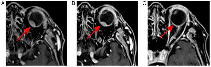
Uveal melanoma (UM) is the most common primary intraocular malignancy worldwide. Surgical intervention and radiation therapy (RT) are the primary treatment options. Given the complexity and cosmetic discomfort associated with eye enucleation, this method is less frequently used. As a result, RT, including photon therapy, proton therapy and brachytherapy, has become the treatment of choice. Traditionally, plaque brachytherapy has been the most commonly used in clinical practice. However, the question of which type of radiation therapy is the most effective, safe, commonly available and cost-effective remains open. The present study provided a follow-up analysis of a patient with UM who was treated using the image-guided volumetric modulated arc therapy (IG-VMAT) technique. A complete response without complications and symptom relief were noted one and a half years after treatment. The present findings suggest that photon external beam radiotherapy using the IG-VMAT technique may offer a viable and safe alternative for the management of UM. This approach potentially sidesteps the complex and morbid aspects of surgical intervention and plaque brachytherapy. Owing to the limited sample size, a more robust understanding of the efficacy and safety of this treatment will require the analysis of additional cases. Further research with a larger cohort is essential to validate these preliminary observations.
Keywords: radiotherapy; stereotactic body radiation therapy; uveal melanoma.
Copyright © 2023, Spandidos Publications.
Conflict of interest statement
The authors have no competing interests to disclose.
Figures
Similar articles
-
Uveal melanomas. Conservation treatment.Hematol Oncol Clin North Am. 2001 Apr;15(2):389-402. doi: 10.1016/s0889-8588(05)70219-7. Hematol Oncol Clin North Am. 2001. PMID: 11370500 Review.
-
Linear accelerator-based stereotactic fractionated photon radiotherapy as an eye-conserving treatment for uveal melanoma.Radiat Oncol. 2018 Aug 2;13(1):140. doi: 10.1186/s13014-018-1088-9. Radiat Oncol. 2018. PMID: 30071857 Free PMC article.
-
Clinical management of uveal melanoma: a comprehensive review with a treatment algorithm.Radiat Oncol J. 2020 Sep;38(3):162-169. doi: 10.3857/roj.2020.00318. Epub 2020 Jul 9. Radiat Oncol J. 2020. PMID: 33012143 Free PMC article.
-
Photon-based High-dose Single-fraction Radiosurgery, an Effective Treatment Modality for Large and Posterior Uveal Melanoma.Anticancer Res. 2022 Apr;42(4):1965-1972. doi: 10.21873/anticanres.15674. Anticancer Res. 2022. PMID: 35347016
-
Deterioration of Visual Acuity after Brachytherapy and Proton Therapy of Uveal Melanoma, and Methods of Counteracting This Complication Based on Recent Publications.Medicina (Kaunas). 2023 Jun 12;59(6):1131. doi: 10.3390/medicina59061131. Medicina (Kaunas). 2023. PMID: 37374335 Free PMC article. Review.
References
Publication types
LinkOut - more resources
Full Text Sources
Research Materials