Review
. 2024 Apr;22(2):290-298.
doi: 10.1007/s11914-024-00859-1. Epub 2024 Feb 15.

Interfacial Tissue Regeneration with Bone

Affiliations
Review

Interfacial Tissue Regeneration with Bone

Stephanie S Steltzer et al. Curr Osteoporos Rep. 2024 Apr.

Abstract

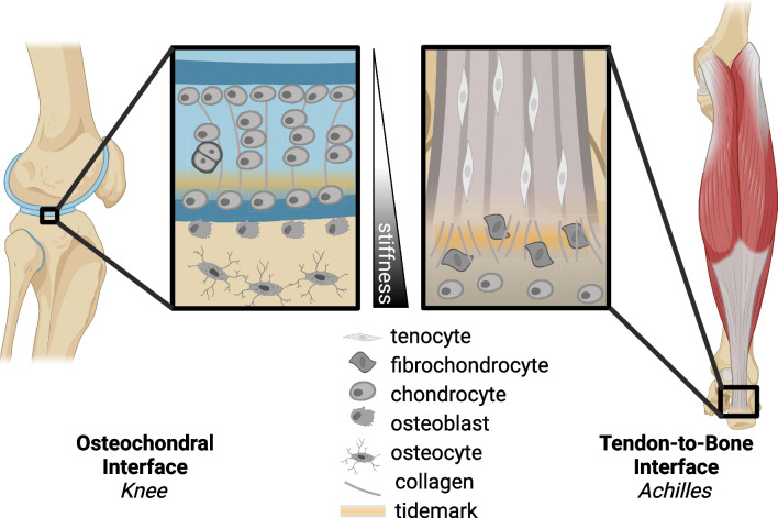
Purpose of review: Interfacial tissue exists throughout the body at cartilage-to-bone (osteochondral interface) and tendon-to-bone (enthesis) interfaces. Healing of interfacial tissues is a current challenge in regenerative approaches because the interface plays a critical role in stabilizing and distributing the mechanical stress between soft tissues (e.g., cartilage and tendon) and bone. The purpose of this review is to identify new directions in the field of interfacial tissue development and physiology that can guide future regenerative strategies for improving post-injury healing.

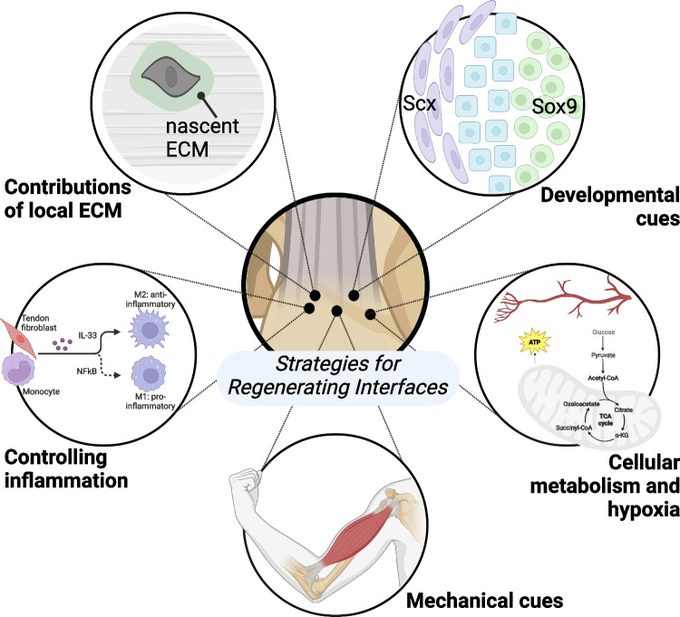
Recent findings: Cues from interfacial tissue development may guide regeneration including biological cues such as cell phenotype and growth factor signaling; structural cues such as extracellular matrix (ECM) deposition, ECM, and cell alignment; and mechanical cues such as compression, tension, shear, and the stiffness of the cellular microenvironment. In this review, we explore new discoveries in the field of interfacial biology related to ECM remodeling, cellular metabolism, and fate. Based on emergent findings across multiple disciplines, we lay out a framework for future innovations in the design of engineered strategies for interface regeneration. Many of the key mechanisms essential for interfacial tissue development and adaptation have high potential for improving outcomes in the clinic.

Keywords: Cellular microenvironment; Enthesis; Extracellular matrix; Mechanical loading; Osteochondral interface.


Conflict of interest statement

The authors have no competing interests of financial or personal nature. Authors’ contributions: SSS and MLK drafted the manuscript. SSS, ACA, and MLK revised the submitted manuscript. All authors approved the final version

Figures

Fig. 1
Fig. 1
Schematics of the osteochondral interface (e.g., within the knee) and the tendon-to-bone interface (e.g., Achilles enthesis), which highlight the stiffness gradients (from bone to tendon), tidemark between mineralized and unmineralized fibrocartilage, variations in collagen alignment and fibril size, and cell type distribution inclusive of tendon fibroblast, fibrochondrocyte, and chondrocytes/osteoblasts
Fig. 2
Fig. 2
Strategies for regenerating interfacial tissues like the osteochondral interface and tendon-bone enthesis include: leveraging developmental cues to promote resident and progenitor cell remodeling of the interfacial tissues (e.g., Sox9 + /Scx + bi-fated cells, shown in blue, residing between Scx + cells in purple and Sox9 + cells in green); identifying factors that influence formation of nascent ECM in native tissues during remodeling and repair; promoting a regenerative, rather than destructive, inflammatory response; controlling the mechanical environment by increasing or decreasing applied loads (e.g., from skeletal muscle); and understanding and controlling interfacial cell metabolism and physiological response and sensitivity to their environment, such as hypoxia

References

    1. Vos T, Lim SS, Abbafati C, et al. Global burden of 369 diseases and injuries in 204 countries and territories, 1990–2019: A systematic analysis for the Global Burden of Disease Study 2019. The Lancet. 2020;396:1204–1222. doi: 10.1016/S0140-6736(20)30925-9. - DOI - PMC - PubMed
    1. Peniche Silva C, Müller S, Quirk N, De la Vega R, Coenen M, Evans C, Balmayor E, van Griensven M. Enthesis: Not the same in each localisation – A molecular, histological and biomechanical study. eCM. 2022;44:43–55. doi: 10.22203/eCM.v044a03. - DOI - PubMed
    1. Brown ME, Puetzer JL. Driving native-like zonal enthesis formation in engineered ligaments using mechanical boundary conditions and β-tricalcium phosphate. Acta Biomater. 2022;140:700–716. doi: 10.1016/j.actbio.2021.12.020. - DOI - PubMed
    1. Puetzer JL, Ma T, Sallent I, Gelmi A, Stevens MM. Driving hierarchical collagen fiber formation for functional tendon, ligament, and meniscus replacement. Biomaterials. 2021;269:120527. doi: 10.1016/j.biomaterials.2020.120527. - DOI - PMC - PubMed
    1. Ganji E, Leek C, Duncan W, Patra D, Ornitz DM, Killian ML. Targeted deletion of Fgf9 in tendon disrupts mineralization of the developing enthesis. FASEB J. 2023;37:e22777. doi: 10.1096/fj.202201614R. - DOI - PMC - PubMed

Publication types