. 2024 Feb 15;20(2):e1012022.
doi: 10.1371/journal.ppat.1012022. eCollection 2024 Feb.

Enterovirus A71 does not meet the uncoating receptor SCARB2 at the cell surface

Affiliations

Enterovirus A71 does not meet the uncoating receptor SCARB2 at the cell surface

Yorihiro Nishimura et al. PLoS Pathog. .

Abstract

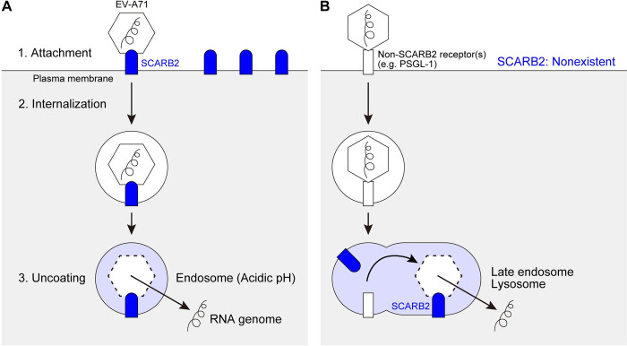
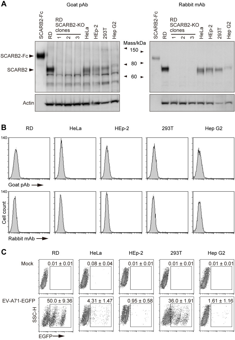
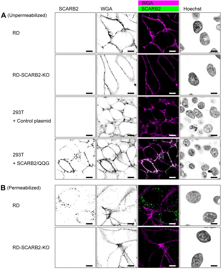
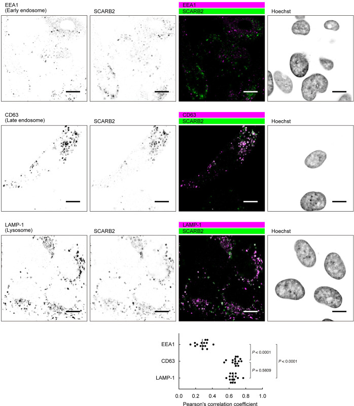
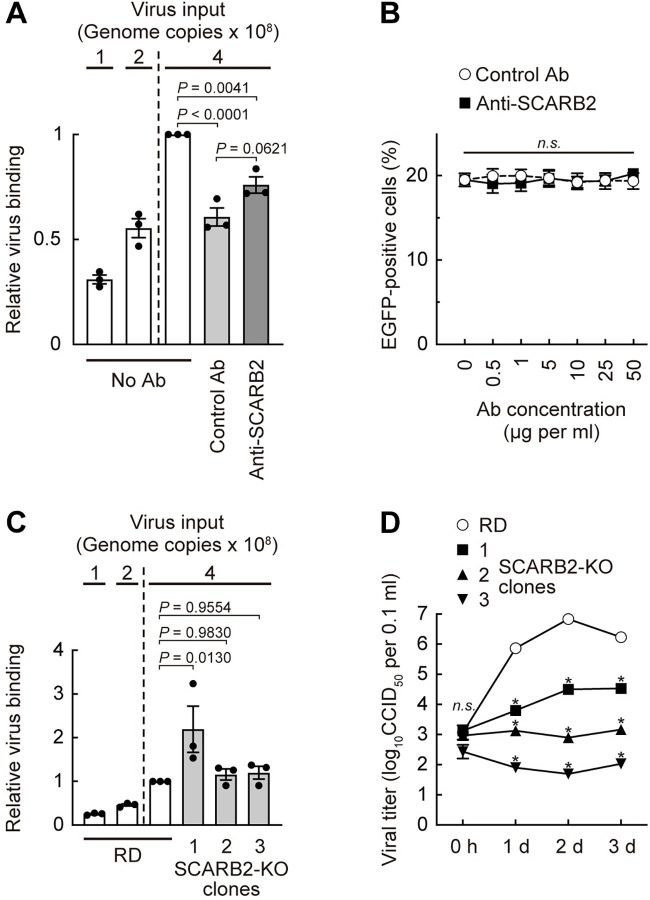
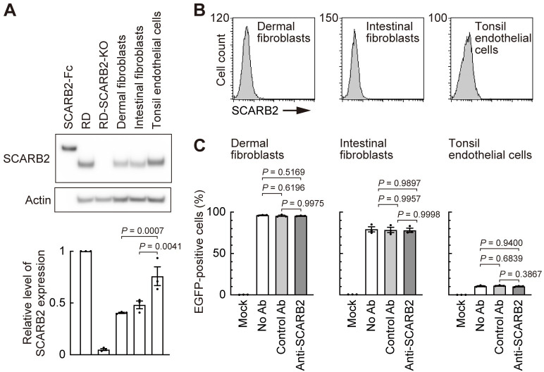
Enterovirus A71 (EV-A71) infection involves a variety of receptors. Among them, two transmembrane protein receptors have been investigated in detail and shown to be critical for infection: P-selectin glycoprotein ligand-1 (PSGL-1) in lymphocytes (Jurkat cells), and scavenger receptor class B member 2 (SCARB2) in rhabdomyosarcoma (RD) cells. PSGL-1 and SCARB2 have been reported to be expressed on the surface of Jurkat and RD cells, respectively. In the work reported here, we investigated the roles of PSGL-1 and SCARB2 in the process of EV-A71 entry. We first examined the expression of SCARB2 in Jurkat cells, and detected it within the cytoplasm, but not on the cell surface. Further, using PSGL-1 and SCARB2 knockout cells, we found that although both PSGL-1 and SCARB2 are essential for virus infection of Jurkat cells, virus attachment to these cells requires only PSGL-1. These results led us to evaluate the cell surface expression and the roles of SCARB2 in other EV-A71-susceptible cell lines. Surprisingly, in contrast to the results of previous studies, we found that SCARB2 is absent from the surface of RD cells and other susceptible cell lines we examined, and that although SCARB2 is essential for infection of these cells, it is dispensable for virus attachment. These results indicate that a receptor other than SCARB2 is responsible for virus attachment to the cell and probably for internalization of virions, not only in Jurkat cells but also in RD cells and other EV-A71-susceptible cells. SCARB2 is highly concentrated in lysosomes and late endosomes, where it is likely to trigger acid-dependent uncoating of virions, the critical final step of the entry process. Our results suggest that the essential interactions between EV-A71 and SCARB2 occur, not at the cell surface, but within the cell.


Conflict of interest statement

The authors have declared that no competing interests exist.

Figures

Fig 1
Fig 1. Two models of the EV-A71 entry.
(A) The previous model suggested by Yamayoshi et al. [9,23]. SCARB2 is highly expressed on the cell surface. Surface SCARB2 binds to EV-A71 at the south rim of the canon [24] and initiates viral entry in endosomal vesicles. SCARB2 induces uncoating when the vesicle lumen becomes acidic. (B) Our new model. SCARB2 is absent from the cell surface. A non-SCARB2 receptor mediates EV-A71 attachment, most likely interacting with the viral five-fold vertex [20,21]. Virus is internalized and transported through the endosomal system, meeting SCARB2 within late endosomes or lysosomes, where uncoating occurs.
Fig 2
Fig 2. Jurkat cells do not express SCARB2 on the cell surface.
(A) Flow cytometric analysis of cell-surface SCARB2 using goat pAb and rabbit mAb (clone 12H5L1). To confirm both Abs are applicable to flow cytometry, 293T cells overexpressing SCARB2 or mutant SCARB2 with the three amino acid substitutions to enhance cell-surface expression (SCARB2/QQG) and Jurkat cells were stained in parallel. The solid line and the shaded area represent staining with anti-SCARB2 Ab and control IgG, respectively, followed by Alexa Fluor 488-tagged secondary Ab. The figure is representative of three independent experiments. (B) A large amount of overexpressed SCARB2 existed inside the 293T cells. Western blotting analysis with anti-SCARB2 mAb (clone 12H5L1). Recombinant SCARB2-Fc (5 ng) was loaded as a positive control. RD and RD-SCARB2-KO (clone No.3) cells were loaded as positive and negative controls, respectively. The figure is representative of three independent experiments.
Fig 3
Fig 3. Anti-SCARB2 pAb does not inhibit EV-A71 binding to and replication in Jurkat cells.
Jurkat cells were pre-treated with anti-PSGL-1 mAb (10 μg ml-1), anti-SCARB2 pAb (50 μg ml-1), or control Abs on ice for 1 h, followed by infection with EV-A71 at 1 CCID50 per cell on ice for 1 h. Cells were then washed (0 h), cultured without Abs, and harvested at 5 days (5 d) post-infection. Results are indicated as the mean and s.e. for triplicate samples.
Fig 4
Fig 4. SCARB2 is not involved in EV-A71 binding to Jurkat cells, but necessary for viral replication.
PSGL-1 or SCARB2 were knocked out by CRISPR/Cas9 in Jurkat cells, and two each of clones were established. (A) Cells were infected with EV-A71 at 1 CCID50 per cell on ice for 1 h, then washed (0 h), cultured, and harvested at 3 days (3 d) post-infection. (B) Cells were infected with EV-A71-EGFP at 10 CCID50 per cell and cultured for 18 h. Infected cells were identified by flow cytometry to detect EGFP expression. Results are indicated as the mean and s.e. for triplicate samples.
Fig 5
Fig 5. SCARB2 is absent from the cell surface, irrespective of EV-A71 susceptibility.
RD, HeLa, HEp-2, 293T, and Hep G2 were obtained from the ATCC specifically for this study and used after limited passage. (A) Western blotting analysis by anti-SCARB2 pAb (left) and mAb (right, clone 12H5L1). Recombinant SCARB2-Fc (1 ng for pAb, 5 ng for mAb) was loaded as a positive control. RD-SCARB2-KO clones were loaded as negative controls. The figure is representative of three independent experiments. (B) Flow cytometric analysis by anti-SCARB2 pAb (top panels) and mAb (bottom panels), followed by Alexa Fluor 488-tagged secondary Ab. The solid line and the shaded area represent staining with anti-SCARB2 Ab and control Ab, respectively. Note that the solid line and the border of the shaded area are almost completely overlapped, indicating the absence of SCARB2 on the cell surface. Representative results with the following passage numbers after receiving from the ATCC; for pAb: RD, 3; HeLa, 3; HEp-2, 3; 293T, 3; Hep G2, 5; for mAb: RD, 13; HeLa, 5; HEp-2, 4; 293T, 4; Hep G2, 8. As a positive control of SCARB2 staining, cells expressing surface SCARB2 were always stained and analyzed in parallel. The figure is representative of three independent experiments. (C) EGFP expression in cells infected with EV-A71-EGFP. Cells were infected with EV-A71-EGFP at 10 CCID50 per cell and cultured for 18 h. Then EGFP expression was measured by flow cytometry. The EGFP-negative cells are not infected. The majority of EGFP-dim cells were infected early in the incubation period, and are dying and losing EGFP expression; some may have just been infected and are starting to express EGFP. The EGFP-bright cells were infected late in the incubation period are actively producing EGFP. The number indicates the percentage of EGFP-positive cells (mean and s.e. for three independent experiments).
Fig 6
Fig 6. SCARB2 is absent from the cell surface and localized in the cytoplasm of RD cells.
(A) Cells were stained with anti-SCARB2 mAb (clone 22H6L14) followed by Alexa Fluor 488-tagged secondary Ab and WGA conjugated with Alexa Fluor 633 without permeabilization. WGA was used to visualize the plasma membrane. Then the cells were fixed and observed under a confocal microscope. RD-SCARB2-KO cells (clone No.3) and 293T cells transfected with a control plasmid were used as negative controls. 293T cells expressing SCARB2/QQG on the cell surface were used as positive control. The figure is representative of three independent experiments. (B) RD and RD-SCARB2-KO (clone No.3) cells were stained with WGA, fixed, permeabilized, and stained with anti-SCARB2 mAb (clone 22H6L14) followed by Alexa Fluor 488-tagged secondary Ab. The figure is representative of three independent experiments. Scale bars, 10 μm.
Fig 7
Fig 7. SCARB2 co-localizes with the markers of late endosomes and lysosomes in RD cells.
RD cells were fixed, permeabilized, and stained with anti-SCARB2 mAb (clone 22H6L14) and mAb against either EEA1 (early endosome), CD63 (late endosome), or LAMP-1 (lysosome), followed by Alexa Fluor-tagged secondary Ab. Then the cells were observed under a confocal microscope. The figure is representative of three independent experiments. In each experiment, five pairs of images were analyzed. The graph shows colocalization between the markers and SCARB2 expressed as Pearson’s correlation coefficient. The vertical line indicates the mean value. Scale bars, 10 μm.
Fig 8
Fig 8. SCARB2 is not involved in EV-A71 binding to RD cells, but necessary for viral replication.
(A) EV-A71 binding to RD cells in the presence of anti-SCARB2 pAb. RD cells pretreated with the anti-SCARB2 pAb (50 μg ml-1) were reacted with EV-A71 (4 × 10⁸ genome copies) on ice for 30 min. Then the cells were washed, and cellular and viral nucleotides were extracted. EV-A71 bound to the cell were analyzed by real-time RT-PCR by ΔΔCt method using ATP5F1 mRNA as an endogenous control. As a technical control of detection of reduced copy number, quarter (1 × 10⁸ genome copies) and half (2 × 10⁸ genome copies) amount of EV-A71 was tested in parallel. The relative virus binding of RD cells reacted with 4 × 10⁸ genome copies of EV-A71 without Ab was expressed as 1. (B) EGFP expression in cells infected with EV-A71-EGFP in the presence of anti-SCARB2 pAb at 18 h post-infection. (C) EV-A71 binding to RD and RD-SCARB2-KO clones. EV-A71 bound to the cell were analyzed as in (A). The relative virus binding of RD cells reacted with 4 × 10⁸ genome copies of EV-A71 was expressed as 1. (D) Replication kinetics of EV-A71 in RD and RD-SCARB2-KO clones. Statistical significance was measured for each time point. Results are indicated as the mean and s.e. for three independent experiments (A, B, C) or triplicate analyses (D). Asterisks indicate P < 0.0001.
Fig 9
Fig 9. EV-A71 enters human primary cells in a SCARB2-independent manner.
Human dermal fibroblasts (neonatal), intestinal fibroblasts, and tonsil endothelial cells were examined as cells presumed to be involved in the in vivo pathogenesis of EV-A71 infection. (A) Western blotting analysis with anti-SCARB2 mAb (clone 12H5L1). Recombinant SCARB2-Fc (5 ng) was loaded as a positive control. RD and RD-SCARB2-KO (clone No.3) cells were loaded as positive and negative controls, respectively. The figure is representative of three independent experiments. The graph displays the relative level of SCARB2 expression normalized by actin. The relative amount of SCARB2 in RD cells was expressed as 1. (B) Flow cytometric analysis by anti-SCARB2 pAb, followed by Alexa Fluor 488-tagged secondary Ab. The solid line and the shaded area represent staining with anti-SCARB2 pAb and control Ab, respectively. Note that the solid line and the border of the shaded area are almost completely overlapped, indicating the absence of SCARB2 on the cell surface. Representative results of cells passaged twice after receiving from the company. As a positive control of SCARB2 staining, cells expressing surface SCARB2 were always stained and analyzed in parallel. The figure is representative of three independent experiments. (C) EGFP expression in cells infected with EV-A71-EGFP in the presence of anti-SCARB2 pAb at 18 h post-infection. Cells with the following passage numbers after receiving from the company were used; dermal fibroblasts, 4; intestinal fibroblasts, 4; tonsil endothelial cells, 2. Results are indicated as the mean and s.e. for three independent experiments (A, C).

Similar articles

Cited by

References

    1. Kinobe R, Wiyatno A, Artika IM, Safari D. Insight into the Enterovirus A71: A review. Rev Med Virol. 2022:e2361. Epub 20220505. doi: 10.1002/rmv.2361 . - DOI - PubMed
    1. Chang LY, Lin HY, Gau SS, Lu CY, Hsia SH, Huang YC, et al.. Enterovirus A71 neurologic complications and long-term sequelae. J Biomed Sci. 2019;26(1):57. Epub 20190808. doi: 10.1186/s12929-019-0552-7 ; PubMed Central PMCID:PMC6688366. - DOI - PMC - PubMed
    1. Tee HK, Zainol MI, Sam IC, Chan YF. Recent advances in the understanding of enterovirus A71 infection: a focus on neuropathogenesis. Expert Rev Anti Infect Ther. 2021;19(6):733–47. Epub 20210217. doi: 10.1080/14787210.2021.1851194 . - DOI - PubMed
    1. Huang SW, Cheng D, Wang JR. Enterovirus A71: virulence, antigenicity, and genetic evolution over the years. J Biomed Sci. 2019;26(1):81. Epub 20191021. doi: 10.1186/s12929-019-0574-1 ; PubMed Central PMCID:PMC6802317. - DOI - PMC - PubMed
    1. Puenpa J, Wanlapakorn N, Vongpunsawad S, Poovorawan Y. The history of enterovirus A71 outbreaks and molecular epidemiology in the Asia-Pacific region. J Biomed Sci. 2019;26(1):75. Epub 20191018. doi: 10.1186/s12929-019-0573-2 ; PubMed Central PMCID:PMC6798416. - DOI - PMC - PubMed

MeSH terms

Substances