. 2024 Apr:306:122505.
doi: 10.1016/j.biomaterials.2024.122505. Epub 2024 Feb 7.

Biomimetic nanodrug targets inflammation and suppresses YAP/TAZ to ameliorate atherosclerosis

Affiliations

Biomimetic nanodrug targets inflammation and suppresses YAP/TAZ to ameliorate atherosclerosis

Hui-Chun Huang et al. Biomaterials. 2024 Apr.

Abstract

Atherosclerosis, a chronic inflammatory disease, is the primary cause of myocardial infarction and ischemic stroke. Recent studies have demonstrated that dysregulation of yes-associated protein (YAP) and transcriptional coactivator with PDZ-binding domain (TAZ) contributes to plaque development, making YAP/TAZ potential therapeutic targets. However, systemic modulation of YAP/TAZ expression or activities risks serious off-target effects, limiting clinical applicability. To address the challenge, this study develops monocyte membrane-coated nanoparticles (MoNP) as a targeted delivery system for activated and inflamed endothelium lining the plaque surface. The MoNP system is used to deliver verteporfin (VP), aimed at inhibiting YAP/TAZ specifically within arterial regions prone to atherosclerosis. The results reveal that MoNP significantly enhance payload delivery to inflamed endothelial cells (EC) while avoiding phagocytic cells. When administered in mice, MoNP predominantly accumulate in intima of the atheroprone artery. MoNP-mediated delivery of VP substantially reduces YAP/TAZ expression, thereby suppressing inflammatory gene expression and macrophage infiltration in cultured EC and mouse arteries exposed to atherogenic stimuli. Importantly, this targeted VP nanodrug effectively decreases plaque development in mice without causing noticeable histopathological changes in major organs. Collectively, these findings demonstrate a lesion-targeted and pathway-specific biomimetic nanodrug, potentially leading to safer and more effective treatments for atherosclerosis.

Keywords: Atherosclerosis; Inflammation; Nanomedicine; Targeted delivery; YAP/TAZ.


Conflict of interest statement

Declaration of competing interest The authors declare the following financial interests/personal relationships which may be considered as potential competing interests: Kuei-Chun Wang reports financial support was provided by National Institutes of Health and American Heart Association.

Figures
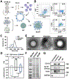
Figure 1.
Figure 1.. Formulation and characterization of MoNP.
(A) A schematic illustrating the preparation of MoNP loaded with a therapeutic payload. (B) Flow cytometric analysis of Mo isolated from differentiated mouse BMC. (C-D) DLS results of MoNP, NP, and Mo vesicles showing (C) hydrodynamic size, (D) PDI, and surface charge. Graphic data in (D): n = 3, *p < 0.05 vs. NP and #p < 0.05 vs. Mo vesicles. (E) Representative TEM images of MoNP, NP, and Mo vesicles. Scale bar = 100 nm. (F) SDS-PAGE analysis of membrane protein profiles of MoNP and Mo vesicles. (G) Western blot analysis of the membrane proteins of MoNP and Mo vesicles.
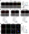
Figure 2.
Figure 2.. MoNP enhanced endothelial uptake and lysosomal escape.
(A-D) Representative fluorescent images showing (A) the cellular uptake of MoNP-DiD or NP-DiD by EC, TNFα-pretreated EC, TNFα-/anti-VCAM1-pretreated EC, (B) the cellular uptake of MoNP-DiD by EC under low or high SS, and (C-D) the cellular uptake of MoNP-DiD or NP-DiD by (C) Mo and (D) macrophages. Scale bar = 100 μm. The intracellular DiD signal was quantified. Graphic data in (A), (C), and (D): n = 3, *p < 0.05 vs. NP-DiD, #p < 0.05 vs. MoNP-DiD, $p < 0.05 vs. MoNP-DiD/TNFα-stimulated EC. Graphic data in (B): n = 3, *p < 0.05 vs. low SS. (E) Representative confocal images of EC incubated with MoNP-DiD or NP-DiD (red), followed by the staining of lysotracker (green) and nuclei (blue). Scale bar = 25 μm. (F) Correlation analysis of MoNP-DiD or NP-DiD with lysosomes. Thirteen cells were randomly selected from fluorescent images acquired in three biological repeats. *p < 0.05 vs. NP-DiD.
Figure 3.
Figure 3.. MoNP enabled active targeting of atheroprone arterial regions.
(A) A schematic showing the experimental design of MoNP-DiD or NP-DiD administration. (B) Representative fluorescent images of the arterial tissues isolated from ApoE−/− mice receiving MoNP-DiD or NP-DiD, with quantification of the fluorescent intensity measured in LCA, RCA, AA, and DA. Red: MoNP-DiD or NP-DiD. (C) Representative fluorescent images of RCA and LCA cross-sections from the mouse receiving MoNP-DiD. Red: MoNP-DiD, green: CD31 and elastic layer autofluorescence, and blue: nuclei. The graph indicates the intensity of DiD signal of the crosslines (yellow). Scale bar = 200 μm. (D) The fluorescent intensity of the major organs isolated from ApoE−/− mice receiving MoNP-DiD or NP-DiD. n = 6 for MoNP-DiD, and n = 4 for NP-DiD, *p < 0.05 vs. the NP-DiD group.
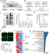
Figure 4.
Figure 4.. MoNP-VP treatment alleviated the inflammatory response in EC.
(A) A schematic showing the experimental design of MoNP-VP treatment in EC. (B) Physicochemical characterization of MoNP-VP. (C) Western blot analysis of the TNFα-induced expression of VCAM1, ICAM1, YAP/TAZ, and CTGF in EC treated with MoNP-VP or MoNP. (D) qRT-PCR analysis of the TNFα-induced expression of VCAM1, ICAM1, and CTGF in EC treated with MoNP-VP or MoNP. For (C-D), the data are normalized to its respective loading controls and the MoNP group. n = 3, *p < 0.05 vs. TNFα/MoNP. (E) Representative images of fluorescently labeled Mo (green) attached to EC monolayers treated with MoNP-VP or MoNP. Scale bar = 500 μm. n = 3, *p < 0.05 vs. TNFα/MoNP. (F) An RNA-Seq heatmap displaying the differences in expression of atheroprone, atheroprotective, and YAP/TAZ-associated genes in TNFα-stimulated EC pretreated with MoNP-VP compared to those with MoNP. The genes are ranked based on their z-scores. (G) KEGG pathway enrichment analysis of DEGs in response to MoNP-VP vs. MoNP in TNFα-stimulated EC. The inflammatory-related pathways and Hippo signaling pathway are highlighted in red. padj. < 0.05.
Figure 5.
Figure 5.. MoNP-VP treatment suppressed the PL-induced inflammatory response in vivo.
(A) A schematic showing the experimental design of MoNP-VP treatment (2 mg/kg) in ApoE−/− mice. The arterial tissues were harvested 7 days after the PL procedure. (B) Western blot analysis of the expressions of YAP/TAZ and CTGF in response to MoNP-VP or MoNP treatment. The band intensity is normalized to its respective loading controls. n = 3, *p < 0.05 vs. MoNP. (C) Immunofluorescence staining of YAP/TAZ, VCAM1, and CD68-positive cells in the arterial wall. Red: YAP/TAZ, VCAM1, or CD68, green: elastic layer autofluorescence, and blue: nuclei. White asterisks indicate the lumen. The intensity of YAP/TAZ and VCAM1 in the intimal layer of LCA and the number of infiltrated CD68-positive cells were quantified. n = 4, *p < 0.05 vs. MoNP.
Figure 6.
Figure 6.. MoNP-VP treatment attenuated plaque development in ApoE−/− mice.
(A) A schematic showing the long-term treatment of MoNP-VP in mouse carotid atherosclerosis. ApoE−/− mice were subjected to the PL procedure, followed by intravenous administration of MoNP-VP, free VP, MoNP, or saline after PL, and every 72 hours afterward, for a total of 6 injections. The lesion in the LCA was assessed on day 28 after PL. (B) Representative images of the arterial tissues from carotid bifurcation to DA of various treatment groups. (C) Representative images of the distal, middle, and proximal segments of the partially ligated LCA cross-sections from various treatment groups stained with Oil Red O and hematoxylin. (D-E) Quantification of (D) the Oil Red O-positive area and (E) the degree of luminal stenosis of the LCA. (F) Measurement of the level of total cholesterol in mouse serum samples. (G) The changes in body weight of the mice throughout the experiment. (H) Histological analysis of major organs isolated from ApoE−/− mice subjected to various treatments. n = 7 each for MoNP-VP, free VP, and MoNP, and n = 5 for saline, *p < 0.05 vs. saline, #p < 0.05 vs. free VP, and $p < 0.05 vs. MoNP.

References

    1. Libby P, Ridker PM, Hansson GK, Progress and challenges in translating the biology of atherosclerosis, Nature 473 (2011) 317–325. 10.1038/nature10146. - DOI - PubMed
    1. Bäck M, Yurdagul A, Tabas I, Öörni K, Kovanen PT, Inflammation and its resolution in atherosclerosis: mediators and therapeutic opportunities, Nat Rev Cardiol 16 (2019) 389–406. 10.1038/s41569-019-0169-2. - DOI - PMC - PubMed
    1. Ridker PM, Everett BM, Thuren T, MacFadyen JG, Chang WH, Ballantyne C, Fonseca F, Nicolau J, Koenig W, Anker SD, Kastelein JJP, Cornel JH, Pais P, Pella D, Genest J, Cifkova R, Lorenzatti A, Forster T, Kobalava Z, Vida-Simiti L, Flather M, Shimokawa H, Ogawa H, Dellborg M, Rossi PRF, Troquay RPT, Libby P, Glynn RJ, CANTOS Trial Group, Antiinflammatory Therapy with Canakinumab for Atherosclerotic Disease, N Engl J Med 377 (2017) 1119–1131. 10.1056/NEJMoa1707914. - DOI - PubMed
    1. Nidorf SM, Fiolet ATL, Mosterd A, Eikelboom JW, Schut A, Opstal TSJ, The SHK, Xu X-F, Ireland MA, Lenderink T, Latchem D, Hoogslag P, Jerzewski A, Nierop P, Whelan A, Hendriks R, Swart H, Schaap J, Kuijper AFM, van Hessen MWJ, Saklani P, Tan I, Thompson AG, Morton A, Judkins C, Bax WA, Dirksen M, Alings M, Hankey GJ, Budgeon CA, Tijssen JGP, Cornel JH, Thompson PL, LoDoCo2 Trial Investigators, Colchicine in Patients with Chronic Coronary Disease, N Engl J Med 383 (2020) 1838–1847. 10.1056/NEJMoa2021372. - DOI - PubMed
    1. Baylis RA, Gomez D, Mallat Z, Pasterkamp G, Owens GK, The CANTOS Trial: One Important Step for Clinical Cardiology but a Giant Leap for Vascular Biology, Arterioscler Thromb Vasc Biol 37 (2017) e174–e177. 10.1161/ATVBAHA.117.310097. - DOI - PMC - PubMed

Publication types

Substances