. 2024 Feb 15;26(1):28.
doi: 10.1186/s13058-024-01785-x.

Identification of CD160-TM as a tumor target on triple negative breast cancers: possible therapeutic applications

Affiliations

Identification of CD160-TM as a tumor target on triple negative breast cancers: possible therapeutic applications

Claire Scheffges et al. Breast Cancer Res. .

Abstract

Background: Despite major therapeutic advances, triple-negative breast cancer (TNBC) still presents a worth prognosis than hormone receptors-positive breast cancers. One major issue relies in the molecular and mutational heterogeneity of TNBC subtypes that is reinforced by the absence of reliable tumor-antigen that could serve as a specific target to further promote efficient tumor cell recognition and depletion. CD160 is a receptor mainly expressed by NK lymphocytes and presenting two isoforms, namely the GPI-anchored form (CD160-GPI) and the transmembrane isoform (CD160-TM). While CD160-GPI is constitutively expressed on resting cells and involved in the generation of NK cells' cytotoxic activity, CD160-TM is neo-synthesized upon activation and promotes the amplification of NK cells' killing ability.

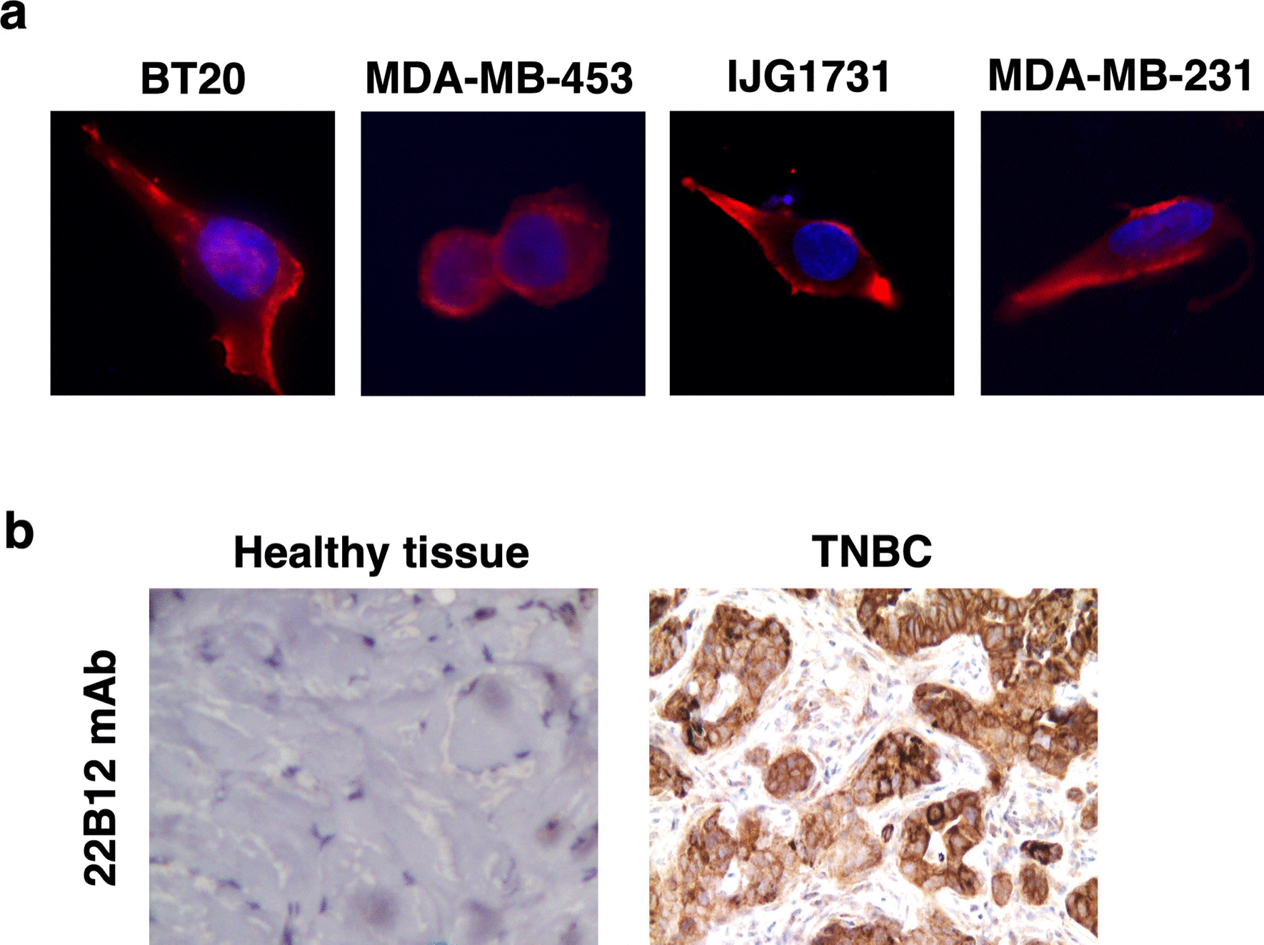
Methods: CD160 expression was assessed by immunohistochemistry (IHC) and flow cytometry on TNBC patient biopsies or cell lines, respectively. Antibody (Ab)-mediated tumor depletion was tested in vitro by performing antibody-dependent cell cytotoxicity (ADCC) and phagocytosis (ADCP) assays, and in vivo on a TNBC mouse model.

Results: Preliminary data obtained by IHC on TNBC patients' tumor biopsies revealed an unconventional expression of CD160 by TNBC tumor cells. By using a specific but conformation-dependent anti-CD160-TM Ab, we established that CD160-TM, but not CD160-GPI, was expressed by TNBC tumor cells. A conformation-independent anti-CD160-TM mAb (22B12; muIgG2a isotype) was generated and selected according to pre-defined specificity and functional criterions. In vitro functional assays demonstrated that ADCC and ADCP could be induced in the presence of 22B12, resulting in TNBC cell line apoptosis. The ability of 22B12 to exert an in vivo anti-tumor activity was also demonstrated on a TNBC murine model.

Conclusions: Our data identify CD160-TM as a tumor marker for TNBC and provide a rational for the use of anti-CD160-TM antibodies as therapeutic tools in this tumor context.

Keywords: Antibody-based therapy; CD160-TM; TNBC; Tumor antigen.


Conflict of interest statement

A.M-C, J.G and A.B are founding members of Alderaan Biotechnology. AF has ownership interest (including patents) in Alderaan Biotechnology. No conflicts of interest were disclosed by the other authors.

Figures

Fig. 1
Fig. 1
Expression of CD160 by TNBC tumor cells. a Healthy (n = 3) or tumor (n = 3) tissue from TNBC patients was subjected to immunostaining using anti-CD160 mAb H3. Displayed images are from representative samples. b After adhesion on glass slides and fixation, TNBC cell lines were incubated with anti-CD160 mAb H3 followed by AlexaFluor594-coupled anti-mouse Igs antibodies. Slides were mounted with a Dapi-containing medium for nuclei counterstaining (blue). Images were acquired on a Nikon Ni fluorescence microscope. Magnification: 60×
Fig. 2
Fig. 2
Expression of CD160-TM isoform by TNBC cell lines. a Total RNAs were extracted from TNBC (BT20, MDA-MB-453, IJG1731 and MDA-MB-453) and NK92 cell lines and subjected to reverse transcription. PCR were realized using a pair of primers corresponding to the 5′ and 3′ ends of CD160-GPI or CD160-TM reported coding sequence. Amplification of β-actin cDNA was performed in parallel as an internal control. b Analysis of CD160-GPI expression by TNBC cells. Cells were either left untreated (top panels) or subjected to a fixation/permeabilization step prior to labeling (bottom panels). Staining was performed using a PE-coupled anti-CD160-GPI mAb (BY55; grey histograms) or isotype control Igs (muIgM; white histograms). Cells were then analyzed by flow cytometry. c Analysis of CD160-TM expression by TNBC cells. Cells were labeled using APC-coupled anti-CD160-TM Ab (A12; grey histograms) or the corresponding isotype control (huIgG1; white histograms) and analyzed by flow cytometry
Fig. 3
Fig. 3
Validation of 22B12 mAb for detection of CD160-TM in TNBC tumor cells. a After adhesion on glass slides and fixation, TNBC cell lines were incubated with anti-CD160 mAb 22B12 followed by AlexaFluor594-coupled anti-mouse Igs antibodies. Control labeling with muIgG2a Ig was performed in parallel. Slides were mounted with a Dapi-containing medium for nuclei counterstaining (blue). Images were acquired on a Nikon Ni fluorescence microscope. Magnification: 60×. b Healthy (n = 4) or tumor (n = 10) tissue from TNBC patients was subjected to single immunostaining using 22B12 mAb. Shown are images from a representative sample (full set of images available in Additional file 1: Figure S3)
Fig. 4
Fig. 4
Promotion of ADCP and ADCC by 22B12 mAb. a CFSE-labeled TNBC cell line (MDA-MB231 or IJG1731) was mixed together with THP-1 at an E/T ratio of 1/1 together with isotype control or 22B12 mAb. The anti-EGFR Ab Cetuximab (huIgG1 isotype) was used as positive control. After 2.5 h of cell contact, phagocytosis was evaluated by flow cytometry by detecting the % of THP-1 cells that became CFSE+. Results are expressed as the mean ± SD of 3 independent experiments. *p < 0.05; **p < 0.01. b Single spheroids were obtained from IJG1731 cells stably transfected with GFP. After 4 days of growth (arrow), PBMC were added together with a mix of IL2 + IL15 and 22B12 mAb, Cetuximab, or their corresponding isotype-matched Abs. The green fluorescence associated to each spheroid was monitored every 6 h for 4 additional days in a live cell imaging system. Left panels: images of a representative spheroid immediately after addition of PBMC and the indicated Abs (96 h) and at the end of the assay (192 h). Right panels: quantification of the green fluorescence associated to each spheroid over time. Results are expressed as the mean ± SD of triplicates. ***p < 0.001
Fig. 5
Fig. 5
In vivo anti-tumor efficacy of 22B12 mAb. Mice were engrafted intra-mammary with BT20 cell line. After tumor growth, five mice per group received either 22B12 mAb or its corresponding isotype control (100 µg per injection twice weekly for two weeks). a Individual tumor growth in mice from the control (left) or 22B12 (right) group. b Same data as in (a) but expressed as the mean ± SD of each group over time. Arrows: mAb injections. Statistical comparison was performed with a Mann–Whitney t test. **p = 0.008 when compared to the control group

Similar articles

Cited by

References

    1. Sung H, Ferlay J, Siegel RL, Laversanne M, Soerjomataram I, Jemal A, et al. Global Cancer Statistics 2020: GLOBOCAN estimates of incidence and mortality worldwide for 36 cancers in 185 countries. CA Cancer J Clin. 2021;71:209–249. doi: 10.3322/caac.21660. - DOI - PubMed
    1. Eliyatkin N, Yalcin E, Zengel B, Aktas S, Vardar E. Molecular classification of breast carcinoma: from traditional, old-fashioned way to a new age, and a new way. J Breast Health. 2015;11:59–66. doi: 10.5152/tjbh.2015.1669. - DOI - PMC - PubMed
    1. Lukasiewicz S, Czeczelewski M, Forma A, Baj J, Sitarz R, Stanislawek A. Breast Cancer-epidemiology, risk factors, classification, prognostic markers, and current treatment strategies-an updated review. Cancers (Basel). 2021;13. - PMC - PubMed
    1. Cardoso F, Kyriakides S, Ohno S, Penault-Llorca F, Poortmans P, Rubio IT, et al. Early breast cancer: ESMO Clinical Practice Guidelines for diagnosis, treatment and follow-updagger. Ann Oncol. 2019;30:1194–1220. doi: 10.1093/annonc/mdz173. - DOI - PubMed
    1. Bauer KR, Brown M, Cress RD, Parise CA, Caggiano V. Descriptive analysis of estrogen receptor (ER)-negative, progesterone receptor (PR)-negative, and HER2-negative invasive breast cancer, the so-called triple-negative phenotype: a population-based study from the California cancer Registry. Cancer. 2007;109:1721–1728. doi: 10.1002/cncr.22618. - DOI - PubMed

Publication types

LinkOut - more resources