. 2024 Feb 15;22(1):129.
doi: 10.1186/s12964-024-01508-x.

Targeting cancer-derived extracellular vesicles by combining CD147 inhibition with tissue factor pathway inhibitor for the management of urothelial cancer cells

Affiliations

Targeting cancer-derived extracellular vesicles by combining CD147 inhibition with tissue factor pathway inhibitor for the management of urothelial cancer cells

Vijay Kumar Boddu et al. Cell Commun Signal. .

Abstract

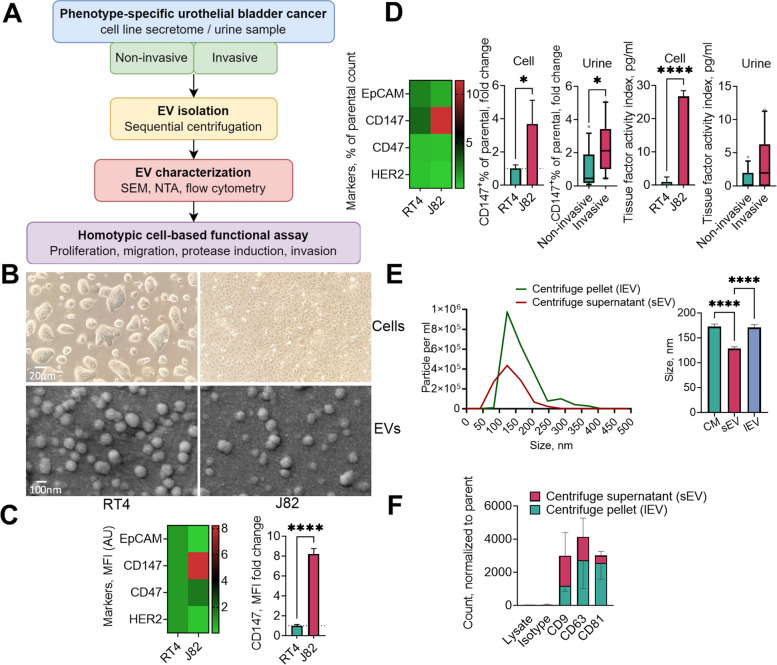
Background: Extracellular vesicles (EVs), including microvesicles, hold promise for the management of bladder urothelial carcinoma (BLCA), particularly because of their utility in identifying therapeutic targets and their diagnostic potential using easily accessible urine samples. Among the transmembrane glycoproteins highly enriched in cancer-derived EVs, tissue factor (TF) and CD147 have been implicated in promoting tumor progression. In this in vitro study, we explored a novel approach to impede cancer cell migration and metastasis by simultaneously targeting these molecules on urothelial cancer-derived EVs.

Methods: Cell culture supernatants from invasive and non-invasive bladder cancer cell lines and urine samples from patients with BLCA were collected. Large, microvesicle-like EVs were isolated using sequential centrifugation and characterized by electron microscopy, nanoparticle tracking analysis, and flow cytometry. The impact of urinary or cell supernatant-derived EVs on cellular phenotypes was evaluated using cell-based assays following combined treatment with a specific CD147 inhibitor alone or in combination with a tissue factor pathway inhibitor (TFPI), an endogenous anticoagulant protein that can be released by low-molecular-weight heparins.

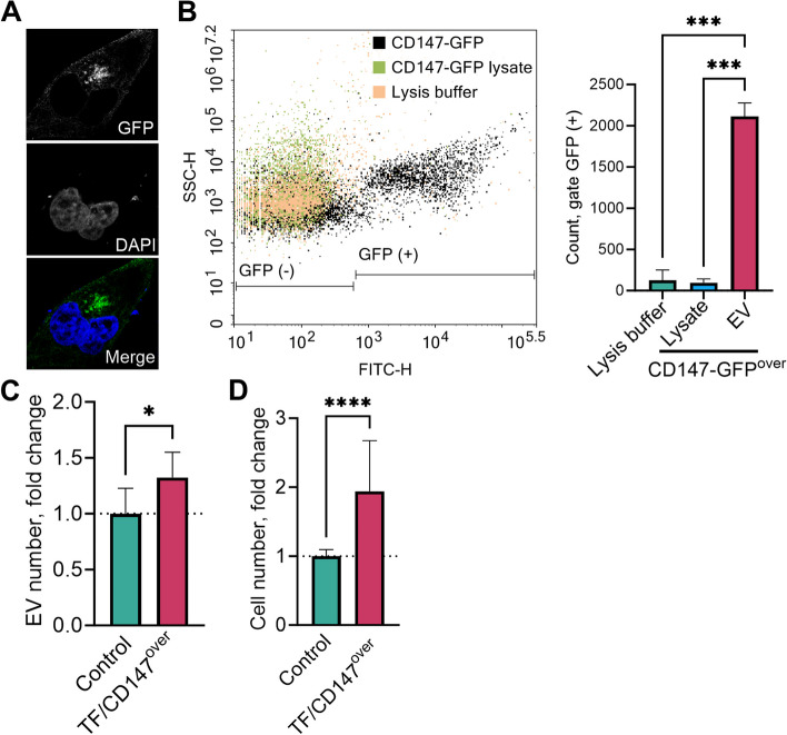
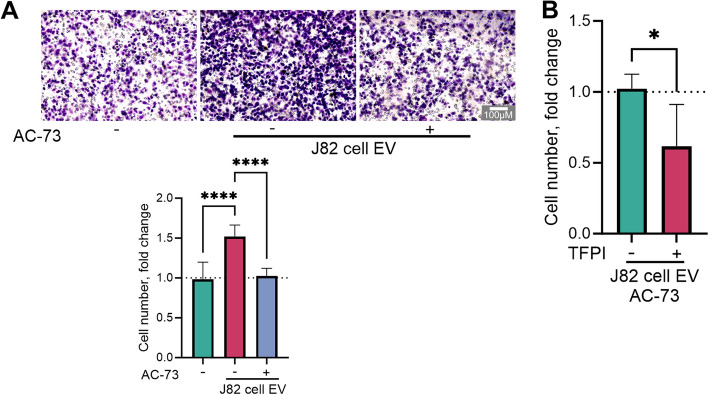
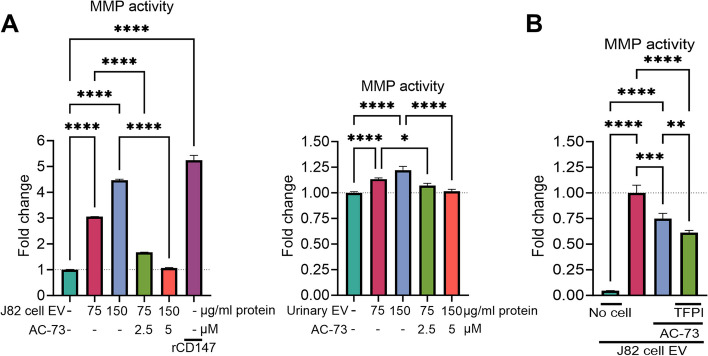
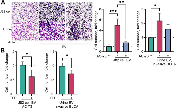
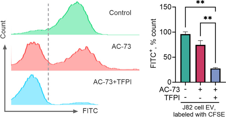
Results: We observed that EVs obtained from the urine samples of patients with muscle-invasive BLCA and from the aggressive bladder cancer cell line J82 exhibited higher TF activity and CD147 expression levels than did their non-invasive counterparts. The shedding of GFP-tagged CD147 into isolated vesicles demonstrated that the vesicles originated from plasma cell membranes. EVs originating from invasive cancer cells were found to trigger migration, secretion of matrix metalloproteinases (MMPs), and invasion. The same induction of MMP activity was replicated using EVs obtained from urine samples of patients with invasive BLCA. EVs derived from cancer cell clones overexpressing TF and CD147 were produced in higher quantities and exhibited a higher invasive potential than those from control cancer cells. TFPI interfered with the effect when used in conjunction with the CD147 inhibitor, further suppressing homotypic EV-induced migration, MMP production, and invasion.

Conclusions: Our findings suggest that combining a CD147 inhibitor with low molecular weight heparins to induce TFPI release may be a promising therapeutic approach for urothelial cancer management. This combination can potentially suppress the tumor-promoting actions of cancer-derived microvesicle-like EVs, including collective matrix invasion.

Keywords: Bladder carcinoma; CD142; EMMPRIN; Microvesicles.

Plain language summary

Small particles or vesicles released by cancer cells into their surroundings have the potential to stimulate the spread and growth of cancer cells. In this study, we focused on two specific molecules presented by these cancer cell-derived vesicles that could play a role in promoting the dissemination of cancer cells: a protein related to blood clotting and a protein on the cell surface.We found that large vesicles from bladder cancer cells that have the ability to spread had higher levels of these proteins than vesicles from nonspreading cancer cells. We also found that the former could make cancer cells move about more, produce more of a substance that helps cancer cells spread, and invade other tissues.To counteract the cancer-promoting actions of these vesicles, we examined the impact of combining a naturally occurring anticlotting protein that can be released by medications derived from heparin with an inhibitor targeting the cancer cell surface protein. We found that this combination stopped the vesicles from helping cancer cells move about more, produce more of the spreading substance, and invade other tissues.This approach of simultaneously targeting the two protein molecules present on cancer cell-derived vesicles might be a new way to treat bladder cancer.


Conflict of interest statement

This work was supported by the LEO Pharma Investigator-Initiated Studies fund. VKB received a scholarship from the LEO Pharma, and TS from the Ad Infinitum Foundation. The funders had no role in the study design, data collection, analysis, decision to publish, or preparation of the manuscript.

Figures

Fig. 1
Fig. 1
Workflow of sample processing and experimental design. Conditioned media were collected over a 24 h incubation period from two human cell lines, RT4 and J82, grown in monolayer culture. Urine samples were collected from patients with bladder urothelial carcinoma. Extracellular vesicles (EVs) were isolated and characterized using scanning electron microscopy (SEM), nanoparticle tracking analysis (NTA), and high-resolution flow cytometry. The homotypic effects of EVs were evaluated via cell-based functional assays with different readouts of cellular phenotypes (A). Cell and EV morphology were visualized using light microscopy and SEM, respectively (B). Cells (C) and cell- and urine-derived Evs (D) were analyzed for surface markers. The particle number and size distribution of the EVs were analyzed in unfractionated conditioned medium, centrifugation supernatant containing small EVs (sEVs), and centrifugation pellet containing large, microvesicle-like EVs (lEVs) by NTA (E), and the presence of tetraspanin EV markers was analyzed by flow cytometry (F) of both the EV pellet and centrifuge supernatant derived from muscle-invasive J82 cells. The parameters of the urine-derived EVs are presented as medians with 10th and 90th percentiles and were compared using the non-parametric Mann–Whitney U test. MFI, mean fluorescence intensity
Fig. 2
Fig. 2
Association of tissue factor (TF) and CD147 overexpression with extracellular vesicle (EV) production and invasion potential. The expression of green fluorescent protein (GFP)-fusion CD147 on the plasma membrane of J82 cells was observed using confocal microscopy (A). To assess the vesicular export of CD147, GFP-labeled vesicles were derived from J82 cells transfected with the CD147-GFP construct and analyzed using flow cytometry (B). The lysates of the EVs were prepared using 0.1% Triton™ X-100. EVs were derived from human bladder urothelial carcinoma J82 cells co-transfected with TF and CD147 plasmids. The EVs were quantified using NTA in the 48-h culture supernatant of J82 cells overexpressing CD147 and TF versus that of the control transfection condition (C). Invasion potency was measured by a Transwell assay, with 6–8 images of the cells that had passed through the coated microporous membrane after 24 h per experiment (D)
Fig. 3
Fig. 3
Impact of the combined inhibitors treatment on extracellular vesicle (EV)-induced migration potential. J82 human bladder urothelial carcinoma cells were exposed to 150 µg/ml protein from J82 cell-derived EVs and treated with either 5 µM CD147 inhibitor AC-73 alone (A) or a combination of 2.5 µM AC-73 and tissue factor pathway inhibitor (TFPI, 300 ng/ml, B) for 16 h. Relative migration was quantified via a Transwell assay utilizing 6–8 images of cells that traversed the microporous membrane per experiment
Fig. 4
Fig. 4
Impact of treatment with the combined inhibitors on extracellular vesicle (EV)-induced matrix metalloproteinases (MMPs) release. J82 human bladder urothelial carcinoma cells were treated with J82 cell-derived EVs or with a pool of isolated EVs from urine samples collected from patients with muscle-invasive bladder cancer (A), along with either 2.5 µM or 5 µM of the CD147 inhibitor AC-73, in the absence or presence of tissue factor pathway inhibitor (TFPI, 300 ng/ml, B) for 24 h. Recombinant CD147 (rCD147) was used as a positive control to induce MMP activity. The induction of MMPs was measured using colorimetric enzymatic methods
Fig. 5
Fig. 5
Impact of treatment with the combined inhibitors on extracellular vesicle (EV)-induced invasion potential. J82 human bladder urothelial carcinoma cells were exposed to 150 µg/ml protein from J82 cell-derived EVs or with a pool of isolated EVs from urine samples collected from patients with muscle-invasive bladder cancer, and treated with either 5 µM of the CD147 inhibitor AC-73 alone (A) or a combination of 2.5 µM AC-73 and tissue factor pathway inhibitor (TFPI, 300 ng/ml, B) for 24 h. Invasion potency was measured via a Transwell assay utilizing 6–8 images of cells that traversed the microporous membrane per experiment
Fig. 6
Fig. 6
Impact of treatment with the combined inhibitors on the extracellular vesicle (EV) binding capability. J82 human bladder urothelial carcinoma cells were exposed in suspension to carboxyfluorescein succinimidyl ester fluorescent dye (CFSE)-labeled J82 cell-derived EVs and treated with either 5 µM CD147 inhibitor AC-73 alone or a combination of 2.5 µM AC-73 and tissue factor pathway inhibitor (TFPI, 300 ng/ml). The cells were analyzed for green fluorescence in the FITC channel using flow cytometry

References

    1. Baumann Z, der AufMaur P, Bentires-Alj M. Feed-forward loops between metastatic cancer cells and their microenvironment-the stage of escalation. EMBO Mol Med. 2022;14(6):e14283. doi: 10.15252/emmm.202114283. - DOI - PMC - PubMed
    1. Cocucci E, Racchetti G, Meldolesi J. Shedding microvesicles: artefacts no more. Trends Cell Biol. 2009;19(2):43–51. doi: 10.1016/j.tcb.2008.11.003. - DOI - PubMed
    1. Ratajczak MZ, Ratajczak J. Extracellular microvesicles/exosomes: discovery, disbelief, acceptance, and the future? Leukemia. 2020;34(12):3126–3135. doi: 10.1038/s41375-020-01041-z. - DOI - PMC - PubMed
    1. Singer SJ. Intercellular communication and cell-cell adhesion. Science. 1992;255(5052):1671–1677. doi: 10.1126/science.1313187. - DOI - PubMed
    1. Raposo G, Stoorvogel W. Extracellular vesicles: exosomes, microvesicles, and friends. J Cell Biol. 2013;200(4):373–383. doi: 10.1083/jcb.201211138. - DOI - PMC - PubMed

Publication types

MeSH terms