Review
. 2024 Feb 15;22(1):124.
doi: 10.1186/s12964-023-01346-3.

Autophagy-modulating biomaterials: multifunctional weapons to promote tissue regeneration

Affiliations
Review

Autophagy-modulating biomaterials: multifunctional weapons to promote tissue regeneration

Yan Wu et al. Cell Commun Signal. .

Abstract

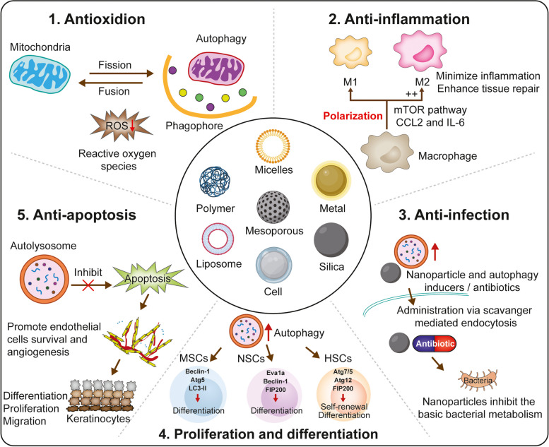
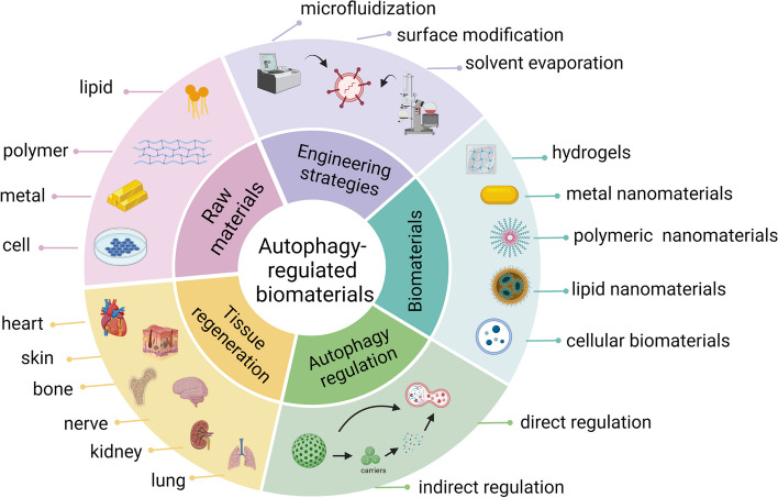
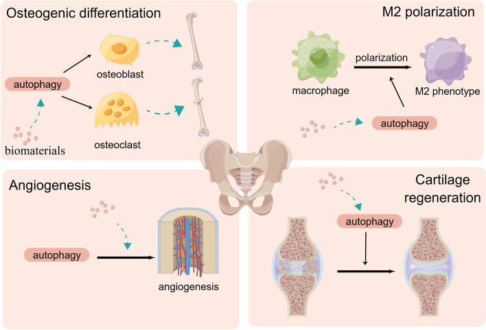
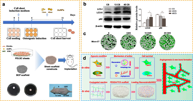
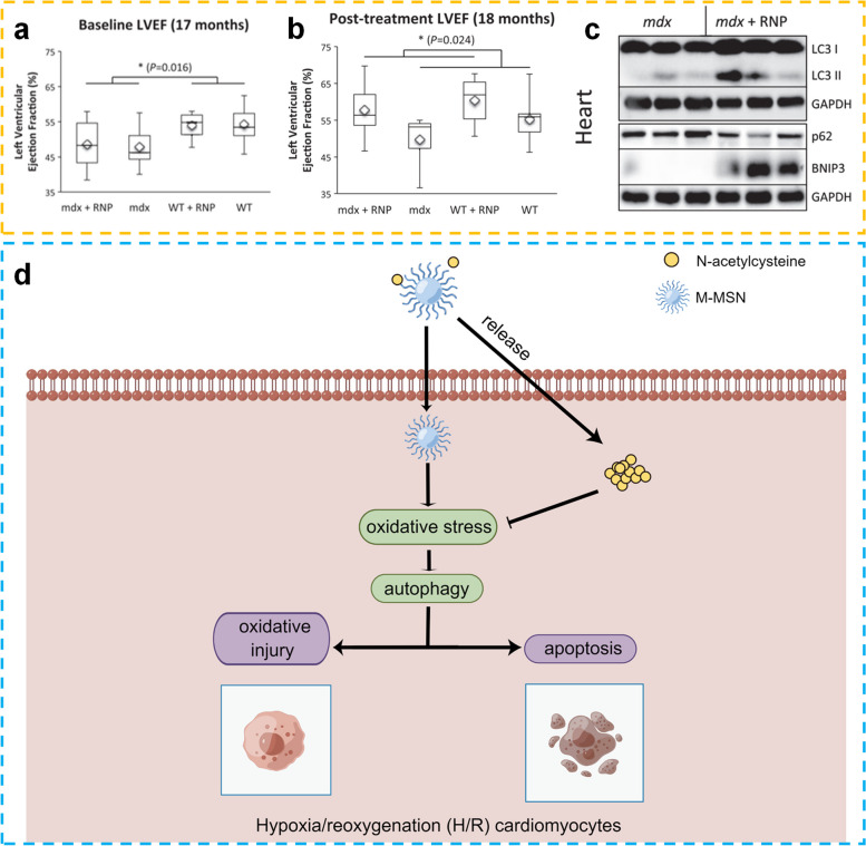
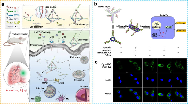
Autophagy is a self-renewal mechanism that maintains homeostasis and can promote tissue regeneration by regulating inflammation, reducing oxidative stress and promoting cell differentiation. The interaction between biomaterials and tissue cells significantly affects biomaterial-tissue integration and tissue regeneration. In recent years, it has been found that biomaterials can affect various processes related to tissue regeneration by regulating autophagy. The utilization of biomaterials in a controlled environment has become a prominent approach for enhancing the tissue regeneration capabilities. This involves the regulation of autophagy in diverse cell types implicated in tissue regeneration, encompassing the modulation of inflammatory responses, oxidative stress, cell differentiation, proliferation, migration, apoptosis, and extracellular matrix formation. In addition, biomaterials possess the potential to serve as carriers for drug delivery, enabling the regulation of autophagy by either activating or inhibiting its processes. This review summarizes the relationship between autophagy and tissue regeneration and discusses the role of biomaterial-based autophagy in tissue regeneration. In addition, recent advanced technologies used to design autophagy-modulating biomaterials are summarized, and rational design of biomaterials for providing controlled autophagy regulation via modification of the chemistry and surface of biomaterials and incorporation of cells and molecules is discussed. A better understanding of biomaterial-based autophagy and tissue regeneration, as well as the underlying molecular mechanisms, may lead to new possibilities for promoting tissue regeneration. Video Abstract.

Keywords: Anti-apoptosis; Anti-infection; Anti-inflammation; Autophagy; Biomaterials; Proliferation and differentiation; Tissue regeneration.


Conflict of interest statement

The authors declare no competing interests.

Figures

Fig. 1
Fig. 1
Mechanisms through which biomaterials regulate tissue regeneration via autophagy: Inhibition of oxidative stress, inflammation, infection, and apoptosis and promotion of cell proliferation and differentiation. (Created with adobe illustrate)
Fig. 2
Fig. 2
Summary of autophagy-modulating biomaterials in tissue regeneration. Various raw materials such as cells, metals, and polymers can be used to produce different types of biomaterials through engineering strategies including solvent evaporation, surface modification, and microfluidization. Biomaterials promote tissue and organ repair by regulating autophagy directly or indirectly. (Created with biorender.com)
Fig. 3
Fig. 3
Schematic diagram of the regulation of bone and cartilage regeneration through autophagy. Autophagy promotes bone regeneration by modulating osteogenic differentiation, inducing M2 polarization, promoting angiogenesis, and improving cartilage regeneration. (Created with figdraw.com)
Fig. 4
Fig. 4
Autophagy-modulating biomaterials promote bone regeneration. a Design scheme and fabrication. b) Western blotting for quantification of LC3 and p62. c Representative micro-CT images for examining bone regeneration promoted by gold nanoparticles (Reproduced with permission [102]; Copyright 2021, Dove Medical Press Ltd.). d Schematic illustration of the synergistic therapeutic effects of Res and ANG2 on bone defects under hypoxic conditions (Reproduced with permission [139]; Copyright 2021, Frontiers Media S.A.
Fig. 5
Fig. 5
Role of autophagy in cutaneous wound healing. Wound healing is regulated by autophagy in three stages: proliferation, hemostasis, and remodeling. Autophagy enhances the survival, proliferation, and migration of neutrophils, macrophages, endothelial cells, keratinocytes, and fibroblasts, which are essential processes for the biological functions of cells and aid in wound healing. NET, neutrophil extracellular trap. (Reproduced with permission [80]; Copyright 2022, Oxford University Press)
Fig. 6
Fig. 6
Autophagy-modulating biomaterials promote skin regeneration. a Comparison of the appearance of PEG and SBMA hydrogels. b Zwitterionic SBMA hydrogels inhibit the PI3K/Akt and mTOR signaling pathways to enhance autophagy. c Images of skin wounds stained with H&E on days 7 and 14 of treatment with PEG or SBMA hydrogels. (Reproduced with permission [118]; Copyright 2021, Frontiers Media S.A.) (d) Autophagy regulates ERK phosphorylation, which increases the release of VEGF from MSCs, and VEGF further promotes the vascularization of endothelial cells. e Assessment of capillary number in the wounded skin after 2 weeks of subcutaneous and intravenous injections of MSCs. f MSCs (red) and LC3-positive cells (green) 24 h after infusion (Reproduced with permission [147]; Copyright 2018, Nature Portfolio). g SEM images and diameter distribution. h Representative bands for LC3II/I, P62, Beclin-1, ATG5, and ATG7. i Representative images of wound healing on days 0, 4, 8, 12, and 16 of treatment with the nanocomposite films CP and CPD (Reproduced with permission [28]; Copyright 2022, Elsevier)
Fig. 7
Fig. 7
Schematic illustration of the mechanisms through which autophagy regulates nerve regeneration (drawn using Figdraw). Autophagy promotes nerve regeneration by clearing protein aggregates, promoting myelinophagy, and inducing the differentiation of neural stem cells. (Created with figdraw.com)
Fig. 8
Fig. 8
Autophagy-modulating biomaterials promote nerve regeneration. a Graphic illustration of the autophagy pathway and its impairment in CRND8 glial cells at two levels. b Western blotting for the quantification of LC3I and LC3II. c Immunoblots of pro-CatD and matu-CatD and their quantification (Reproduced with permission [90]; Copyright 2014, American Chemical Society). d Graphic illustration of NanoCA for TFEB-regulated cellular clearance of α-syn in experimental models of PD. e Schematic illustration of the effects of NanoCA on TFEB-mediated exosome release for α-syn clearance. f Western blotting of LC3II, TFEB, and LAMP1 in Neuro-2a cells after treatment with NanoCA (Reproduced with permission [150]; Copyright 2020, American Chemical Society). g XRD pattern of EuIII(OH)3 nanorods. h TEM image of EuIII(OH)3 nanorods. i Western blotting of soluble and insoluble GFP-Htt. j Western blotting of LC3I and LC3II in three types of cells treated with EuIII(OH)3 nanorods (Reproduced with permission [154]; Copyright 2014, Elsevier)
Fig. 9
Fig. 9
Role of autophagy in cardiac diseases. Autophagy facilitates regeneration in cardiac diseases and protects cardiomyocytes from injury. (Reproduced with permission [227]; Copyright 2021, John Wiley and Sons)
Fig. 10
Fig. 10
Autophagy-modulated biomaterials promote heart regeneration. a Baseline left ventricular ejection fraction of mdx and wild-type mice before treatment. b Improvement in cardiac function after treatment with rapamycin-loaded nanoparticles. c Western blotting of LC3I, LC3II, p62 and BNIP3 in the hearts of mdx mice treated with rapamycin-loaded nanoparticles. Reproduced with permission. [156] Copyright 2014, John Wiley and Sons. d Schematic illustration of the role of M-MSN@NAC in reducing oxidative injury and apoptosis by downregulating autophagy in cardiomyocytes stimulated with hypoxia and reoxygenation [155]
Fig. 11
Fig. 11
Illustration of the role of autophagy in chronic kidney disease. DN, diabetic nephropathy; LNm lupus nephropathy; APCKD, adult polycystic kidney disease. (Reproduced with permission [230]; Copyright 2019, MDPI)
Fig. 12
Fig. 12
Autophagy-modulating biomaterials promote kidney regeneration. a Preparation of magnetic Fe3O4 nanoparticles encapsulated with albumin (Fe3O4@BSA) and illustration of the protective effects of Fe3O4@BSA on albumin-induced tubulointerstitial fibrosis. b Imaging of the structure of the kidney and autophagic vacuoles in the kidneys of mice after treatment. c Western blotting for the quantification of LC3II/LC3I levels in the kidneys of mice (Reproduced with permission [157]; Copyright 2021, Elsevier). d Schematic illustration of the role of KIM-1-Res NPs in ameliorating chronic kidney disease [27]. e Schematic illustration of the role of Nano-TiO2 in inducing autophagy to protect against cell death [75]
Fig. 13
Fig. 13
Underlying mechanism of autophagy in lung tissue. Nutrient deprivation and stress can trigger autophagy. ATP, adenosine triphosphate; α-KG, alpha-ketoglutarate; PPP, pentose-phosphate pathway; ROS, reactive oxygen species; TCA, tricarboxylic acid cycle. (Reproduced with permission [246]; Copyright 2021, Springer Nature)
Fig. 14
Fig. 14
Autophagy-modulating biomaterials promote lung regeneration. a Schematic illustration of the synergistic anti-inflammatory effects of TETSpd-mTOR on ALI (Reproduced with permission [17]; Copyright 2022, John Wiley and Sons). b Schematic illustration demonstrating self-assembled mTOR siRNA-loaded nanotubes that stimulate PASMC proliferation and autophagy. c Fluorescence imaging demonstrating autophagy in pulmonary arterial smooth muscle cells incubated with mTOR siRNA under hypoxic conditions (Reproduced with permission [159]; Copyright 2015, Elsevier)

References

    1. Xie Z, Klionsky DJ. Autophagosome formation: core machinery and adaptations. Nat Cell Biol. 2007;9:1102–9. doi: 10.1038/ncb1007-1102. - DOI - PubMed
    1. Klionsky DJ. Autophagy: from phenomenology to molecular understanding in less than a decade. Nat Rev Mol Cell Biol. 2007;8:931–7. doi: 10.1038/nrm2245. - DOI - PubMed
    1. Antonioli M, Di Rienzo M, Piacentini M, Fimia GM. Emerging mechanisms in initiating and terminating Autophagy. Trends Biochem Sci. 2017;42:28–41. doi: 10.1016/j.tibs.2016.09.008. - DOI - PubMed
    1. Levine B, Kroemer G. Biological Functions of Autophagy genes: a Disease Perspective. Cell. 2019;176:11–42. doi: 10.1016/j.cell.2018.09.048. - DOI - PMC - PubMed
    1. Li L, Tan J, Miao Y, Lei P, Zhang Q. ROS and Autophagy: interactions and Molecular Regulatory Mechanisms. Cell Mol Neurobiol. 2015;35:615–21. doi: 10.1007/s10571-015-0166-x. - DOI - PMC - PubMed

MeSH terms

Substances

LinkOut - more resources