. 2024 Feb 15;10(1):79.
doi: 10.1038/s41420-024-01845-3.

Antibody and antibody fragments site-specific conjugation using new Q-tag substrate of bacterial transglutaminase

Affiliations

Antibody and antibody fragments site-specific conjugation using new Q-tag substrate of bacterial transglutaminase

Meddy El Alaoui et al. Cell Death Discov. .

Abstract

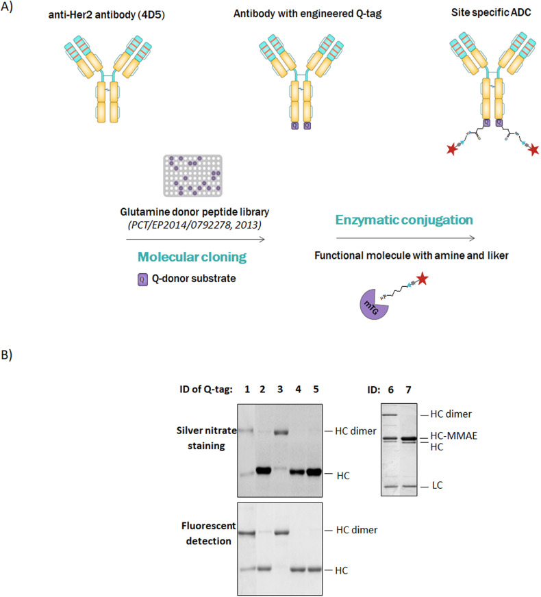
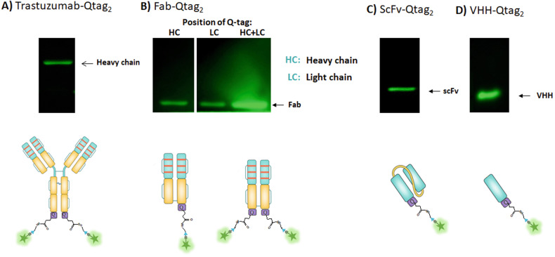
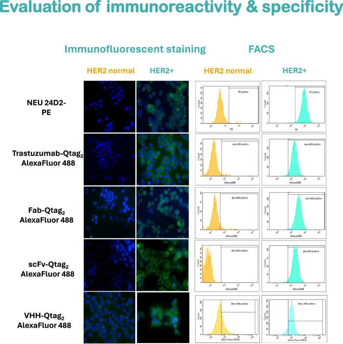
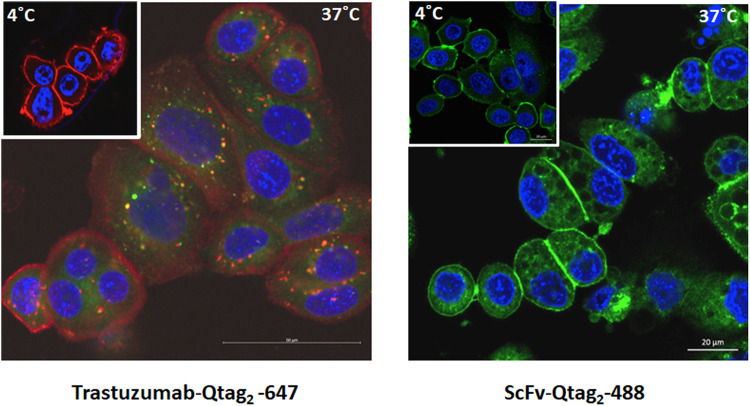
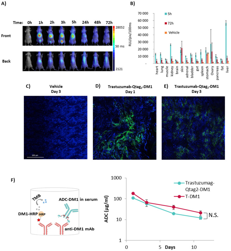
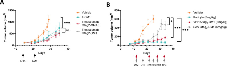
During the last few years Antibody-Drug Conjugates (ADCs) have become one of the most active and very promising therapeutic weapons. Lessons learned from the traditional chemical conjugations (via lysine or cysteine residues of the antibodies) and the clinical studies of the developed ADCs have recently paved the way to the improvement of the conjugation technologies. Use of site-specific conjugation is considered as the promising path for improving the design and development of homogeneous ADCs with controlled Drug-Antibody ratio (DAR). Moreover, some of these conjugations can be applied to antibody fragments such as Fab, scfv and VHH for which random and chemical conjugation showed significant limitations. In this study, we identified a novel small peptide substrate (Q-tag) with high affinity and specificity of bacterial transglutaminase which can be genetically fused to different formats of antibodies of interest for the development of enzymatic site-specific conjugation we named "CovIsolink" platform. We describe the synthesis of chemically defined drugs conjugation in which the site and stoichiometry of conjugation are controlled using a genetically encoded Q-tag peptide with specific amino acids which serves as a substrate of bacterial transglutaminase. This approach has enabled the generation of homogeneous conjugates with DAR 1,7 for full IgG and 0,8 drug ratio for Fab, scfv and VHH antibody fragments without the presence of significant amounts of unconjugated antibody and fragments. As a proof of concept, Q-tagged anti Her-2 (human IgG1 (Trastuzumab) and the corresponding fragments (Fab, scfv and VHH) were engineered and conjugated with different aminated-payloads. The corresponding Cov-ADCs were evaluated in series of in vitro and in vivo assays, demonstrating similar tumor cell killing potency as Trastuzumab emtansine (Kadcyla®) even with lower drug-to-antibody ratio (DAR).


Conflict of interest statement

The authors declare no competing interests.

Figures

Fig. 1
Fig. 1. Microbial transglutaminase mediated site-specific conjugation of antibody drug conjugates.
A Schematic representation of antibody with engineered glutamine tag (Q-Tag) and microbial transglutaminase mediated conjugation. B SDS-PAGE analysis of AlexaFluor488-cadaverine or MMAE conjugated engineered Trastuzumab-Qtag antibodies: 1-LLQG, 2-Tag2, 3-Tag3, 4-Tag4, 5-Tag5. ID 6: Trastuzumab-Qtag2-MMAE; ID 7: TrastuzumabK453del-Qtag2-MMAE. The reaction products were separated by 10% SDS-PAGE, then visualized by fluorescent detection and silver-nitrate staining. LC light chain, HC heavy chain, HC dimer heavy chain dimer.
Fig. 2
Fig. 2. Microbial transglutaminase mediated site-specific conjugation of antibody drug conjugates.
SDS-PAGE analysis and fluorescence detection of the incorporation of alexa488-cadaverine (amine donor) into Her2 trastuzumabQtag2 (A), Fab- Qtag2g (B), scfv- Qtag2 (C) and VHH- Qtag2 (D) using mTG.
Fig. 3
Fig. 3. Immunofluorescent staining (left panel) and fluorescent activated cell sorting (FACS) analysis (right panel) on SKBR3 (HER2/neu positive) and MCF7 (HER2/neu status normal) cells.
Trastuzumab-Qtag2, Fab-Qtag2, scFv-Qtag2 and VHH-Qtag2 were conjugated with AlexaFluor488-cadaverine and the antigen binding profile was compared to the commercially available Neu-antibody(24D2)-PE. Green, AlexaFluor488 or PE signal; blue, DAPI (nucleus).
Fig. 4
Fig. 4. Immunofluorescent staining of breast cancer cell lines: SKBR3 (HER2/neu positive).
Evaluation of intracellular internalization of AlexaFluor674 conjugated Trastuzumab-Qtag2 (left panel) or scfv-Qtag2 (right panel) at 4 °C or 37 °C using fluorescent microscopy. Red, AlexaFluor674 signal; green, lysotracker; blue, DAPI (nucleus). AlexaFluor 488 scfv (right panel) at 4 °C or 37 °C using fluorescent microscopy. Green, AlexaFluor 488 – scfv; blue, DAPI.
Fig. 5
Fig. 5. Biodistribution of ADC.
A 2D in vivo fluorescent images of NMRI Nudes mice and B ex vivo fluorescent signal of organs after intravenous injection of AlexaFluor 680 labeled Trastuzumab-Qtag2-MMAE (Mean ± SEM (n = 4)). Immunofluorescent analysis of BT-474 tumor sections after intraperitoneal injection of C vehicle control or Trastuzumab-Qtag2-DM1 D on day 1 and E day 3. The green signal represents the homing of ADC using AlexaFluor488 labeled anti-DM1 mouse monoclonal antibody. Blue, DAPI (nucleus). F Quantitation of the ADCs concentrations in mouse serum (15 mg/kg of ADC, BT-474-Her2 positive tumor) was performed by competition ELISA assay. 96-well plates were coated with anti-DM1 mAb to capture ADC-DM1 and HRP-labeled DM1. The detected absorbance is inversely proportional to the amount of ADC-DM1 conjugate in the test sample (Mean ± SEM (n = 3). Statistical significance was evaluated using GraphPad Prism 7.0b (GraphPad Software Inc., San Diego, CA, USA) software using unpaired one tailed t-test, N.S. non significative.
Fig. 6
Fig. 6. In vivo evaluation of ADCs in a mouse ectopic xenograft model.
The in vivo efficacy was investigated in SCID mice implanted subcutaneous with BT-474 HER2-positive mammary tumors. A Tumor response was evaluated using single administration of 3 mg/kg dose (trastuzumab ADC or T-DM1) and tumor volume was measured at different time intervals. B Tumor response was evaluated using several administrations of various dose of antibody fragments (trastuzumab -Qtag2 -DM1 or T-DM1) and tumor volume was measured at different time intervals Mean ± SEM (n = 6). Tumor size was followed over 3 weeks. Statistical significance was calculated by unpaired one tailed t-test using GraphPad Prism 7.0b (GraphPad Software Inc., San Diego, CA, USA) software;. n.s : non significative, *P < 0.05 ***P < 0.001.

Similar articles

Cited by

References

    1. Strebhardt K, Ullrich A. Paul Ehrlich’s magic bullet concept: 100 years of progress. Nat Rev Cancer. 2008;8:473–80. doi: 10.1038/nrc2394. - DOI - PubMed
    1. Schrama D, Reisfeld RA, Becker JC. Antibody targeted drugs as cancer therapeutics. Nat Rev Drug Discov. 2006;5:147–59. doi: 10.1038/nrd1957. - DOI - PubMed
    1. Fu Z, Li S, Han S, Shi C, Zhang Y. Antibody drug conjugate: the “biological missile” for targeted cancer therapy. Sig Transduct Target Ther. 2022;7:1–25. doi: 10.1038/s41392-022-00947-7. - DOI - PMC - PubMed
    1. Alley SC, Okeley NM, Senter PD. Antibody–drug conjugates: targeted drug delivery for cancer. Curr Opin Chem Biol. 2010;14:529–37. doi: 10.1016/j.cbpa.2010.06.170. - DOI - PubMed
    1. Junutula JR, Raab H, Clark S, Bhakta S, Leipold DD, Weir S, et al. Site-specific conjugation of a cytotoxic drug to an antibody improves the therapeutic index. Nat Biotechnol. 2008;26:925–32. doi: 10.1038/nbt.1480. - DOI - PubMed