. 2024 Feb 15;14(1):3845.
doi: 10.1038/s41598-024-54502-1.

Enhanced visualization in endoleak detection through iterative and AI-noise optimized spectral reconstructions

Affiliations

Enhanced visualization in endoleak detection through iterative and AI-noise optimized spectral reconstructions

Wojciech Kazimierczak et al. Sci Rep. .

Abstract

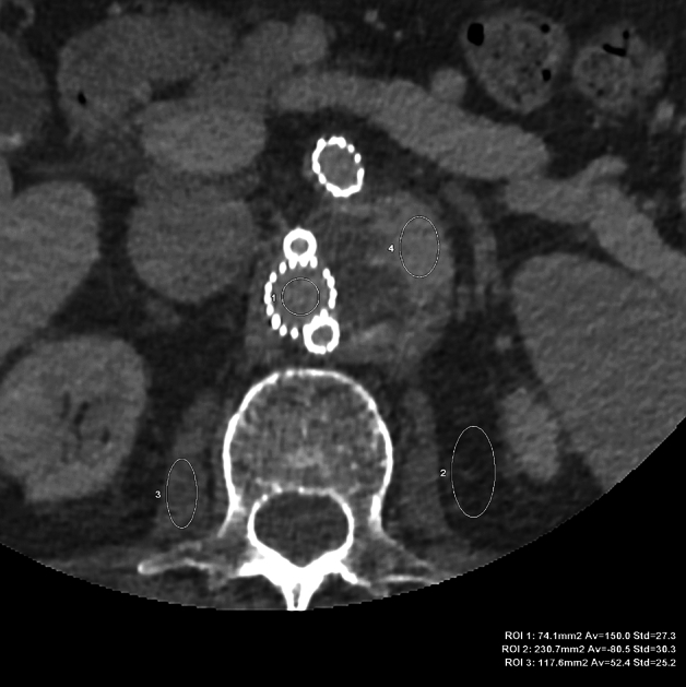
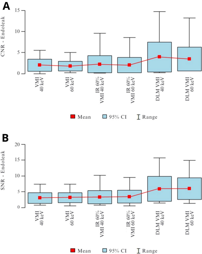
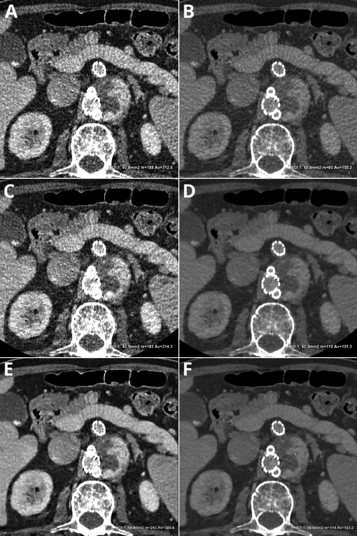
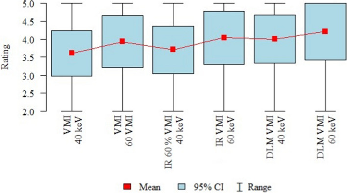
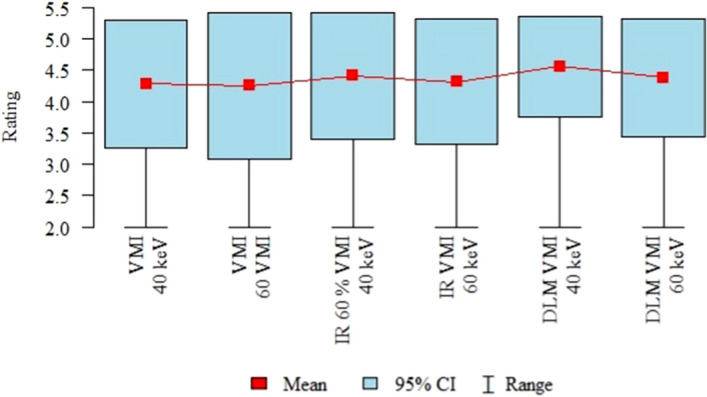
To assess the image quality parameters of dual-energy computed tomography angiography (DECTA) 40-, and 60 keV virtual monoenergetic images (VMIs) combined with deep learning-based image reconstruction model (DLM) and iterative reconstructions (IR). CT scans of 28 post EVAR patients were enrolled. The 60 s delayed phase of DECTA was evaluated. Objective [noise, contrast-to-noise ratio (CNR), signal-to-noise ratio (SNR)] and subjective (overall image quality and endoleak conspicuity - 3 blinded readers assessment) image quality analyses were performed. The following reconstructions were evaluated: VMI 40, 60 keV VMI; IR VMI 40, 60 keV; DLM VMI 40, 60 keV. The noise level of the DLM VMI images was approximately 50% lower than that of VMI reconstruction. The highest CNR and SNR values were measured in VMI DLM images. The mean CNR in endoleak in 40 keV was accounted for as 1.83 ± 1.2; 2.07 ± 2.02; 3.6 ± 3.26 in VMI, VMI IR, and VMI DLM, respectively. The DLM algorithm significantly reduced noise and increased lesion conspicuity, resulting in higher objective and subjective image quality compared to other reconstruction techniques. The application of DLM algorithms to low-energy VMIs significantly enhances the diagnostic value of DECTA in evaluating endoleaks. DLM reconstructions surpass traditional VMIs and IR in terms of image quality.

Keywords: Abdominal aortic aneurysms; Adaptive statistical iterative reconstruction; Dual-energy computed tomography angiography; Endoleak; Endovascular aneurysm repair; Image reconstruction, deep learning model; Virtual monoenergetic images.


Conflict of interest statement

The authors declare no competing interests.

Figures

Figure 1
Figure 1
VMI 60 keV axial image for objective image quality evaluation. ROIs placed in the main stentgraft module (ROI 1 – 150 HU, SD 27.3), adipose tissue (ROI 2 – Av. − 80.5 HU, SD 30.3), psoas muscle (ROI 3 – Av. 52.4 HU, SD 25.2), endoleak (ROI 4 – Av. 145.2 HU, SD 27.3). The same window settings (W 800, L 100), patient one month after EVAR.
Figure 2
Figure 2
(A,B) CNR (A) and SNR (B) in the endoleaks in VMI, VMI IR, and VMI DLM reconstructions.
Figure 3
Figure 3
Sample ROI measurements in type III endoleak in: VMI 40 keV – Av. 312.0 HU, SD 94.9 (A), VMI 60 keV – Av. 155.2 HU, SD 50.6 (B), IR 60% VMI 40 keV – Av. 314.3 HU, SD 85.6 (C), IR 60% VMI 60 keV – Av. 151.3 HU, SD 42.0 (D), DLM VMI 40 keV – Av. 309.9 HU, SD 76.9 (E), DLM VMI 60 keV – Av. 153.2 HU, SD 39.7 (F). The same window settings (W 800, L 100), patient one month after EVAR.
Figure 4
Figure 4
Results of the overall image quality assessment (mean values, 95% confidence intervals (CI), ranges).
Figure 5
Figure 5
Results of the endoleak conspicuity assessment (mean values, 95% confidence intervals (CI), ranges).
Figure 6
Figure 6
Sample of extremally high image noise level in reconstructions: VMI 40 keV – SD 105.5 (A); VMI 60 keV – SD 52.6 (B); DLM VMI 40 keV – SD 66.1 (C); DLM VMI 60 keV – SD 32.6 (D).

Similar articles

Cited by

References

    1. McPhee JT, Hill JS, Eslami MH. The impact of gender on presentation, therapy, and mortality of abdominal aortic aneurysm in the United States, 2001–2004. J. Vasc. Surg. 2007;45:891. doi: 10.1016/j.jvs.2007.01.043. - DOI - PubMed
    1. Wanhainen A, et al. Editor’s Choice—European Society for Vascular Surgery (ESVS) 2019 Clinical Practice Guidelines on the Management of Abdominal Aorto-iliac Artery Aneurysms. Eur. J. Vasc. Endovasc. Surg. 2019;57:8. doi: 10.1016/j.ejvs.2018.09.020. - DOI - PubMed
    1. Glebova NO, et al. Fenestrated endovascular repair of abdominal aortic aneurysms is associated with increased morbidity but comparable mortality with infrarenal endovascular aneurysm repair. J. Vasc. Surg. 2015;61:604. doi: 10.1016/j.jvs.2014.10.025. - DOI - PubMed
    1. White GH, Yu W, May J, Chaufour X, Stephen MS. Endoleak as a complication of endoluminal grafting of abdominal aortic aneurysms: Classification, incidence, diagnosis, and management. J. Endovasc. Surg. 1997 doi: 10.1583/1074-6218(1997)004<0152:EAACOE>2.0.CO;2. - DOI - PubMed
    1. Chaikof EL, et al. The Society for Vascular Surgery practice guidelines on the care of patients with an abdominal aortic aneurysm. J. Vasc. Surg. 2018;67:2. doi: 10.1016/j.jvs.2017.10.044. - DOI - PubMed

MeSH terms