. 2024 Feb 15;14(1):3834.
doi: 10.1038/s41598-024-54075-z.

Ripa-56 protects retinal ganglion cells in glutamate-induced retinal excitotoxic model of glaucoma

Affiliations

Ripa-56 protects retinal ganglion cells in glutamate-induced retinal excitotoxic model of glaucoma

Lemeng Feng et al. Sci Rep. .

Abstract

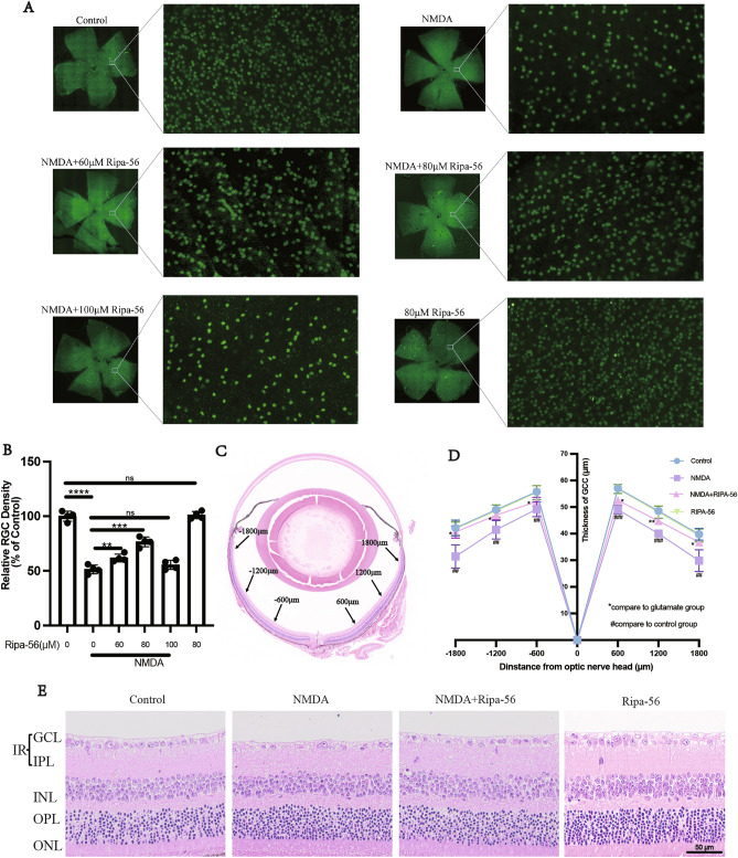
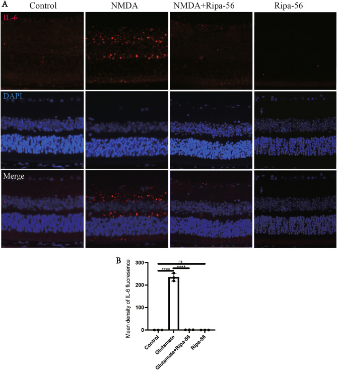
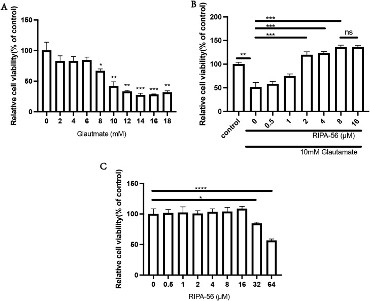
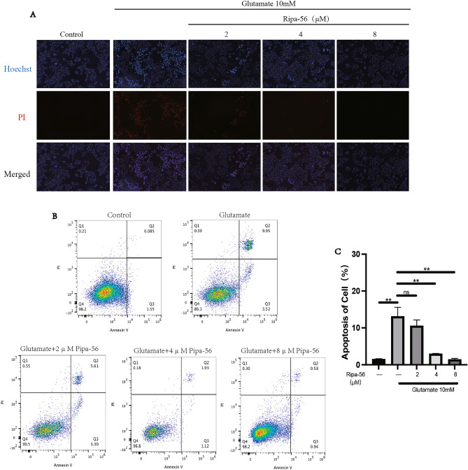
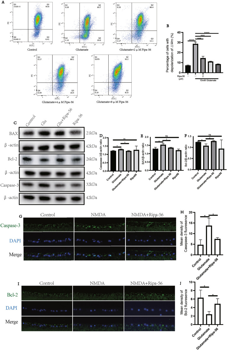
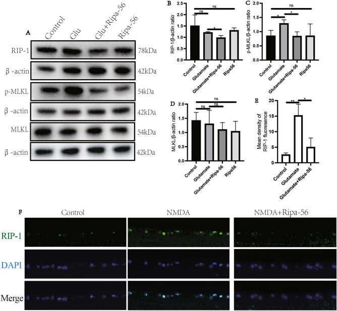
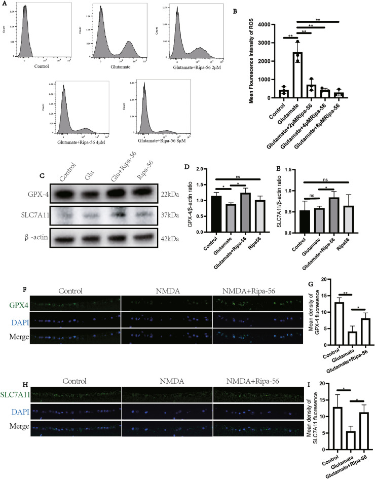
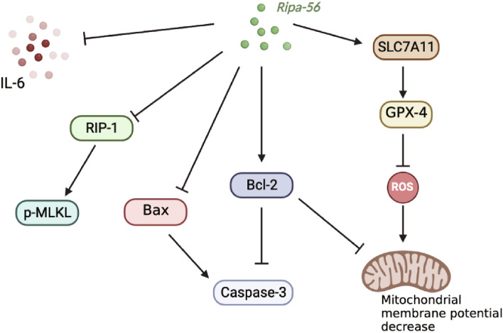
Glaucoma is a prevalent cause of blindness globally, characterized by the progressive degeneration of retinal ganglion cells (RGCs). Among various factors, glutamate excitotoxicity stands out as a significant contributor of RGCs loss in glaucoma. Our study focused on Ripa-56 and its protective effect against NMDA-induced retinal damage in mice, aiming to delve into the potential underlying mechanism. The R28 cells were categorized into four groups: glutamate (Glu), Glu + Ripa-56, Ripa-56 and Control group. After 24 h of treatment, cell death was assessed by PI / Hoechst staining. Mitochondrial membrane potential changes, apoptosis and reactive oxygen species (ROS) production were analyzed using flow cytometry. The alterations in the expression of RIP-1, p-MLKL, Bcl-2, BAX, Caspase-3, Gpx4 and SLC7A11 were examined using western blot analysis. C57BL/6j mice were randomly divided into NMDA, NMDA + Ripa-56, Ripa-56 and control groups. Histological changes in the retina were evaluated using hematoxylin and eosin (H&E) staining. RGCs survival and the protein expression changes of RIP-1, Caspase-3, Bcl-2, Gpx4 and SLC7A11 were observed using immunofluorescence. Ripa-56 exhibited a significant reduction in the levels of RIP-1, p-MLKL, Caspase-3, and BAX induced by glutamate, while promoting the expression of Bcl-2, Gpx-4, and SLC7A1 in the Ripa-56-treated group. In our study, using an NMDA-induced normal tension glaucoma mice model, we employed immunofluorescence and H&E staining to observe that Ripa-56 treatment effectively ameliorated retinal ganglion cell loss, mitigating the decrease in retinal ganglion cell layer and bipolar cell layer thickness caused by NMDA. In this study, we have observed that Ripa-56 possesses remarkable anti- necroptotic, anti-apoptotic and anti-ferroptosis properties. It demonstrates the ability to combat not only glutamate-induced excitotoxicity in R28 cells, but also NMDA-induced retinal excitotoxicity in mice. Therefore, Ripa-56 could be used as a potential retinal protective agent.

Keywords: Apoptosis; Ferroptosis; Glaucoma; Glutamate; Necroptosis; Ripa-56.


Conflict of interest statement

The authors declare no competing interests.

Figures

Figure 1
Figure 1
Protective effect of Ripa-56 in NMDA-induced mice retinal injury model. (A) Effects of Ripa-56 on NMDA-induced RGCs injury in mice model (n = 4). NMDA and Ripa-56 were injected into the vitreous cavity. 3 days later, we made retinal slices. RGCs were fluorescently labeled with Brn3a antibody. (B) Relative RGCs density in Control group (100 ± 4.57%), NMDA group (51.36 ± 4.00%), NMDA + 60 µM Ripa-56 group (61.76 ± 3.54%), NMDA + 80 µM Ripa-56 group (76.52 ± 4.41%), NMDA + 100 µM Ripa-56 group (55.49 ± 4.19%), 80 µM Ripa-56 group (101.02 ± 3.33%) (n = 4). (C,D) Thickness of the retinal ganglion cell body complex (GCC) at 600, 1200, and 1800 μm from the optic disc 3 days after intravitreal injection of NMDA and Ripa-56. (E) Effects of Ripa-56 on retinal morphology in NMDA model rats. Hematoxylin and eosin staining was performed 3 days after intravitreal injection of NMDA and Ripa-56. INL, inner nuclear layer; ONL, outer nuclear layer; GCL, ganglion cell layer; IPL, inner plexiform layer. IR consists of GCL and IPL. The results were recorded as mean ± SD from at least three independent experiments. Data analyzed via one-way ANOVA and Tukey’s post-test. *p < 0.05,**p < 0.01, ***p < 0.001, ****p < 0.0001, ns p > 0.05. #p < 0.05, ##p < 0.01, ###p < 0.001 , ####p < 0.0001.
Figure 2
Figure 2
Effect of Ripa-56 on retinal inflammation in NMDA model mice. (A,B) Three days after intravitreal injection of NMDA and Ripa-56, paraffin sections were collected and processed for immunofluorescence experiments to measure the fluorescence intensity of IL-6 of the retinal sections in Control group (0.075 ± 0.025), NMDA group (235.23 ± 17.42), NMDA + Ripa-56 group (0.82 ± 0.14) and Ripa-56 group (0.055 ± 0.032) under a fluorescence microscope (n = 3). DAPI was used to label the cell nucleus.
Figure 3
Figure 3
Ripa-56 protect against glutamate induced damage to R28 cell line. (A) The effect of 0 mM (100.00 ± 13.64%), 2 mM (82.64 ± 8.75%), 4 mM (82.64 ± 7.62%), 6 mM (84.21 ± 5.11%), 8 mM (66.60 ± 3.46%), 10 mM (41.93 ± 7.01%), 12 mM (32.89 ± 2.20%), 14 mM (27.21 ± 3.11%), 16 mM (28.02 ± 1.10%) and 18 mM (31.57 ± 2.75%) of glutamate on R28 cells (n = 3). (B) Protection of Ripa-56 against glutamate-induced excitotoxicity in R28 cells (n = 3). The relative cell viability in control group (100.00 ± 4.03%), 0 µM Ripa-56 group (51.40 ± 10.01%), 0.5 µM Ripa-56 group (58.04 ± 5.67%), 1 µM Ripa-56 group (74.49 ± 4.89%), 2 µM Ripa-56 group (119.47 ± 6.80%), 4 µM Ripa-56 group (123.50 ± 3.87%), 8 µM Ripa-56 group (135.50 ± 4.75%) and 16 µM Ripa-56 group (135.84 ± 3.40%). (C) The effect of 0 µM (100.00 ± 8.41%), 0.5 µM (101.30 ± 5.84%), 1 µM (102.04 ± 9.64%), 2 µM (100.48 ± 4.84%), 4 µM (103.34 ± 5.12%), 8 µM (103.69 ± 7.08%), 16 µM (108.38 ± 4.07%) 32 µM (84.35 ± 2.36%) and 64 µM (56.49 ± 2.57%) of Ripa-56 treatment for 24 h on the R28 cell viability (n = 3). The results were recorded as mean ± SD from at least three independent experiments. Data analyzed via one-way ANOVA and Tukey’s post-test. *p < 0.05, **p < 0.01, ***p < 0.001 , ****p < 0.0001, ns p > 0.05.
Figure 4
Figure 4
Ripa-56 protect R28 cells against glutamate induced apoptosis. (A) The effect of Ripa-56 on apoptosis was observed using Hoechst/PI staining kit and observed under a fluorescence microscope. (B) After Annexin-V-FITC/PI staining, detected the effect of Ripa-56 on apoptosis in R28 cells by flow cytometry (n = 3). (C) Effects of Ripa-56 on the apoptosis percentage in control group (1.52 ± 0.11%), glutamate group (13.12 ± 2.46%), glutamate + 2 µM Ripa-56 group (10.57 ± 1.59%), glutamate + 4 µM Ripa-56 group (2.95 ± 0.11%) and glutamate + 8 µM Ripa-56 group (1.43 ± 0.30%) (n = 3). The results were recorded as mean ± SD from at least three independent experiments. Data analyzed via one-way ANOVA and Tukey’s post-test. *p < 0.05, **p < 0.01, ***p < 0.001 , ****p < 0.0001, ns p > 0.05.
Figure 5
Figure 5
Mechanism of Ripa-56 preventing glutamate-induced apoptosis in R28 cells. (A) The effect of Ripa-56 on mitochondrial membrane potential of R28 cell line treated with 10 mM glutamate for 24 h was detected using the flow cytometry after JC-1 staining (n = 3). (B) Effect of Ripa-56 on the percentage of mitochondrial membrane potential depolarized R28 cells in control group (7.08 ± 0.58%), glutamate group (28.47 ± 1.96%), glutamate + 2 µM Ripa-56 group (14.37 ± 1.99%), glutamate + 4 µM Ripa-56 group (10.97 ± 0.51%) and glutamate + 8 µM Ripa-56 group (8.40 ± 0.40%) (n = 3). (C) Effects of Ripa-56 on Caspase-3, BAX and Bcl-2 protein relative expression (n = 3). (D) Relative expression of Caspase-3 protein in control group (1.20 ± 0.042), glutamate group (1.29 ± 0.036), glutamate + Ripa-56 group (1.19 ± 0.047) and Ripa-56 group (1.26 ± 0.24). (E) Relative expression of BAX protein in control group (1.28 ± 0.10), glutamate group (1.53 ± 0.10), glutamate + Ripa-56 group (1.35 ± 0.039) and Ripa-56 group (1.19 ± 0.084). (F) Relative expression of Bcl-2 protein in control group (1.23 ± 0.030), glutamate group (1.06 ± 0.09), glutamate + Ripa-56 group (1.27 ± 0.036) and Ripa-56 group (0.93 ± 0.40). (G–J) Three days after intravitreal injection of NMDA and Ripa-56, paraffin sections were collected and processed for immunofluorescence experiments to measure the fluorescence intensity of Casepase-3 under a fluorescence microscope (G,H) in control group (4.67 ± 2.46), glutamate group (13.70 ± 3.55) and glutamate + Ripa-56 group (7.43 ± 0.72), and Bcl-2 (I, J) in control group (6.29 ± 1.82), glutamate group (2.38 ± 0.70) and glutamate + Ripa-56 group (4.86 ± 1.24) (n = 3). The results were recorded as mean ± SD from at least three independent experiments. Data analyzed via one-way ANOVA and Tukey’s post-test. *p < 0.05, **p < 0.01, ***p < 0.001 , ****p < 0.0001, ns p > 0.05.
Figure 6
Figure 6
Mechanism of Ripa-56 preventing glutamate-induced apoptosis in R28 cells. (A) Effects of Ripa-56 on RIP-1, MLKL and p-MLKL protein relative expression (n = 3). (B) Relative expression of RIP-1 protein in control group (1.51 ± 0.48), glutamate group (1.21 ± 0.038), glutamate + Ripa-56 group (0.98 ± 0.098) and Ripa-56 group (1.30 ± 0.11). (C) Relative expression of p-MLKL protein in control group (0.85 ± 0.20), glutamate group (1.29 ± 0.13), glutamate + Ripa-56 group (0.84 ± 0.15) and Ripa-56 group (0.85 ± 0.42). (D) Relative expression of MLKL protein in control group (1.42 ± 0.29), glutamate group (1.30 ± 0.48), glutamate + Ripa-56 group (1.10 ± 0.24) and Ripa-56 group (1.04 ± 0.35). (E,F) Three days after intravitreal injection of NMDA and Ripa-56, paraffin sections were collected and processed for immunofluorescence experiments to measure the fluorescence intensity of RIP-1 of the retinal sections in control group (2.62 ± 0.55), glutamate group (15.22 ± 3.62) and glutamate + Ripa-56 group (5.05 ± 2.92) under a fluorescence microscope (n = 3). The results were recorded as mean ± SD from at least three independent experiments. Data analyzed via one-way ANOVA and Tukey’s post-test. *p < 0.05, **p < 0.01, ***p < 0.001 , ****p < 0.0001, ns p > 0.05.
Figure 7
Figure 7
Ripa-56 attenuates glutamate-induced ferroptosis in glaucoma model. (A,B) Flow cytometric technique to detect the ROS generation in R28 cells (n = 3). (C) Effects of Ripa-56 on GPX-4 and SLC7A11 protein relative expression (n = 3). (D) Relative expression of GPX-4 protein in control group (1.13 ± 0.12), glutamate group (0.88 ± 0.048), glutamate + Ripa-56 group (1.23 ± 0.16) and Ripa-56 group (1.00 ± 0.14). (E) Relative expression of SLC7A11 protein in control group (0.53 ± 0.23), glutamate group (0.58 ± 0.053), glutamate + Ripa-56 group (0.83 ± 0.15) and Ripa-56 group (0.64 ± 0.27). (F–H) Three days after intravitreal injection of NMDA and Ripa-56, paraffin sections were collected and processed for immunofluorescence experiments to measure the fluorescence intensity of GPX-4 (F,G) in control group (12.93 ± 1.46), glutamate group (4.03 ± 1.77) and glutamate + Ripa-56 group (8.02 ± 1.74), and SLC7A11 (H,I) in control group (12.80 ± 3.84), glutamate group (5.49 ± 1.59) and glutamate + Ripa-56 group (11.20 ± 2.35) under a fluorescence microscope (n = 3). The results were recorded as mean ± SD from at least three independent experiments. Data analyzed via one-way ANOVA and Tukey’s post-test. *p < 0.05, **p < 0.01, ***p < 0.001 , ****p < 0.0001, ns p > 0.05.
Figure 8
Figure 8
Protective effect of Ripa-56 on RGCs.

Similar articles

Cited by

References

    1. Weinreb RN, Aung T, Medeiros FA. The pathophysiology and treatment of glaucoma: A review. Jama. 2014;311(18):1901–1911. doi: 10.1001/jama.2014.3192. - DOI - PMC - PubMed
    1. The Advanced Glaucoma Intervention Study (AGIS): 7. The relationship between control of intraocular pressure and visual field deterioration. The AGIS Investigators. Am. J. Ophthalmol.130(4), 429–440 (2000). - PubMed
    1. Kass MA, Heuer DK, Higginbotham EJ, Johnson CA, Keltner JL, Miller JP, Parrish RK, Wilson MR, Gordon MO. The Ocular Hypertension Treatment Study: A randomized trial determines that topical ocular hypotensive medication delays or prevents the onset of primary open-angle glaucoma. Arch. Ophthalmol. 2002;120(6):701–713. doi: 10.1001/archopht.120.6.701. - DOI - PubMed
    1. Calkins DJ. Critical pathogenic events underlying progression of neurodegeneration in glaucoma. Prog. Retin. Eye Res. 2012;31(6):702–719. doi: 10.1016/j.preteyeres.2012.07.001. - DOI - PMC - PubMed
    1. Tanito M, Kaidzu S, Takai Y, Ohira A. Status of systemic oxidative stresses in patients with primary open-angle glaucoma and pseudoexfoliation syndrome. PLoS One. 2012;7(11):e49680. doi: 10.1371/journal.pone.0049680. - DOI - PMC - PubMed