. 2024 Feb 1:15:1328707.
doi: 10.3389/fimmu.2024.1328707. eCollection 2024.

Specificities and redundancies in the NEL family of bacterial E3 ubiquitin ligases of Salmonella enterica serovar Typhimurium

Affiliations

Specificities and redundancies in the NEL family of bacterial E3 ubiquitin ligases of Salmonella enterica serovar Typhimurium

Andrea Bullones-Bolaños et al. Front Immunol. .

Abstract

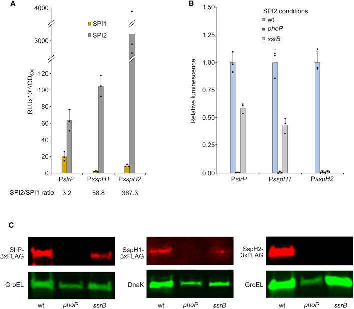
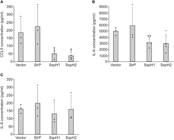
Salmonella enterica serovar Typhimurium expresses two type III secretion systems, T3SS1 and T3SS2, which are encoded in Salmonella pathogenicity island 1 (SPI1) and SPI2, respectively. These are essential virulent factors that secrete more than 40 effectors that are translocated into host animal cells. This study focuses on three of these effectors, SlrP, SspH1, and SspH2, which are members of the NEL family of E3 ubiquitin ligases. We compared their expression, regulation, and translocation patterns, their role in cell invasion and intracellular proliferation, their ability to interact and ubiquitinate specific host partners, and their effect on cytokine secretion. We found that transcription of the three genes encoding these effectors depends on the virulence regulator PhoP. Although the three effectors have the potential to be secreted through T3SS1 and T3SS2, the secretion of SspH1 and SspH2 is largely restricted to T3SS2 due to their expression pattern. We detected a role for these effectors in proliferation inside fibroblasts that is masked by redundancy. The generation of chimeric proteins allowed us to demonstrate that the N-terminal part of these proteins, containing the leucine-rich repeat motifs, confers specificity towards ubiquitination targets. Furthermore, the polyubiquitination patterns generated were different for each effector, with Lys48 linkages being predominant for SspH1 and SspH2. Finally, our experiments support an anti-inflammatory role for SspH1 and SspH2.

Keywords: E3 ubiquitin ligase; Salmonella enterica; SlrP; SspH1; SspH2; type III secretion systems.


Conflict of interest statement

The authors declare that the research was conducted in the absence of any commercial or financial relationships that could be construed as a potential conflict of interest.

Figures

Figure 1
Figure 1
Expression and regulation of slrP, sspH1, and sspH2. DNA fragments containing the promoters of slrP, sspH1, or sspH2 were cloned into plasmid pSB377 to generate luxCDABE transcriptional fusions. (A) Luminescence was measured in cultures of S. enterica serovar Typhimurium strain 14028 carrying these plasmids grown to stationary phase in LB 0.3 M NaCl (SPI1-inducing conditions) and LPM (SPI2-inducing conditions). (B) Luminescence was measured under SPI2-inducing conditions in different genetic backgrounds: wild-type (wt), phoP null mutant or ssrB null mutant. RLU, relative light units. Means and standard deviations of three independent measurements are represented. Dots represent individual values. (C) Protein extracts of derivatives of S. enterica serovar Typhimurium strains producing 3xFLAG-tagged SlrP, SspH1, or SspH2 grown under SPI2-inducing conditions were resolved by SDS-PAGE. Immunoblotting was performed with monoclonal anti-FLAG antibodies. Anti-GroEL or anti-DNAK antibodies were used as loading control. Replicate experiments for this panel are shown in Supplementary Material .
Figure 2
Figure 2
Expression of slrP, sspH1, and sspH2 during host cells infection. S. enterica serovar Typhimurium carrying plasmids expressing PslrP::luxCDABE (A, D, G), PsspH1:luxCDABE (B, E, H) or PsspH2::luxCDABE (C, F, I) were grown under invasive conditions (16 h in LB with 0.3 M NaCl without aeration) to infect HeLa cells (A–C) and NRK cells (G–I), or non-invasive conditions (24 h in LB at 37°C with aeration) to infect RAW264.7 macrophages (D–F). Luminescence produced by intracellular bacteria was measured 2, 4, and 8 h p.i.. Dots represent individual values.
Figure 3
Figure 3
Translocation of SlrP, SspH1 and SspH2 into mammalian cells. Human epithelial HeLa cells (A, B), RAW264.7 murine macrophages (C, D), and NRK rat kidney fibroblasts (E, F) were infected with derivatives of S. enterica serovar Typhimurium 14028 (wild-type, wt, prgH, ssaV, and prgH ssaV strains) carrying a plasmid expressing SlrP-CyaA’, SspH1-CyaA’, or SspH2-CyaA’ fusions from a constitutive promoter (A, C, E) or chromosomal fusions expressed under native promoters (B, D, F). Bacteria were grown under SPI1-inducing conditions (invasive conditions) except for infections of RAW264.7 cells for 8 h (non-invasive conditions to prevent early cell death). Levels of cAMP were measured as and indication of translocation. For each effector and cell type, these levels were relativized to the level obtained for the wild-type infection at 2 h or 8 h, that was set to 1. Means and standard deviations from triplicate experiments are represented. Dots represent individual values.
Figure 4
Figure 4
Effect of SlrP, SspH1, and SspH2 on host cell invasion and proliferation. (A) Analysis of invasion and intracellular proliferation of the triple mutant slrP sspH1 sspH2 in mixed infections with a trg::MudJ mutant used as the wild-type strain. (B) Analysis of intracellular proliferation in NRK cells of the indicated mutants in mixed infections with a trg::MudJ mutant used as the control strain. Competitive indices are the means of three infections. The error bars represent the standard deviations. Dots represent individual values. Asterisks denote that the indices are significantly different from 1 for a t-test: *P-value < 0.05, **P-value < 0.01.
Figure 5
Figure 5
Design of clones for the expression of chimeric NEL effectors. The represented N- and C-terminal fragments of SlrP, SspH1 and SspH2 were cloned into the vectors pLEX10 (for yeast two-hybrid experiments) and pQE80L (for 6His fusions generation) using the indicated restriction enzymes.
Figure 6
Figure 6
Interaction of SlrP, SspH1 and SspH2 with host proteins in the yeast two-hybrid system. Derivatives of pLEX10 and pGAD1318 (pGAD) were introduced into yeast strain L40 by transformation. Transformants were selected in media lacking tryptophan and leucine. (A) Study of the interaction of SlrP or derivatives in which the C-terminal part was replaced by that of SspH1 or SspH2 with SNRPD2 (D2). (B) Study of the interaction of SspH1 or derivatives in which the C-terminal part was replaced by that of SlrP or SspH2 with PKN1. (C) Study of the interaction of SspH2 or derivatives in which the C-terminal part was replaced by that of SlrP or SspH1 with NOD1.
Figure 7
Figure 7
Ubiquitination of SNRPD2 and PKN1 by SlrP, SspH1, SspH2 and chimeric effectors. The ubiquitination of GST-SNRPD2 or GST-PKN1 bound to glutathione-agarose beads was tested in the presence of HA-ubiquitin, E1, E2, and a Salmonella effector fused to 6His. The beads were washed prior to immunoblot analysis. The sizes in kDa of the molecular weight markers are shown on the left. Replicate experiments for this figure are shown in Supplementary Material .
Figure 8
Figure 8
Analysis of the specific type of polyubiquitination catalyzed by SlrP, SspH1, and SspH2. HA-ubiquitin and its derivatives with the indicated mutations replacing lysines by arginines (K48R, K63R, K48R/K63R) were used in ubiquitination assays with 6His fusions of SlrP, SspH1, and SspH2. (A) No additional substrate was added and the whole reaction was analyzed to detect polyubiquitination of ubiquitin. (B) GST-SNRPD2 or GST-PKN1 bound to glutathione-agarose beads were added as substrates and beads were washed prior to immunoblot analysis with anti-HA antibodies. The sizes in kDa of the molecular weight markers are shown on the left. Replicate experiments for this figure are shown in Supplementary Material .
Figure 9
Figure 9
Effect of SlrP, SspH1 and SspH2 on the secretion of cytokines by mammalian cells. HeLa cells were transfected with a plasmid expressing SlrP, SspH1 or SspH2 or with the empty vector and the concentration of CCL5 (A), IL-6 (B), and IL-8 (C) was measured in the supernatants of cell cultures 24 h after transfection. Data are presented as mean values + standard deviations of at least three biological replicates. Dots represent individual values. * p < 0.05, ** p < 0.01, for a two-tailed Student’s t-test comparing cells expressing each effector with cells transfected with the empty vector.

Similar articles

Cited by

References

    1. Teklemariam AD, Al-Hindi RR, Albiheyri RS, Alharbi MG, Alghamdi MA, Filimban AAR, et al. . Human salmonellosis: A continuous global threat in the farm-to-fork food safety continuum. Foods Basel Switz (2023) 12:1756. doi: 10.3390/foods12091756 - DOI - PMC - PubMed
    1. Pais SV, Kim E, Wagner S. Virulence-associated type III secretion systems in Gram-negative bacteria. Microbiol Read Engl (2023) 169:1328. doi: 10.1099/mic.0.001328 - DOI - PMC - PubMed
    1. Chen P, Goldberg MB. Recent insights into type-3 secretion system injectisome structure and mechanism of human enteric pathogens. Curr Opin Microbiol (2023) 71:102232. doi: 10.1016/j.mib.2022.102232 - DOI - PMC - PubMed
    1. De Ryck J, Van Damme P, Goormachtig S. From prediction to function: Current practices and challenges towards the functional characterization of type III effectors. Front Microbiol (2023) 14:1113442. doi: 10.3389/fmicb.2023.1113442 - DOI - PMC - PubMed
    1. Fattinger SA, Sellin ME, Hardt W-D. Salmonella effector driven invasion of the gut epithelium: breaking in and setting the house on fire. Curr Opin Microbiol (2021) 64:9–18. doi: 10.1016/j.mib.2021.08.007 - DOI - PubMed

Publication types

Substances