

Unraveling the link: white matter damage, gray matter atrophy and memory impairment in patients with subcortical ischemic vascular disease
- PMID: 38362024
- PMCID: PMC10867202
- DOI: 10.3389/fnins.2024.1355207
Unraveling the link: white matter damage, gray matter atrophy and memory impairment in patients with subcortical ischemic vascular disease
Abstract
Introduction: Prior MRI studies have shown that patients with subcortical ischemic vascular disease (SIVD) exhibited white matter damage, gray matter atrophy and memory impairment, but the specific characteristics and interrelationships of these abnormal changes have not been fully elucidated.
Materials and methods: We collected the MRI data and memory scores from 29 SIVD patients with cognitive impairment (SIVD-CI), 29 SIVD patients with cognitive unimpaired (SIVD-CU) and 32 normal controls (NC). Subsequently, the thicknesses and volumes of the gray matter regions that are closely related to memory function were automatically assessed using FreeSurfer software. Then, the volume, fractional anisotropy (FA), mean diffusivity (MD), amplitude of low-frequency fluctuation (ALFF) and regional homogeneity (ReHo) values of white matter hyperintensity (WMH) region and normal-appearing white matter (NAWM) were obtained using SPM, DPARSF, and FSL software. Finally, the analysis of covariance, spearman correlation and mediation analysis were used to analyze data.
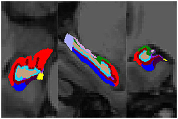
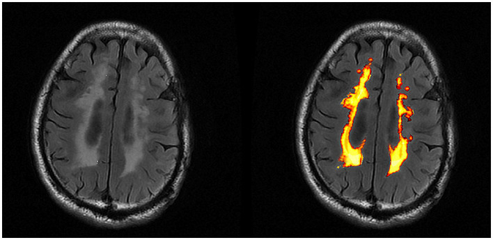
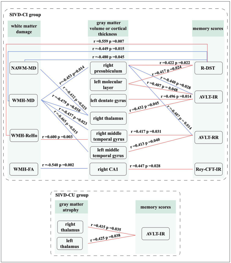
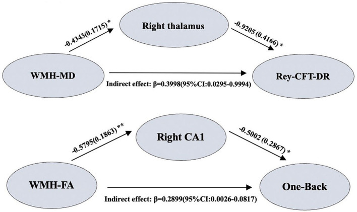
Results: Compared with NC group, patients in SIVD-CI and SIVD-CU groups showed significantly abnormal volume, FA, MD, ALFF, and ReHo values of WMH region and NAWM, as well as significantly decreased volume and thickness values of gray matter regions, mainly including thalamus, middle temporal gyrus and hippocampal subfields such as cornu ammonis (CA) 1. These abnormal changes were significantly correlated with decreased visual, auditory and working memory scores. Compared with the SIVD-CU group, the significant reductions of the left CA2/3, right amygdala, right parasubiculum and NAWM volumes and the significant increases of the MD values in the WMH region and NAWM were found in the SIVD-CI group. And the increased MD values were significantly related to working memory scores. Moreover, the decreased CA1 and thalamus volumes mediated the correlations between the abnormal microstructure indicators in WMH region and the decreased memory scores in the SIVD-CI group.
Conclusion: Patients with SIVD had structural and functional damages in both WMH and NAWM, along with specific gray matter atrophy, which were closely related to memory impairment, especially CA1 atrophy and thalamic atrophy. More importantly, the volumes of some temporomesial regions and the MD values of WMH regions and NAWM may be potentially helpful neuroimaging indicators for distinguishing between SIVD-CI and SIVD-CU patients.
Keywords: gray matter atrophy; magnetic resonance imaging; memory impairment; normal-appearing white matter; subcortical ischemic vascular disease; white matter hyperintensity.
Copyright © 2024 Huang, Cheng, Liu, Chen and Luo.
Conflict of interest statement
The authors declare that the research was conducted in the absence of any commercial or financial relationships that could be construed as a potential conflict of interest.
Figures
Similar articles
-
Study of gray matter atrophy pattern with subcortical ischemic vascular disease-vascular cognitive impairment no dementia based on structural magnetic resonance imaging.Front Aging Neurosci. 2023 Feb 6;15:1051177. doi: 10.3389/fnagi.2023.1051177. eCollection 2023. Front Aging Neurosci. 2023. PMID: 36815175 Free PMC article.
-
Relationships Between Memory Impairments and Hippocampal Structure in Patients With Subcortical Ischemic Vascular Disease.Front Aging Neurosci. 2022 Apr 18;14:823535. doi: 10.3389/fnagi.2022.823535. eCollection 2022. Front Aging Neurosci. 2022. PMID: 35517055 Free PMC article.
-
Loss of Integrity of Corpus Callosum White Matter Hyperintensity Penumbra Predicts Cognitive Decline in Patients With Subcortical Vascular Mild Cognitive Impairment.Front Aging Neurosci. 2021 Feb 18;13:605900. doi: 10.3389/fnagi.2021.605900. eCollection 2021. Front Aging Neurosci. 2021. PMID: 33679371 Free PMC article.
-
[Diffusion characteristics of subcortical structures in patients with subcortical ischemic vascular disease and its correlation to cognitive function].Nan Fang Yi Ke Da Xue Xue Bao. 2011 Oct;31(10):1737-41. Nan Fang Yi Ke Da Xue Xue Bao. 2011. PMID: 22027780 Chinese.
-
A multi-parametric MRI study on changes in the structure, function, and connectivity of thalamic subregions and their relationship with cognitive impairment in patients with subcortical ischemic vascular disease.Brain Res. 2025 Mar 1;1850:149420. doi: 10.1016/j.brainres.2024.149420. Epub 2024 Dec 24. Brain Res. 2025. PMID: 39725375
References
-
- Ahnaou A., Moechars D., Raeymaekers L., Biermans R., Manyakov N. V., Bottelbergs A., et al. . (2017). Emergence of early alterations in network oscillations and functional connectivity in a tau seeding mouse model of Alzheimer's disease pathology. Sci. Rep. 7:14189. doi: 10.1038/s41598-017-13839-6, PMID: - DOI - PMC - PubMed
-
- Caillaud M., Hudon C., Boller B., Brambati S., Duchesne S., Lorrain D., et al. . (2020). Evidence of a relation between hippocampal volume, white matter Hyperintensities, and cognition in subjective cognitive decline and mild cognitive impairment. J. Gerontol. B Psychol. Sci. Soc. Sci. 75, 1382–1392. doi: 10.1093/geronb/gbz120, PMID: - DOI - PMC - PubMed
LinkOut - more resources
Full Text Sources
Miscellaneous