

Case report: First documented case of cerebral angiostrongyliasis caused by Angiostrongylus costaricensis in a free-ranging opossum
- PMID: 38362296
- PMCID: PMC10867154
- DOI: 10.3389/fvets.2024.1294484
Case report: First documented case of cerebral angiostrongyliasis caused by Angiostrongylus costaricensis in a free-ranging opossum
Abstract
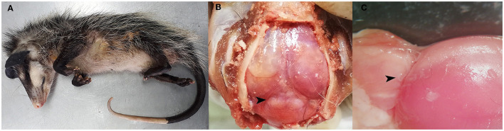
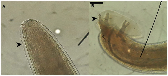
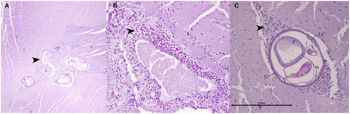
Angiostrongylus costaricensis is a metastrongyloid nematode that primarily infects the mesenteric arteries of wild rodents. This parasite is endemic in several regions of the American continent, and in humans, causes a disease known as abdominal angiostrongyliasis. Despite the important health implications of this nematode, there are limited studies investigating the involvement of wild animals in its life cycle. In this study, we present the clinical manifestations, pathologic findings, and molecular diagnosis, to the best of our current knowledge, of the first documented onset of cerebral angiostrongyliasis because of A. costaricensis infection in a juvenile free-ranging opossum (Didelphis marsupialis). Histopathological findings stress the presence of eosinophilic meningoencephalitis with nematodes present within the lesions, and PCR was positive for cox1 and ITS1 reactions. The obtained sequences for a 279 bp fragment of ITS1 were 100% identical to A. costaricensis from Costa Rica. This case highlights the substantial difficulties in diagnosing neuroangiostrongyliasis, yet underscores the importance of considering A. costaricensis as a potential culprit behind neurological conditions in wild marsupials. It acts as an urgent call to action to improve surveillance programs tracking infectious and parasitic diseases causing mortality in wildlife populations.
Keywords: angiostrongyliasis; case report; parasitic diseases; wildlife reservoir; zoonosis.
Copyright © 2024 Solorzano-Scott, Aguilar-Vargas, Cordero-Salas, Conejo, Rojas and Baldi.
Conflict of interest statement
The authors declare that the research was conducted in the absence of any commercial or financial relationships that could be construed as a potential conflict of interest. The reviewer GD is currently organizing a Research Topic with the author AR.
Figures
Similar articles
-
The white-nosed coati (Nasua narica) is a naturally susceptible definitive host for the zoonotic nematode Angiostrongylus costaricensis in Costa Rica.Vet Parasitol. 2016 Sep 15;228:93-95. doi: 10.1016/j.vetpar.2016.08.017. Epub 2016 Aug 26. Vet Parasitol. 2016. PMID: 27692339
-
Angiostrongyliasis cantonensis (eosinophilic meningitis, Alicata's disease).Contemp Neurol Ser. 1975;12:133-64. Contemp Neurol Ser. 1975. PMID: 1095293 Review.
-
Abdominal angiostrongyliasis in the Americas: fifty years since the discovery of a new metastrongylid species, Angiostrongylus costaricensis.Parasit Vectors. 2021 Jul 22;14(1):374. doi: 10.1186/s13071-021-04875-3. Parasit Vectors. 2021. PMID: 34294132 Free PMC article. Review.
-
Pulmonary angiostrongyliasis: Two cases of atypical manifestations of Angiostrongylus costaricensis in Guatemala.Rev Esp Patol. 2025 Jul-Sep;58(3):100821. doi: 10.1016/j.patol.2025.100821. Epub 2025 Apr 8. Rev Esp Patol. 2025. PMID: 40203530
-
A practical guide for the diagnosis of abdominal angiostrongyliasis caused by the nematode Angiostrongylus costaricensis.Parasit Vectors. 2023 Apr 29;16(1):155. doi: 10.1186/s13071-023-05757-6. Parasit Vectors. 2023. PMID: 37120597 Free PMC article. Review.
Cited by
-
A Scoping Review of Angiostrongyliasis and Other Diseases Associated with Terrestrial Mollusks, Including Lissachatina fulica: An Overview of Case Reports and Series.Pathogens. 2024 Oct 2;13(10):862. doi: 10.3390/pathogens13100862. Pathogens. 2024. PMID: 39452733 Free PMC article.
References
-
- Rojas A, Maldonado-Junior A, Mora J, Morassutti A, Rodriguez R, Solano-Barquero A, et al. . Abdominal angiostrongyliasis in the Americas: fifty years since the discovery of a new metastrongylid species, Angiostrongylus costaricensis. Parasit Vectors. (2021) 14:374. 10.1186/s13071-021-04875-3 - DOI - PMC - PubMed
-
- Graeff-Teixeira C, Rodriguez R. Abdominal Angiostrongyliasis. In: Hunter's Tropical Medicine and Emerging Infectious Diseases. London: Elsevier; (2020). p. 895–897.
Publication types
LinkOut - more resources
Full Text Sources
Miscellaneous