

Potassium-selective channelrhodopsins
- PMID: 38362336
- PMCID: PMC10865875
- DOI: 10.2142/biophysico.bppb-v20.s011
Potassium-selective channelrhodopsins
Abstract
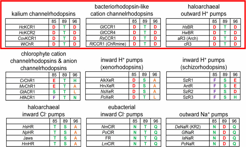
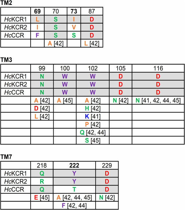
Since their discovery 21 years ago, channelrhodopsins have come of age and have become indispensable tools for optogenetic control of excitable cells such as neurons and myocytes. Potential therapeutic utility of channelrhodopsins has been proven by partial vision restoration in a human patient. Previously known channelrhodopsins are either proton channels, non-selective cation channels almost equally permeable to Na+ and K+ besides protons, or anion channels. Two years ago, we discovered a group of channelrhodopsins that exhibit over an order of magnitude higher selectivity for K+ than for Na+. These proteins, known as "kalium channelrhodopsins" or KCRs, lack the canonical tetrameric selectivity filter found in voltage- and ligand-gated K+ channels, and use a unique selectivity mechanism intrinsic to their individual protomers. Mutant analysis has revealed that the key residues responsible for K+ selectivity in KCRs are located at both ends of the putative cation conduction pathway, and their role has been confirmed by high-resolution KCR structures. Expression of KCRs in mouse neurons and human cardiomyocytes enabled optical inhibition of these cells' electrical activity. In this minireview we briefly discuss major results of KCR research obtained during the last two years and suggest some directions of future research.
Keywords: ion channels; ion selectivity; optogenetics; photocurrent.
2023 THE BIOPHYSICAL SOCIETY OF JAPAN.
Conflict of interest statement
The authors declare no conflict of interest.
Figures
References
-
- Emiliani, V., Entcheva, E., Hedrich, R., Hegemann, P., Konrad, K. R., Lüscher, C., et al. . Optogenetics for light control of biological systems. Nature Reviews Methods Primers 2, 55 (2022). https://doi.org/10.1038/s43586-022-00136-4 - PMC - PubMed
-
- Deisseroth, K., Feng, G., Majewska, A. K., Miesenböck, G., Ting, A., Schnitzer, M. J.. Next-generation optical technologies for illuminating genetically targeted brain circuits. J. Neurosci. 26, 10380–10386 (2006). https://doi.org/10.1523/JNEUROSCI.3863-06.2006 - PMC - PubMed
-
- Sahel, J. A., Boulanger-Scemama, E., Pagot, C., Arleo, A., Galluppi, F., Martel, J. N., et al. . Partial recovery of visual function in a blind patient after optogenetic therapy. Nat. Med. 27, 1223–1229 (2021). https://doi.org/10.1038/s41591-021-01351-4 - PubMed
-
- Govorunova, E. G., Sineshchekov, O. A., Liu, X., Janz, R., Spudich, J. L.. Natural light-gated anion channels: A family of microbial rhodopsins for advanced optogenetics. Science 349, 647–650 (2015). https://doi.org/10.1126/science.aaa7484 - PMC - PubMed
-
- Mardinly, A. R., Oldenburg, I. A., Pegard, N. C., Sridharan, S., Lyall, E. H., Chesnov, K., et al. . Precise multimodal optical control of neural ensemble activity. Nat. Neurosci. 21, 881–893 (2018). https://doi.org/10.1038/s41593-018-0139-8 - PMC - PubMed