Review
. 2024 Jan;85(1):95-108.
doi: 10.3348/jksr.2023.0079. Epub 2024 Jan 26.

CT Findings of Azygos Venous System: Congenital Variants and Acquired Structural Changes

Review

CT Findings of Azygos Venous System: Congenital Variants and Acquired Structural Changes

Ha Neul Kim et al. J Korean Soc Radiol. 2024 Jan.

Abstract

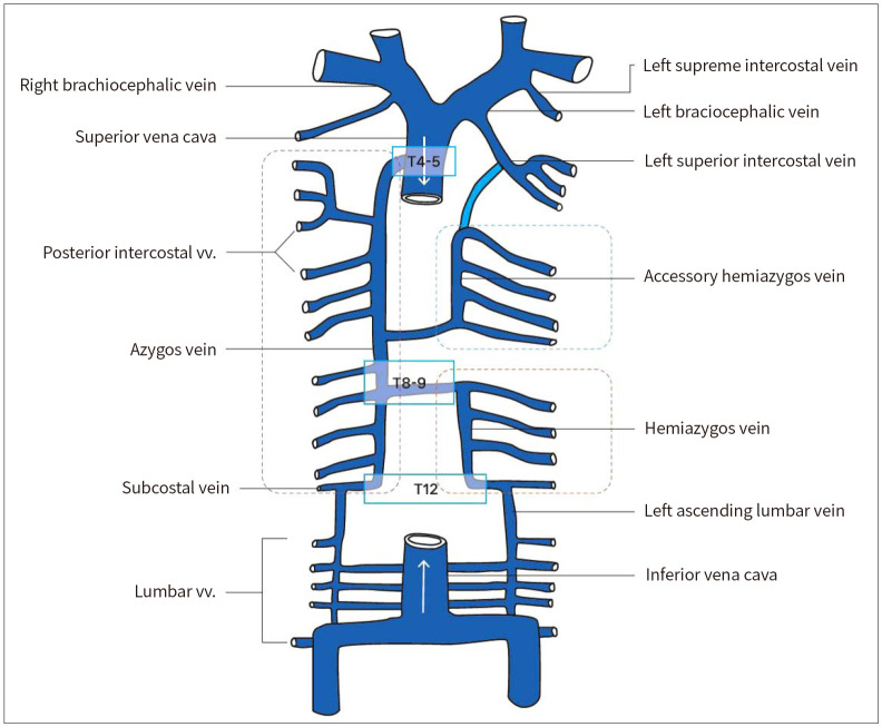
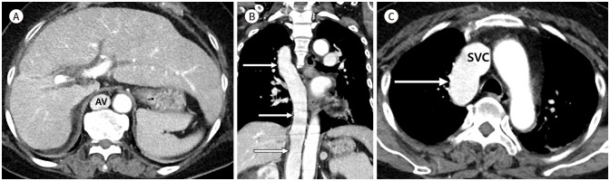
The azygos venous system is a crucial conduit of the posterior thorax and potentially vital collateral pathway. However, it is often overlooked clinically and radiologically. This pictorial essay reviews the normal azygos venous anatomy and CT findings of congenital variations and structural changes associated with acquired pathologies.

기정맥계는 후방 흉부의 중요한 부속 정맥이며 측부순환으로서 중대한 역할을 한다. 그러나, 그 중요성에도 불구하고 임상적 혹은 영상의학적으로 종종 간과된다. 본 임상화보에서는, 기정맥계의 정상 해부학에 대해 알아보고, 기정맥계에서 볼 수 있는 다양한 선천 변이와 후천적 질환에 따른 구조 변화의 CT 소견에 대하여 검토하고자 한다.

Keywords: Azygos Vein; Collateral Pathway; Inferior Vena Cava; Left Superior Intercostal Vein; Superior Vena Cava.


Conflict of interest statement

Conflicts of Interest: The authors have no potential conflicts of interest to disclose.

Figures

Fig. 1
Fig. 1. Normal azygos venous system.
The azygos vein arises at the junction between the right ascending lumbar vein and right subcostal vein around the T12 level. It ascends along the right side of the vertebral column and drains into the superior vena cava at the T4–T5 level. The hemiazygos vein, serving as a counterpart to the lower azygos vein, ascends along the left side of the vertebral column, crosses behind the aorta and esophagus, and drains into the azygos vein at the T8–T9 level. At this level, the hemiazygos vein communicates with the accessory hemiazygos vein, which corresponds to the upper azygos vein. The left superior intercostal vein communicates with the accessory hemiazygos vein in approximately 75% of patients, draining into the left brachiocephalic vein.
Fig. 2
Fig. 2. Absence of azygos vein with isolated persistent left superior vena cava.
A. Anterior view of the three-dimensional volume-rendered image shows the isolated persistent left SVC (arrows). Without the right SVC, the confluence of the right internal jugular and right subclavian vein drains into the persistent left SVC (arrows) via the bridging vein (curved arrow). B. Axial CT image shows the prominent CS, which drains the persistent left SVC. The hemiazygos vein (arrow) ascends left to the descending thoracic aorta. C, D. Left cranial view of a three-dimensional volume-rendered image (C) and an oblique axial CT image (D). The hemiazygos vein continues as an accessory hemiazygos vein (black arrows) and drains into the isolated persistent left SVC (white arrows) via the prominent left superior intercostal vein (asterisks). CS = coronary sinus, SVC = superior vena cava
Fig. 3
Fig. 3. Absence of azygos vein with left-sided double inferior vena cava and a persistent left superior vena cava.
A. At the level of the infrarenal IVC, the axial CT image shows left-sided double IVC (asterisks) positioned left to the abdominal Ao. B. At the level of the renal veins, the axial CT image shows left-sided double IVC becoming a single left-sided IVC (asterisk). The bilateral renal veins (arrows) drain into the left-sided IVC. C. At the aortic hiatus level, the axial CT image shows the left-sided IVC (asterisk) continuing as the hemiazygos vein. There is no intrahepatic IVC. D-F. Axial (D, E) and coronal (F) CT images show an enlarged hemiazygos vein (asterisks) ascending along the left side of the descending thoracic Ao, continuing as an accessory hemiazygos vein, and draining into the persistent left SVC (white arrows) via the LSIV (curved arrow). Note the absence of the azygos vein that normally drains into the right SVC (blank arrows). G. Coronal CT image shows a double SVC without a bridging vein. The persistent left SVC (white arrows) drains into the right atrium through the coronary sinus. Note the right SVC (blank arrows). Ao = aorta, IVC = inferior vena cava, LSIV = left superior intercostal vein, SVC = superior vena cava
Fig. 4
Fig. 4. Azygos continuation of inferior vena cava.
A. Axial CT image shows the engorged AV without the intrahepatic IVC. B, C. Coronal (B) and axial (C) CT images show that the IVC continues as a diffusely engorged AV (arrows) and drains into the SVC. AV = azygos vein, IVC = inferior vena cava, SVC = superior vena cava
Fig. 5
Fig. 5. Hemiazygos and azygos continuation of the left-sided inferior vena cava (polysplenia syndrome).
A, B. Axial CT images show that the left-sided IVC (white arrow) continues as the hemiazygos vein (asterisk) in the absence of the intrahepatic IVC. Small multiple splenunculi (dashed-line circles) are noted. A congenitally short pancreas (P) and preduodenal portal vein (blank arrow) located anterior to the duodenal bulb (black arrows) are also noted. C, D. Coronal maximum intensity projection image (C) and axial CT image (D) show the enlarged hemiazygos vein (black asterisks) draining into the enlarged AV (white asterisks) and finally into the SVC. AV = azygos vein, IVC = inferior vena cava, SVC = superior vena cava
Fig. 6
Fig. 6. Azygos lobe and fissure.
A Chest PA shows the azygos fissure (white arrow) as a fine line and the tubular azygos arch (black arrow) in the right upper lobe. The azygos lobe (asterisk) is incompletely separated from the right upper lung.
Fig. 7
Fig. 7. Azygos lobe and fissure.
A-C. Chest PA (A) and axial CT images (B, C) show the azygos lobe (white asterisks) demarcated by the azygos fissure (white arrows). The azygos vein (black arrows) arches anteriorly, crosses the right upper lobe, and drains into the superior vena cava (black asterisk). D, E. Coronal CT images show the relationship between the azygos fissure (white arrows), azygos vein (black arrows), and azygos lobe (asterisks).
Fig. 8
Fig. 8. Idiopathic azygos vein aneurysm.
A. Chest PA shows the enlarged azygos vein aneurysm (arrow) presenting as a right paratracheal mass. B, C. Axial (B) and coronal (C) contrast-enhanced CT images show a well-defined mediastinal mass (arrows) enhanced homogeneously in the venous phase, which is an aneurysm in the azygos arch draining into the SVC. Courtesy of Hye Sun Hwang of Samsung Medical Center. SVC = superior vena cava
Fig. 9
Fig. 9. Liver cirrhosis with a prominent azygos-hemiazygos vein as portosystemic collateral.
A. Chest PA shows an enlarged azygos arch (arrow) as a right paratracheal mass. The retrocardiac mass shadows represent the border of the engorged paraesophageal varices (white arrowheads) and bulging of the descending thoracic Ao line (black arrowheads), in a patient with underlying liver cirrhosis. B, C. Axial CT images show enlarged PV, LGV, and SV. Note the cirrhotic figure of the liver and the obliteration of the PV that ends at the level of the hepatic hilum. D-F. Axial (D, E) and coronal (F) CT images show markedly engorged paresophageal varices (arrowheads) draining into the prominent hemiazygos vein (arrows) at the mid-esophageal level. Ao = aorta, IVC = inferior vena cava, LGV = left gastric vein, PV = portal vein, SV = splenic vein, SVC = superior vena cava
Fig. 10
Fig. 10. Vertebrolumbar-azygos collateral pathway in a patient with Budd-Chiari syndrome.
A. Axial CT image shows a pair of ascending lumbar veins (arrows) arising from the posterior aspect of the bilateral common iliac veins. These ascending lumbar veins drain the reverse venous flow from the inferior vena cava. B, C. Axial CT images show bilateral dilated ascending lumbar veins (white arrows) between the psoas muscle and the lumbar vertebra communicating with multiple engorged lumbar veins (black arrow), a prominent vertebral venous plexus (blank arrows), and a branch of the renal vein (curved arrow). D, E. On the axial CT image (D) and three-dimensional volume–rendered left anterior oblique view image (E), the prominent retroperitoneal collaterals (white arrows) continue as dilated azygos (arrowheads) and hemiazygos veins (blank arrowheads). Finally, the vertebrolumbar azygos pathway empties the venous blood from the lower body into the superior vena cava.
Fig. 11
Fig. 11. Partial obstruction of superior vena cava due to lung cancer with metastatic lymphadenopathy.
A-C. Advanced lung cancer and conglomerated metastatic lymphadenopathy in the mediastinum. Axial (A) and coronal (B, C) CT images show luminal narrowing of the superior vena cava (arrows) due to extrinsic compression and possible direct tumor invasion. A dilated azygos venous system (arrowheads) is noted. D. Sagittal CT image shows an enlarged azygos vein (arrowhead) distal to the impending obstruction site (arrow). In the present case, the flow in the azygos vein was retrograde.

Similar articles

Cited by

References

    1. Demos TC, Posniak HV, Pierce KL, Olson MC, Muscato M. Venous anomalies of the thorax. AJR Am J Roentgenol. 2004;182:1139–1150. - PubMed
    1. Tatar I, Denk CC, Celik HH, Oto A, Karaosmanoglu DA, Ozdemir BM, et al. Anatomy of the azygos vein examined by computerized tomography imaging. Saudi Med J. 2008;29:1585–1588. - PubMed
    1. Dudiak CM, Olson MC, Posniak HV. CT evaluation of congenital and acquired abnormalities of the azygos system. Radiographics. 1991;11:233–246. - PubMed
    1. Shin DS, Sandstrom CK, Ingraham CR, Monroe EJ, Johnson GE. The inferior vena cava: a pictorial review of embryology, anatomy, pathology, and interventions. Abdom Radiol (NY) 2019;44:2511–2527. - PubMed
    1. Piciucchi S, Barone D, Sanna S, Dubini A, Goodman LR, Oboldi D, et al. The azygos vein pathway: an overview from anatomical variations to pathological changes. Insights Imaging. 2014;5:619–628. - PMC - PubMed