

Preoperative Shoulder MRI Findings to Predict Subscapularis Tendon Tear Requiring Surgical Repair
- PMID: 38362403
- PMCID: PMC10864144
- DOI: 10.3348/jksr.2023.0050
Preoperative Shoulder MRI Findings to Predict Subscapularis Tendon Tear Requiring Surgical Repair
Abstract
Purpose: This study aimed to investigate which indirect parameters on preoperative MRI were the principal predictors of subscapularis tendon tears (STTs) requiring surgical repair.
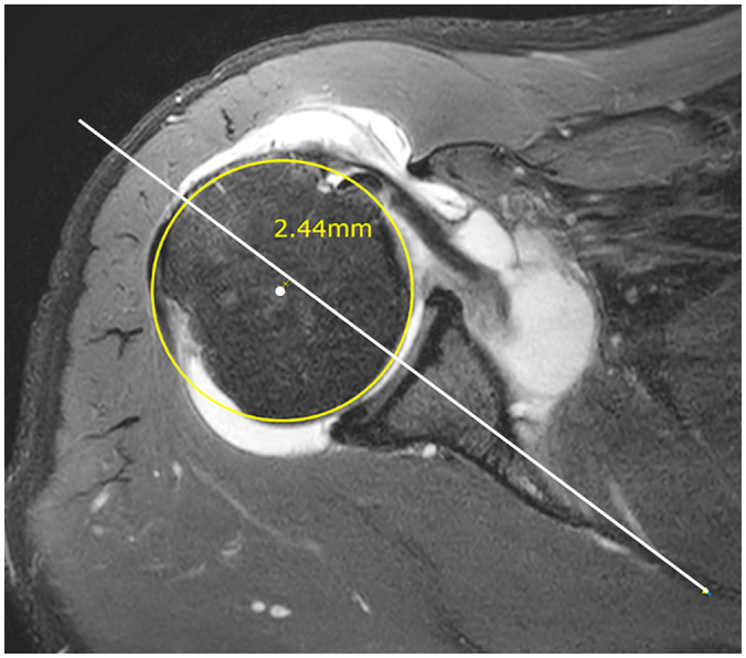
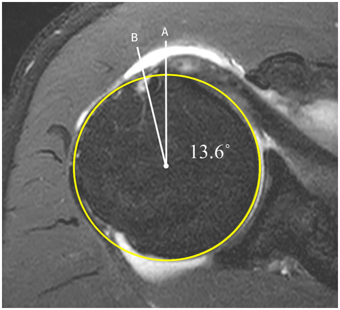
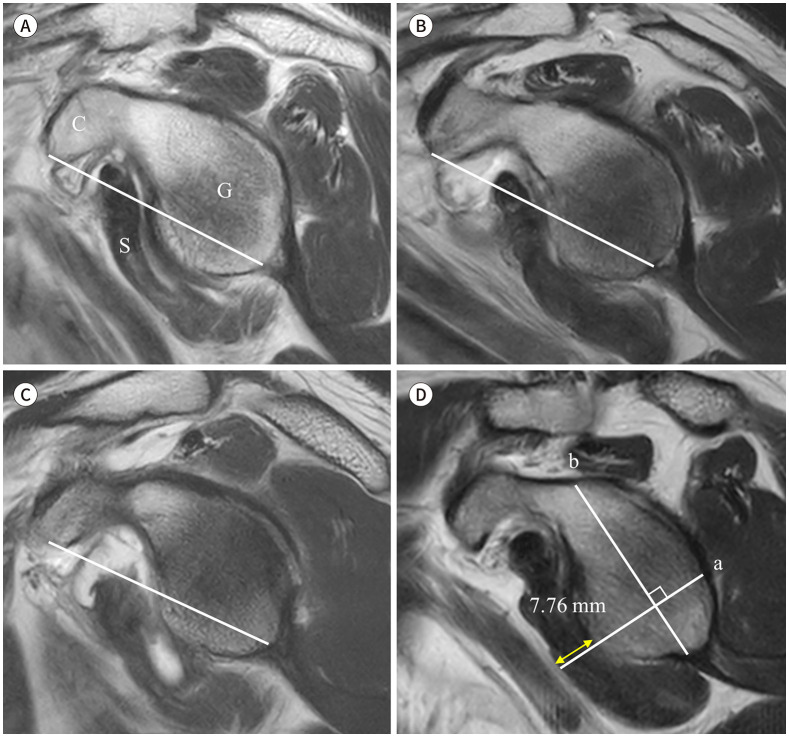
Materials and methods: Preoperative MRI scans of 86 patients were retrospectively reviewed for visual assessment of the STT, pathology of the long head of the biceps tendon (LHBT), posterior decentering (PD) of the humeral head, humeral rotation, fatty degeneration, and subscapularis muscle atrophy. To evaluate atrophy, visual grading using the anatomical line connecting the coracoid tip to the glenoid base, designated as the base-to-tip line (BTL), and thickness measurements were performed in the en-face view.
Results: Arthroscopically, 31 patients (36%) exhibited Lafosse type III or IV STT and underwent surgical repair. LHBT pathology (p = 0.002), PD of the humeral head (p = 0.012), fatty degeneration (p < 0.001), and BTL grade (p = 0.003) significantly correlated with STT. In the multivariate analysis, PD of the humeral head (p = 0.011, odds ratio [OR] = 5.14) and fatty degeneration (p = 0.046, OR = 2.81) were independent predictors of STT.
Conclusion: PD of the humeral head and fatty degeneration of the subscapularis can help to diagnose clinically significant STT. Interpretation of these findings may contribute to the planning of an optimal surgical strategy.
목적: 본 연구의 목적은 수술 전 MRI의 다양한 간접 소견 중 어떤 소견이 외과적 치료가 필요한 견갑하건 파열을 예측하는 데 가장 주요한 것인지 조사하는 것이다.
대상과 방법: 총 86명의 환자를 대상으로 수술 전 MRI 영상을 후향적으로 분석하였다. 견갑하건 파열의 직접평가, 이두박근 장두의 병리, 상완골두의 후방위, 상완골 회전, 견갑하근의 지방변성과 위축을 평가하였다. En-face 보기에서 부리돌기의 끝과 관절오목의 기저를 연결한 base-to-tip line (이하 BTL)을 이용한 육안 등급 및 두께 측정을 통해서 위축을 평가하였다.
결과: 관절경 시술에서 31명(36%)의 환자가 Lafosse type III 또는 IV의 견갑하건 파열을 보여, 재건수술을 받았다. 이두박근 장두의 병리(p = 0.002), 상완골두의 후방위(p = 0.012), 견갑하근의 지방 변성(p < 0.001), BTL 등급(p = 0.003)은 견갑하건 파열과 유의한 상관관계가 있었다. 다변량 분석에서 상완골두의 후방위(p = 0.011, odds ratio [이하 OR] = 5.14)와 견갑하근의 지방변성(p = 0.046, OR = 2.81)은 견갑하건 파열의 독립적인 예측인자였다.
결론: 상완골두의 후방위와 지방변성은 견갑하건 파열 진단에 도움이 될 수 있다. 이러한 결과를 판독하는 것은 최적의 수술 전략을 계획하는 데 기여할 수 있다.
Keywords: Fatty Degeneration; Magnetic Resonance Imaging; Posterior Decentering; Rotator Cuff Tear; Subscapularis.
Copyrights © 2024 The Korean Society of Radiology.
Conflict of interest statement
Conflicts of Interest: The authors have no potential conflicts of interest to disclose.
Figures
Similar articles
-
A novel diagnostic method to predict subscapularis tendon tear with sagittal oblique view magnetic resonance imaging.Knee Surg Sports Traumatol Arthrosc. 2019 Jan;27(1):277-288. doi: 10.1007/s00167-018-5203-0. Epub 2018 Oct 13. Knee Surg Sports Traumatol Arthrosc. 2019. PMID: 30317525
-
Predicting the clinically significant subscapularis tendon tear: malposition and tear of the long head of the biceps tendon on shoulder magnetic resonance imaging.Acta Radiol. 2021 Dec;62(12):1648-1656. doi: 10.1177/0284185120980017. Epub 2020 Dec 16. Acta Radiol. 2021. PMID: 33325726
-
Accuracy of long head of the biceps subluxation as a predictor for subscapularis tears.Arthroscopy. 2015 Apr;31(4):615-9. doi: 10.1016/j.arthro.2014.11.034. Epub 2015 Jan 28. Arthroscopy. 2015. PMID: 25636987
-
Imaging Review of Subscapularis Tendon and Rotator Interval Pathology.Radiol Res Pract. 2022 Jan 11;2022:4009829. doi: 10.1155/2022/4009829. eCollection 2022. Radiol Res Pract. 2022. PMID: 35070451 Free PMC article. Review.
-
Subscapularis tears: hidden and forgotten no more.JSES Open Access. 2018 Mar 1;2(1):74-83. doi: 10.1016/j.jses.2017.11.006. eCollection 2018 Mar. JSES Open Access. 2018. PMID: 30675571 Free PMC article. Review.
References
-
- Lee H, Ahn JM, Kang Y, Oh JH, Lee E, Lee JW, et al. Evaluation of the subscapularis tendon tears on 3T magnetic resonance arthrography: comparison of diagnostic performance of T1-weighted spectral presaturation with inversion-recovery and T2-weighted turbo spin-echo sequences. Korean J Radiol. 2018;19:320–327. - PMC - PubMed
-
- Kappe T, Sgroi M, Reichel H, Daexle M. Diagnostic performance of clinical tests for subscapularis tendon tears. Knee Surg Sports Traumatol Arthrosc. 2018;26:176–181. - PubMed
-
- Shim JW, Pang CH, Min SK, Jeong JY, Yoo JC. A novel diagnostic method to predict subscapularis tendon tear with sagittal oblique view magnetic resonance imaging. Knee Surg Sports Traumatol Arthrosc. 2019;27:277–288. - PubMed
-
- Furukawa R, Morihara T, Arai Y, Ito H, Kida Y, Sukenari T, et al. Diagnostic accuracy of magnetic resonance imaging for subscapularis tendon tears using radial-slice magnetic resonance images. J Shoulder Elbow Surg. 2014;23:e283–e290. - PubMed
-
- Lee SH, Nam DJ, Kim SJ, Kim JW. Comparison of clinical and structural outcomes by subscapularis tendon status in massive rotator cuff tear. Am J Sports Med. 2017;45:2555–2562. - PubMed
LinkOut - more resources
Full Text Sources
Research Materials
