. 2024 Jan 15;15(7):2509-2517.
doi: 10.1039/d3sc06551k. eCollection 2024 Feb 14.

Aldehyde-mediated inhibition of asparagine biosynthesis has implications for diabetes and alcoholism

Affiliations

Aldehyde-mediated inhibition of asparagine biosynthesis has implications for diabetes and alcoholism

Tobias John et al. Chem Sci. .

Abstract

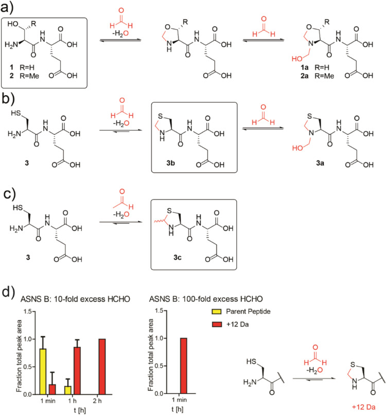
Patients with alcoholism and type 2 diabetes manifest altered metabolism, including elevated aldehyde levels and unusually low asparagine levels. We show that asparagine synthetase B (ASNS), the only human asparagine-forming enzyme, is inhibited by disease-relevant reactive aldehydes, including formaldehyde and acetaldehyde. Cellular studies show non-cytotoxic amounts of reactive aldehydes induce a decrease in asparagine levels. Biochemical analyses reveal inhibition results from reaction of the aldehydes with the catalytically important N-terminal cysteine of ASNS. The combined cellular and biochemical results suggest a possible mechanism underlying the low asparagine levels in alcoholism and diabetes. The results will stimulate research on the biological consequences of the reactions of aldehydes with nucleophilic residues.


Conflict of interest statement

There are no conflicts to declare.

Figures

Fig. 1
Fig. 1. Aldehyde addition decreases asparagine and increases cysteine-derived thiazolidine levels in human cells. (a) Asparagine, thioproline and 2-methylthiazolidine-4-carboxylic acid (MTCA) levels in HEK293T cells treated with aldehydes. Full data set: Fig. S3. (b) Reaction of cysteine with aldehydes/ketones. Structures of the cysteine-HCHO adduct (thioproline) and the cysteine-AcH adduct (MTCA) are shown; the cysteine-MGO adduct was not detected under our conditions. Independent replicates: Fig. S2b. (c) ASNS mRNA levels in HEK293T cells treated with HCHO (150 μM, then a second dose – 225 μM after 24 hours) compared to water (Milli-Q water, MQ). ASNS gene, fc = 0.58, p = 0.298, q = 0.594. Errors: SD of the mean (n = 3). (d) Western blots showing protein levels of ASNS and GAPDH from HEK293T cells treated with the indicated amount of HCHO or water for 14 hours. Independent replicates: Fig. S8. (e) MS analyses of asparagine levels in mouse livers. Full analyses: Fig. S12. (f) Oxidation of ethanol (EtOH) by alcohol dehydrogenase (ADH) and of AcH by aldehyde dehydrogenase 2 (ALDH2).
Fig. 2
Fig. 2. N-Terminal cysteine residues react with formaldehyde and acetaldehyde to form stable thiazolidines. (a) Peptides 1 and 2 react reversibly with HCHO to form N-terminal oxazolidines 1a and 2a, which are unstable under HPLC conditions, or on excess GSH addition (40 : 1 GSH : dipeptide). Full analyses: Fig. S13, S14 and S17a/b. (b) Peptide 3 reacts with HCHO to form thiazolidine 3b and hemiaminal 3a; the latter of is unstable under HPLC conditions. Addition of a 40-fold excess GSH to 3a results in 3b. Full analyses: Fig. S15–S17c. (c) Peptide 3 reacts with AcH to form N-terminal thiazolidine 3c, which is stable under HPLC conditions (Fig. S18†), but which degrades releasing AcH over time. Reactions employed a 10-fold excess of aldehyde (pD 9.4, 12 hours). Boxed structures are the major species isolated after HPLC purification. (d) Reaction of an ASNS B 7-residue N-terminal peptide (NH2-CSIFGVF-NH2) with HCHO (10-fold or 100-fold excess) at pH 7.4 and 50 mM potassium phosphate. HCHO adducts: red (+12 Da). Parent (unreacted) peptide: yellow. Reactions were monitored following addition of HCHO at the indicated times. Integrated areas of peaks were used (Fig. S22†). Errors: SEM (n = 3, technical repeats).
Fig. 3
Fig. 3. HCHO selectively inhibits the N-terminal glutaminase, but not the C-terminal synthetase, partial reaction of ASNS B. (a) Reactions involved in ASNS B catalysis (see also Fig. S1†). The glutamine amide NH2, which is transferred to aspartic acid, is in green. (b) 1H NMR (700 MHz) analysis of the C3 (Asp, Asn) and C4 (Gln, Glu) methylene groups in ASNS B-catalysed conversion of Gln (δH 2.42–2.51 ppm) to Glu (δH 2.55–2.61 ppm) and Asp (δH 3.03–3.13 ppm) to Asn (δH 2.97–3.00 ppm) (panel 2, +ASNS B). The glutaminase reaction, which is dependent on the ASNS B N-terminal cysteine, is inhibited by HCHO (panels 3, 4). Panel 6: the C-terminal synthetase domain of ASNS B is active in the presence of Asp, ATP, NH4Cl and a 103 excess of HCHO, but not when a 104-fold excess HCHO is pre-incubated with ASNS B (panel 7). See Fig. S25 for an independent replicate. (c) Order of hydration equilibrium constants for reactive carbonyl compounds; note glyoxal can undergo two hydrations. (d) ASNS B was pre-incubated with the indicated molar excess of carbonyl compound (or MQ water control) (45 minutes, 37 °C), then added to the assay components, with monitoring by 1H NMR (700 MHz, errors: SD of the mean (n = 2 independent repeats); see Fig. S26 for details). (e) Inhibition of THP-1 acute monocytic leukaemia cell growth by treatment with HCHO (80 μM), the HCHO metabolism inhibitor N6022 (10 μM), and/or ASNase (0.05 U mL−1). Growth inhibition (%) = 100 − (treatment group OD/non-treatment OD) × 100; OD = optical density. (*p value = 0.01 to 0.05, **p value = 0.001 to 0.01, ***p value = 0.0001 to 0.001, ****p value < 0.0001, n = 3 technical repeats. Biological replicates of the experiment shown in Fig. S27,n = 4 biological repeats).

Similar articles

Cited by

References

    1. Pontel L. B. Rosado I. V. Burgos-Barragan G. Garaycoechea J. I. Yu R. Arends M. J. Chandrasekaran G. Broecker V. Wei W. Liu L. Swenberg J. A. Crossan G. P. Patel K. J. Mol. Cell. 2015;60:177–188. doi: 10.1016/j.molcel.2015.08.020. - DOI - PMC - PubMed
    1. Maessen D. E. Hanssen N. M. Scheijen J. L. van der Kallen C. J. van Greevenbroek M. M. Stehouwer C. D. Schalkwijk C. G. Diabetes Care. 2015;38:913–920. doi: 10.2337/dc14-2605. - DOI - PubMed
    1. Kim J. K. Samaranayake M. Pradhan S. Cell. Mol. Life Sci. 2009;66:596–612. doi: 10.1007/s00018-008-8432-4. - DOI - PMC - PubMed
    1. Walport L. J. Hopkinson R. J. Schofield C. J. Curr. Opin. Chem. Biol. 2012;16:525–534. doi: 10.1016/j.cbpa.2012.09.015. - DOI - PubMed
    1. Skrzydlewska E. Toxicol. Mech. Methods. 2003;13:277–293. doi: 10.1080/713857189. - DOI - PubMed