. 2024 Apr;81(4):876-886.
doi: 10.1161/HYPERTENSIONAHA.123.22292. Epub 2024 Feb 16.

Differential 5'-tRNA Fragment Expression in Circulating Preeclampsia Syncytiotrophoblast Vesicles Drives Macrophage Inflammation

Affiliations

Differential 5'-tRNA Fragment Expression in Circulating Preeclampsia Syncytiotrophoblast Vesicles Drives Macrophage Inflammation

William R Cooke et al. Hypertension. 2024 Apr.

Abstract

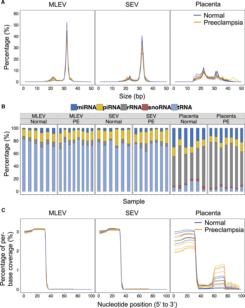
Background: The relationship between placental pathology and the maternal syndrome of preeclampsia is incompletely characterized. Mismatch between placental nutrient supply and fetal demands induces stress in the syncytiotrophoblast, the layer of placenta in direct contact with maternal blood. Such stress alters the content and increases the release of syncytiotrophoblast extracellular vesicles (STB-EVs) into the maternal circulation. We have previously shown 5'-tRNA fragments (5'-tRFs) constitute the majority of small RNA in STB-EVs in healthy pregnancy. 5'-tRFs are produced in response to stress. We hypothesized STB-EV 5'-tRF release might change in preeclampsia.

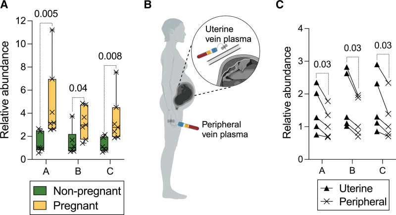
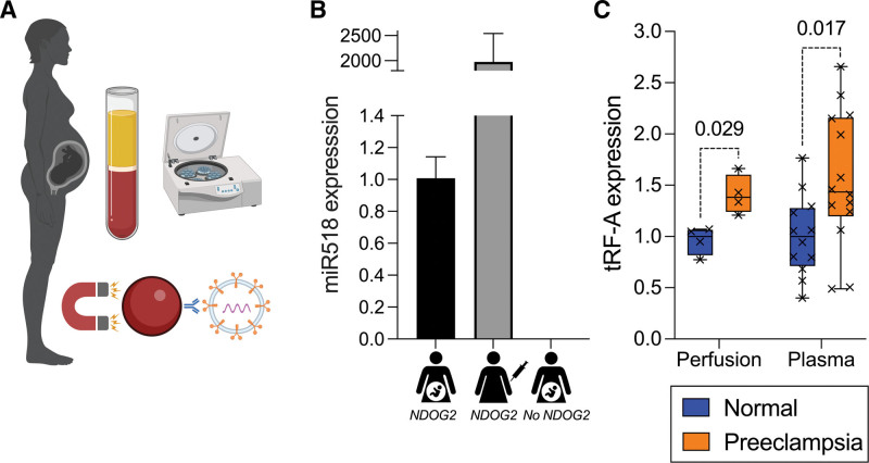
Methods: We perfused placentas from 8 women with early-onset preeclampsia and 6 controls, comparing small RNA expression in STB-EVs. We used membrane-affinity columns to isolate maternal plasma vesicles and investigate placental 5'-tRFs in vivo. We quantified 5'-tRFs from circulating STB-EVs using a placental alkaline phosphatase immunoassay. 5'-tRFs and scrambled RNA controls were added to monocyte, macrophage and endothelial cells in culture to investigate transcriptional responses.

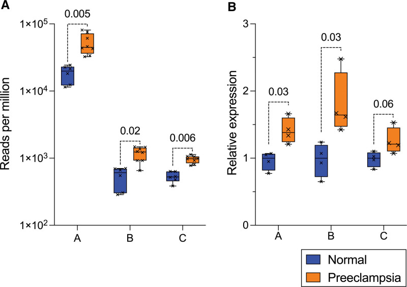
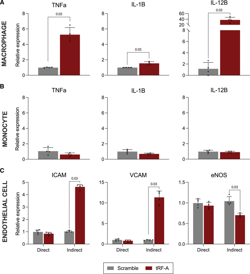
Results: 5'-tRFs constitute the majority of small RNA in STB-EVs from both preeclampsia and normal pregnancies. More than 900 small RNA fragments are differentially expressed in preeclampsia STB-EVs. Preeclampsia-dysregulated 5'-tRFs are detectable in maternal plasma, where we identified a placentally derived load. 5'-tRF-Glu-CTC, the most abundant preeclampsia-upregulated 5'-tRF in perfusion STB-EVs, is also increased in preeclampsia STB-EVs from maternal plasma. 5'-tRF-Glu-CTC induced inflammation in macrophages but not monocytes. The conditioned media from 5'-tRF-Glu-CTC-activated macrophages reduced eNOS (endothelial NO synthase) expression in endothelial cells.

Conclusions: Increased release of syncytiotrophoblast-derived vesicle-bound 5'-tRF-Glu-CTC contributes to preeclampsia pathophysiology.

Keywords: extracellular vesicles; macrophages; preeclampsia; transfer RNA.


Conflict of interest statement

Y.M.D. Lo holds equity in DRA, Insighta, Grail/Illumina and Take2. P. Jiang holds equity in Illumina. P. Jiang is a consultant to Take2. P. Jiang is a Director of Take2, Insighta, DRA and KingMed Future. Y.M.D. Lo , P. Jiang , and L. Ji receive royalties from Illumina, LabCorp, Grail, DRA, Xcelom and Take2.

Figures

Figure 1.
Figure 1.
Assessment of reads <50 base pairs (bps) from medium-large extracellular vesicles (MLEVs), small extracellular vesicles (SEV), and placental samples obtained from placentas in normotensive pregnancy (normal, n=6) and early onset preeclampsia (PE, n=8). A, Sequence length distribution plots showing size in bp for small RNA fragments after removal of adaptors and low-quality reads. B, Mapping of small RNA fragments. C, Coverage plots showing percentage of per-base coverage for mapped tRNA fragments.
Figure 2.
Figure 2.
Expression of 3 target small RNA fragments in early onset preeclampsia and normotensive pregnancy syncytiotrophoblast extracellular vesicles (STB-EVs) obtained by placental perfusion. A, Quantified using RNA sequencing (n=6 normotensive, n=8 preeclampsia; Mann-Whitney U test after Benjamini-Hochberg correction displayed). B, Quantified using stem-loop qPCR (normalized to reference gene TBP, n=4 per group). Boxes show median/interquartile range; whiskers show max/min. Fragments labeled A, B, C for brevity; full sequences in Table S2.
Figure 3.
Figure 3.
Detection of 3 target small RNA fragments in plasma using qPCR. A, Expression in total extracellular vesicles (EVs) isolated from peripheral venous plasma from 7 healthy third trimester pregnancies and 6 female volunteers of reproductive age (boxes show median/interquartile range; whiskers show max/min). B, Diagram demonstrating sampling rationale for uterine and peripheral venous blood. C, Expression in total EVs isolated from uterine and peripheral venous plasma taken simultaneously from 5 healthy term pregnancies (paired 1-tailed Wilcoxon tests displayed). Fragments labeled A, B, C for brevity; full sequences in Table S2.
Figure 4.
Figure 4.
Isolation of syncytiotrophoblast extracellular vesicles (STB-EVs) from venous plasma. A, Diagram summarizing protocol for STB-EV isolation: plasma preparation, 10 000g centrifugation to isolate medium-large extracellular vesicles (MLEVs), immuno-isolation using magnetic beads coated with antibody to placental alkaline phosphatase (NDOG2). B, Relative expression of miR518 in EVs isolated from 3 samples using above technique (from left to right: pregnant plasma using beads coated with NDOG2; nonpregnant plasma spiked with perfusion STB-EVs using beads coated with NDOG2; pregnant plasma using beads without NDOG2 coating). Bars represent median, error bars represent interquartile range. C, Relative expression of 5′-tRF-Glu-CTC (tRF-A) in perfusion-derived STB-EVs (not gestation-matched, reproduced from Figure 2B for comparison) and gestation-matched peripheral venous plasma STB-EVs from women with early onset preeclampsia (n=14) and normotensive pregnancies (n=12).
Figure 5.
Figure 5.
5′-tRF-Glu-CTC (tRF-A) actions in immune and endothelial cells. A, Cytokine expression by THP-1-derived macrophages after treatment with vesicle-encapsulated RNA for 12 hours. B, Cytokine expression by THP-1 monocytes (undifferentiated) after treatment with vesicle-encapsulated RNA for 12 hours. C, Expression of cell adhesion molecule and endothelial NO synthase mRNA in human umbilical vein endothelial cells treated with vesicle-encapsulated RNA (Direct), or the cell culture medium from macrophages pretreated with vesicle-encapsulated RNA (Indirect) for 6 h. Fragments labeled tRF-A and Scramble for brevity; full sequences in Table S2 and Methods; n=4 per condition.

References

    1. Powe CE, Levine RJ, Karumanchi SA. Preeclampsia, a disease of the maternal endothelium: the role of antiangiogenic factors and implications for later cardiovascular disease. Circulation. 2011;123:2856–2869. doi: 10.1161/CIRCULATIONAHA.109.853127 - PMC - PubMed
    1. Magee LA, Nicolaides KH, von Dadelszen P. Preeclampsia. N Engl J Med. 2022;386:1817–1832. doi: 10.1056/NEJMra2109523 - PubMed
    1. Redman CWG, Staff AC, Roberts JM. Syncytiotrophoblast stress in preeclampsia: the convergence point for multiple pathways. Am J Obstet Gynecol. 2022;226:S907–S927. doi: 10.1016/j.ajog.2020.09.047 - PubMed
    1. Levine RJ, Maynard SE, Qian C, Lim KH, England LJ, Yu KF, Schisterman EF, Thadhani R, Sachs BP, Epstein FH, et al. . Circulating angiogenic factors and the risk of preeclampsia. N Engl J Med. 2004;350:672–683. doi: 10.1056/NEJMoa031884 - PubMed
    1. Cindrova-Davies T, Sanders DA, Burton GJ, Charnock-Jones DS. Soluble FLT1 sensitizes endothelial cells to inflammatory cytokines by antagonizing VEGF receptor-mediated signalling. Cardiovasc Res. 2011;89:671–679. doi: 10.1093/cvr/cvq346 - PMC - PubMed