. 2024 Mar 12;92(3):e0056022.
doi: 10.1128/iai.00560-22. Epub 2024 Feb 16.

Drosophila melanogaster Sting mediates Coxiella burnetii infection by reducing accumulation of reactive oxygen species

Affiliations

Drosophila melanogaster Sting mediates Coxiella burnetii infection by reducing accumulation of reactive oxygen species

Rosa M Guzman et al. Infect Immun. .

Abstract

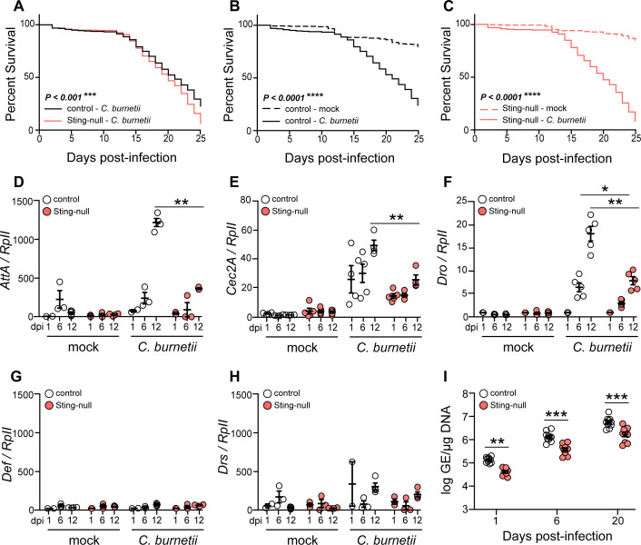
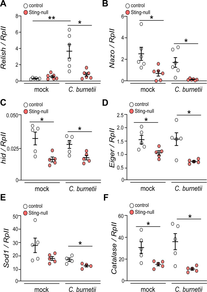
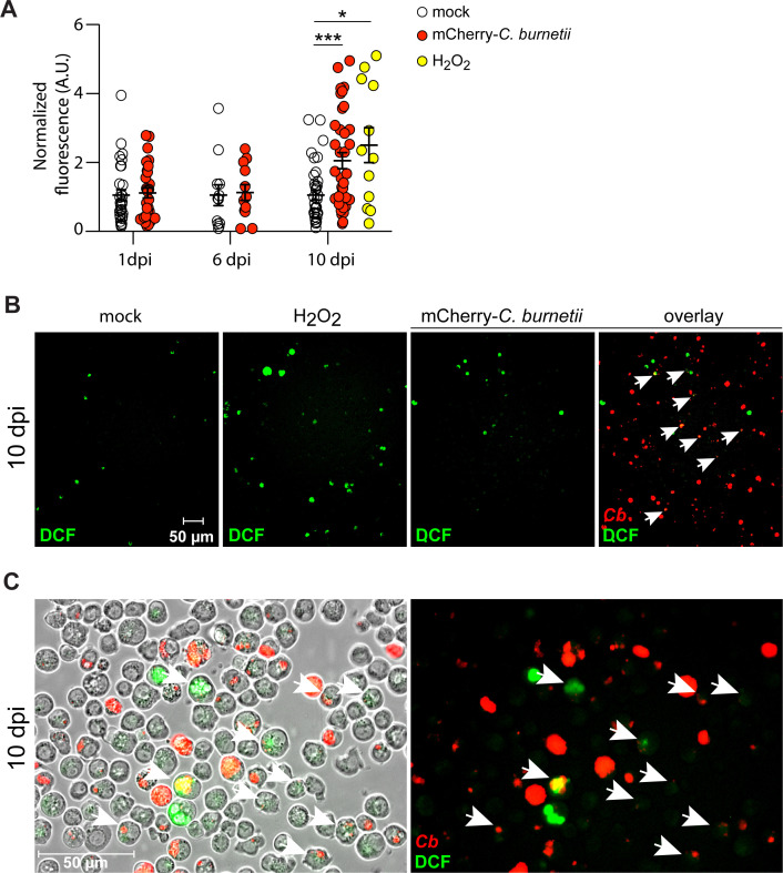
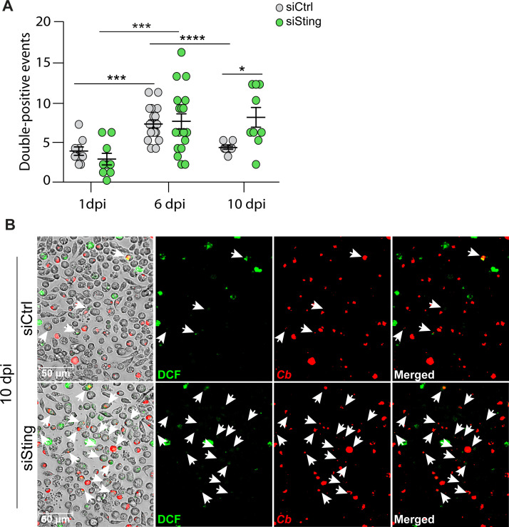
The Gram-negative bacterium Coxiella burnetii is the causative agent of query fever in humans and coxiellosis in livestock. C. burnetii infects a variety of cell types, tissues, and animal species including mammals and arthropods, but there is much left to be understood about the molecular mechanisms at play during infection in distinct species. Human stimulator of interferon genes (STING) induces an innate immune response through the induction of type I interferons (IFNs), and IFN promotes or suppresses C. burnetii replication, depending on tissue type. Drosophila melanogaster contains a functional STING ortholog (Sting) which activates NF-κB signaling and autophagy. Here, we sought to address the role of D. melanogaster Sting during C. burnetii infection to uncover how Sting regulates C. burnetii infection in flies. We show that Sting-null flies exhibit higher mortality and reduced induction of antimicrobial peptides following C. burnetii infection compared to control flies. Additionally, Sting-null flies induce lower levels of oxidative stress genes during infection, but the provision of N-acetyl-cysteine (NAC) in food rescues Sting-null host survival. Lastly, we find that reactive oxygen species levels during C. burnetii infection are higher in Drosophila S2 cells knocked down for Sting compared to control cells. Our results show that at the host level, NAC provides protection against C. burnetii infection in the absence of Sting, thus establishing a role for Sting in protection against oxidative stress during C. burnetii infection.

Keywords: ROS; STING; bacteria; fly; pathogenesis.


Conflict of interest statement

The authors declare no conflict of interest.

Figures

Fig 1
Fig 1
Sting-null flies succumb to C. burnetii infection. (A–C) Adult Drosophila flies were mock-infected or infected with C. burnetii, and survival was monitored for 25 days, comparing the infected flies of each genotype (A) or mock- and C. burnetii-infected flies for each genotype (B and C). Expression of Attacin (D), Cecropin (E), Drosocin (F), Defensin (G), and Drosomycin (H) in control or Sting-null flies were determined at 1, 6, and 12 days post-infection (dpi) by reverse transcriptase quantitative real-time PCR, and the results were normalized to Drosophila RpII transcripts. (I) Bacterial loads were quantified at 1, 6, and 20 dpi by quantitative PCR. Asterisks denote significance: *P < 0.05, **P < 0.01, ***P < 0.001, ****P < 0.0001. Error bars indicate standard error of the mean. Each survival curve represents three independent experiments of 40–50 flies that were combined for a final survival curve and statistical analysis. For qRT-PCR, each circle represents an individual biological replicate of a pooled collection of five flies, and results are representative of three independent experiments.
Fig 2
Fig 2
Sting-null flies cannot induce cell death and oxidative stress genes. Expression of Relish (A), Nazo (B), hid (C), Eiger (D), Sod1 (E), and Catalase (F) at 12 dpi was determined in control and Sting-null flies by reverse transcriptase quantitative real time PCR, and the results were normalized to Drosophila RpII transcripts. Asterisks denote significance: *P < 0.05, **P < 0.01. Error bars indicate standard error of the mean. Each circle represents an individual biological replicate of a pooled collection of five flies, and results are representative of three independent experiments.
Fig 3
Fig 3
ROS is generated during C. burnetii infection in S2 cells. S2 cells were infected with mCherry-C. burnetii (100 GE/cell), and at 1, 6, and 10 dpi, cells were collected for DCFH-DA staining. Cells were imaged, and fluorescence intensity was quantified using ImageJ, where fluorescence of infected cells was normalized to mock (A). H2O2 was used as positive control to induce ROS in non-infected cells. Asterisks denote significance: *P < 0.05, ***P < 0.001. Error bars indicate standard error of the mean. Representative images of data quantification from 10 dpi are shown in panel B, where DCF fluorescence is shown for mock, H2O2, mCherry-C. burnetii, and overlay channels of infected cells, where white arrows point at double-positive cells (×200 magnification), and ×400 magnification (C). Data are representative of six individual images from three independent experiments.
Fig 4
Fig 4
Expression of Sod1 is reduced in Sting-knockdown S2 cells during C. burnetii infection. Schematic of experimental design (A). At the indicated dpi, cells are transfected with control dsRNA (siCtrl) or dsRNA targeting Sting (siSting). Two days post-transfection, the ROS assay is performed. Expression of Sting (B), Sod1 (C), and Catalase (D) at 1, 6, and 10 dpi was determined in control and Sting-knockdown cells by reverse transcriptase quantitative real-time PCR, and the results were normalized to Drosophila RpII transcripts. Asterisks denote significance: *P < 0.05, **P < 0.01, ****P < 0.0001. Error bars indicate standard errors of the mean. Each circle represents an individual biological replicate of a well of cells, and results are representative of three independent experiments.
Fig 5
Fig 5
ROS is higher in Sting-knockdown S2 cells during C. burnetii infection. The number of double-positive S2 cells with both mCherry-C. burnetii and DCF fluorescence was quantified during siCtrl or siSting knockdown at 1, 6, and 10 dpi (A). Asterisks denote significance: *P < 0.05, ***P < 0.001, ****P < 0.0001. Error bars indicate standard error of the mean. Representative images of siCtrl and siSting S2 cells at ×400 magnification at 10 dpi showing (from left to right) phase contrast image overlay, DCF fluorescence, mCherry-C. burnetii fluorescence, and merged panels (B), where white arrows point at double-positive cells. Data are representative of six individual images from three independent experiments.
Fig 6
Fig 6
NAC rescues host survival to C. burnetii infection in Sting-null hosts. Adult flies were mock- or C. burnetii-infected. Following infection, flies were placed on control or NAC-containing food, and survival was monitored for 25 days. Survival curves compare infected NAC-fed flies of both fly genotypes (A), control genotypes under all conditions (B), or Sting-null genotypes under all conditions (C). Asterisks denote significance: *P < 0.05, ****P < 0.0001. Each survival curve represents three independent experiments of 40–50 flies that were combined for a final survival curve and statistical analysis.

Similar articles

Cited by

References

    1. Maurin M, Raoult D. 1999. Q fever. Clin Microbiol Rev 12:518–553. doi:10.1128/CMR.12.4.518 - DOI - PMC - PubMed
    1. Plummer PJ, McClure JT, Menzies P, Morley PS, Van den Brom R, Van Metre DC. 2018. Management of Coxiella burnetii infection in livestock populations and the associated zoonotic risk: a consensus statement. J Vet Intern Med 32:1481–1494. doi:10.1111/jvim.15229 - DOI - PMC - PubMed
    1. Mori M, Roest HJ. 2018. Farming, Q fever and public health: agricultural practices and beyond. Arch Public Health 76:2. doi:10.1186/s13690-017-0248-y - DOI - PMC - PubMed
    1. Waag DM. 2007. Coxiella burnetii: host and bacterial responses to infection. Vaccine 25:7288–7295. doi:10.1016/j.vaccine.2007.08.002 - DOI - PubMed
    1. Duron O, Sidi-Boumedine K, Rousset E, Moutailler S, Jourdain E. 2015. The importance of ticks in Q fever transmission: what has (and has not) been demonstrated? Trends Parasitol 31:536–552. doi:10.1016/j.pt.2015.06.014 - DOI - PubMed

Publication types

MeSH terms

LinkOut - more resources