. 2024 Feb 16;47(1):83.
doi: 10.1007/s10143-024-02311-5.

Fully endoscopic microvascular decompression for hemifacial spasm: a clinical study and analysis

Affiliations

Fully endoscopic microvascular decompression for hemifacial spasm: a clinical study and analysis

Xialin Zheng et al. Neurosurg Rev. .

Abstract

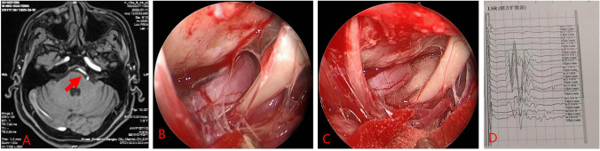
Fully endoscopic microvascular decompression (MVD) of the facial nerve is the main surgical treatment for hemifacial spasm. However, the technique presents distinct surgical challenges. We retrospectively analyzed prior cases to consolidate surgical insights and assess clinical outcomes. Clinical data from 16 patients with facial nerve spasms treated at the Department of Neurosurgery in the First Affiliated Hospital of Bengbu Medical College, between August 2020 and July 2023, were retrospectively examined. Preoperatively, all patients underwent magnetic resonance angiography to detect any offending blood vessels; ascertain the relationship between offending vessels, facial nerves, and the brainstem; and detect any cerebellopontine angle lesions. Surgery involved endoscopic MVD of the facial nerve using a mini Sigmoid sinus posterior approach. Various operative nuances were summarized and analyzed, and clinical efficacy, including postoperative complications and the extent of relief from facial paralysis, was evaluated. Fully endoscopic MVD was completed in all patients, with the offending vessels identified and adequately padded during surgery. The offending vessels were anterior inferior cerebellar artery in 12 cases (75%), vertebral artery in 3 cases (18.75%), and posterior inferior cerebellar artery in 1 case (6.25%). Intraoperative electrophysiological monitoring revealed that the lateral spread response of the facial nerve vanished in 15 cases and remained unchanged in 1 case. Postoperative facial spasms were promptly alleviated in 15 cases (93.75%) and delayed in 1 case (6.25%). Two cases of postoperative complications were recorded-one intracranial infection and one case of tinnitus-both were resolved or mitigated with treatment. All patients were subject to follow-up, with no instances of recurrence or mortality. Fully endoscopic MVD of the facial nerve is safe and effective. Proficiency in endoscopy and surgical skills are vital for performing this procedure.

Keywords: Endoscope; Facial spasm; Microvascular decompression; Neuroendoscopy.


Conflict of interest statement

The authors declare no competing interests.

Figures

Fig. 1
Fig. 1
Procedure for fully endoscopic microvascular decompression of hemifacial spasms. A A curved incision behind the ear is depicted by the black line. B Position the patient in the lateral decubitus position with the mastoid tip at the highest point and secure the head with a three-pin headrest. C Slightly flex the head forward, maintaining a distance of two fingerbreadths between the mandible and the chest wall to avoid excessive flexion of the head. D Perform burr hole drilling. E The exposure range of the bone window is indicated, with the sigmoid sinus marked by a red circle and the transverse sinus by a yellow circle. F Ensure that the sigmoid sinus side of the bone window is fully exposed and level so that this edge aligns with the posterior edge of the petrous bone in a straight line, as indicated by the red dashed line. G The size of the bone flap should be similar to the size of the volar surface of the thumb. H The assistant holding the endoscope is positioned to the dominant left side, the endoscopic screen is placed opposite the surgeon, and the scrub nurse is positioned diagonally opposite the surgeon. I Two-person, three-handed “triangular three-point” microsurgical technique: the assistant holds the apex, while the surgeon uses both hands with the microscope positioned at the two base points for instrument manipulation. J One support point for the assistant holding the endoscope is the edge of the bone window at the sigmoid sinus side (yellow circle). K Another support point for the assistant holding the endoscope (red circle). L Intradermal suturing along the skin eversion line
Fig. 2
Fig. 2
Case 1: male, 51 years old. A Preoperative magnetic resonance tomographic angiography (MRTA) indicates left vertebral artery compression of the facial nerve, with the red arrow pointing to the left vertebral artery. B Intraoperative observation shows the vertebral artery compressing the facial nerve. C Insertion of a spacer. D Laser speckle rheology (LSR) monitoring indicates the disappearance of abnormal waves after spacer insertion
Fig. 3
Fig. 3
Case 2: male, 53 years old. A Preoperative magnetic resonance tomographic angiography (MRTA) suggests the right vertebral artery is exhibiting a climbing pattern compressing the facial nerve, with the red arrow pointing to the left vertebral artery. B Intraoperative observation reveals the right vertebral artery compressing the facial nerve. C Insertion of a spacer
Fig. 4
Fig. 4
Case 3: female, 51 years old. A Preoperative magnetic resonance tomographic angiography (MRTA) suggests compression of the facial nerve root exit zone (REZ) by the right anterior inferior cerebellar artery, with the red arrow pointing to the right anterior inferior cerebellar artery. B Intraoperative observation shows the right anterior inferior cerebellar artery in a climbing pattern compressing the facial nerve. C Insertion of a spacer. D LSR indicates the disappearance of abnormal waves

Similar articles

Cited by

References

    1. Jiang HT et al (2022) Fully endoscopic microvascular decompression for hemifacial spasm. Exp Ther Med 24(1):483. 10.3892/etm.2022.11410 - PMC - PubMed
    1. Wang L et al (2023) Bilateral transient dilated and fixed pupils after microvascular decompression: rare clinical experience. J Craniofac Surg 34(4):1296–1300. 10.1097/SCS.0000000000009293 - PubMed
    1. Guo X et al (2022) Fully endoscopic microvascular decompression for hemifacial spasm using improved retrosigmoid infrafloccular approach: clinical analysis of 81 cases. Oper Neurosurg 23(1):40–45. 10.1227/ons.0000000000000221 - PubMed
    1. Cai Q, Li Z, Guo Q, Wang W, Ji B, Chen Z, Dong H, Mao S (2023) Microvascular decompression using a fully transcranial neuroendoscopic approach. Br J Neurosurg 37(5):1375–1378. 10.1080/02688697.2020.1820943 - PubMed
    1. Matmusaev M et al (2020) Endoscopic microvascular decompression for hemifacial spasm. Asian J Neurosurg 15(4):833–838. 10.4103/ajns.AJNS_152_20 - PMC - PubMed

MeSH terms

LinkOut - more resources