. 2024 Jul 15;119(4):1234-1247.
doi: 10.1016/j.ijrobp.2024.01.224. Epub 2024 Feb 15.

FLASH Proton Radiation Therapy Mitigates Inflammatory and Fibrotic Pathways and Preserves Cardiac Function in a Preclinical Mouse Model of Radiation-Induced Heart Disease

Affiliations

FLASH Proton Radiation Therapy Mitigates Inflammatory and Fibrotic Pathways and Preserves Cardiac Function in a Preclinical Mouse Model of Radiation-Induced Heart Disease

Kyle Kim et al. Int J Radiat Oncol Biol Phys. .

Abstract

Purpose: Studies during the past 9 years suggest that delivering radiation at dose rates exceeding 40 Gy/s, known as "FLASH" radiation therapy, enhances the therapeutic index of radiation therapy (RT) by decreasing normal tissue damage while maintaining tumor response compared with conventional (or standard) RT. This study demonstrates the cardioprotective benefits of FLASH proton RT (F-PRT) compared with standard (conventional) proton RT (S-PRT), as evidenced by reduced acute and chronic cardiac toxicities.

Methods and materials: Mice were imaged using cone beam computed tomography to precisely determine the heart's apex as the beam isocenter. Irradiation was conducted using a shoot-through technique with a 5-mm diameter circular collimator. Bulk RNA-sequencing was performed on nonirradiated samples, as well as apexes treated with F-PRT or S-PRT, at 2 weeks after a single 40 Gy dose. Inflammatory responses were assessed through multiplex cytokine/chemokine microbead assay and immunofluorescence analyses. Levels of perivascular fibrosis were quantified using Masson's Trichrome and Picrosirius red staining. Additionally, cardiac tissue functionality was evaluated by 2-dimensional echocardiograms at 8- and 30-weeks post-PRT.

Results: Radiation damage was specifically localized to the heart's apex. RNA profiling of cardiac tissues treated with PRT revealed that S-PRT uniquely upregulated pathways associated with DNA damage response, induction of tumor necrosis factor superfamily, and inflammatory response, and F-PRT primarily affected cytoplasmic translation, mitochondrion organization, and adenosine triphosphate synthesis. Notably, F-PRT led to a milder inflammatory response, accompanied by significantly attenuated changes in transforming growth factor β1 and α smooth muscle actin levels. Critically, F-PRT decreased collagen deposition and better preserved cardiac functionality compared with S-PRT.

Conclusions: This study demonstrated that F-PRT reduces the induction of an inflammatory environment with lower expression of inflammatory cytokines and profibrotic factors. Importantly, the results indicate that F-PRT better preserves cardiac functionality, as confirmed by echocardiography analysis, while also mitigating the development of long-term fibrosis.


Figures

Fig. 1.
Fig. 1.
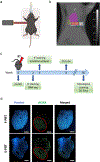
Irradiation setup. (a) Irradiation setting; the proton beam angled at 15o to the vertical (created with BioRender.com). (b) Dose plan in MuriPlan. Frontal view of RT planning and delivery to the cardiac apex at a dose of 40 Gy with a 5-mm diameter circular collimator angled at 15o to the horizontal. Purple and orange areas indicate the unirradiated and irradiated cardiac areas, respectively. (c) Timeline of experiments conducted in this study. (d) phosphorylated form of histone H2AX immunofluorescence staining in optimal cutting temperature-frozen FLASH proton RT or standard (conventional) proton RT-treated cardiac sections. The red dotted circle outlines the irradiated area. Scale bar, 1 mm. Abbreviation: RT = radiation therapy.
Fig. 2.
Fig. 2.
RNA-seq analysis on S-PRT and F-PRT treated hearts (apex) at 2 weeks post-proton RT. (a) Groups of samples (n = 4 for nonirradiated; n = 4 for S-PRT; and n = 3 for F-PRT) analyzed using multidimensional scaling plots. (b) Venn diagram indicating the number of significant (false discovery rate < 0.01) differentially expressed genes across 2 key comparisons (F-PRT, S-PRT) and the overlap between each set of genes. (c, d) Gene ontology enrichment analysis of the differentially expressed genes (upregulated) in the heart of S-PRT treated mice compared with F-PRT treated mice; n = 3–4 per group. Abbreviations: F-PRT = FLASH proton RT; RT = radiation therapy; S-PRT = standard (conventional) proton RT.
Fig. 3.
Fig. 3.
Significant reduction of inflammatory and profibrotic responses in F-PRT treated hearts compared with S-PRT group. (a) Fold change (normalized to nonirradiated, n = 4) of tumor necrosis factor a from heart lysates at 3 weeks post 40 Gy of focal PRT (n = 4 for S-PRT and n = 4 for F-PRT). (b) Quantification of integrated density of TGF-β1 production at 2 weeks and (c) at 3 weeks post-PRT (n = 5 for S-PRT and n = 5 for F-PRT). Each dot represents quantitative value from a ×10 field. (d) Representative images of TGF-β1 staining. (e) Mean TGF-β1 integrated density at various timepoints post-PRT from (b) and (c). (f) Quantification of %α-smooth muscle actin + area at 3 weeks post-PRT (n = 5 for S-PRT and n = 5 for F-PRT). (g) Representative images of aSMA/CD31 costaining. Scale bar, 100 mm. Box and whisker plots. P values calculated with 2-tailed t test. *P < .05, **P < .01, ***P < .001. Abbreviations: F-PRT = FLASH proton RT; n.s.= not significant; PRT = proton RT; RT = radiation therapy; S-PRT = standard (conventional) proton RT; TGF = transforming growth factor.
Fig. 4.
Fig. 4.
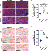
Significant reduction of myocardial fibrosis in F-PRT treated mice at 30 weeks post-PRT. (a) Representative Masson’s trichrome stain images (n = 2) of formalin fixed heart sections irradiated with 40 Gy at 30 weeks post-PRT. (b) Quantification of fibrosis formation by calculating the average muscle layer thickness (n = 3 for nonirradiated; n = 5 for standard [conventional] proton RT; and n = 5 for F-PRT). (c) Representative Picrosirius red stain images (n = 2) of formalin fixed heart sections irradiated with 40 Gy at 30 weeks post-PRT. (d) Quantification of the percentage of collagen area in irradiated heart areas (n = 3 for nonirradiated; n = 5 for standard [conventional] proton RT; and n = 5 for F-PRT). Box and whisker plots. P values calculated with 2-tailed t test. *P < .05, **P < .01, ***P < .001; Scale bar, 200 mm. Abbreviations: F-PRT = FLASH proton RT; n. s. = not significant; PRT = proton RT; RT = radiation therapy.
Fig. 5.
Fig. 5.
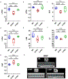
Preservation of heart functionality in F-PRT treated mice. (a-c) Diastolic 2-dimensional echocardiography on S-PRT and F-PRT treated hearts at 8 weeks post-PRT (n = 5 for nonirradiated; n = 5 for S-PRT; and n = 5 for F-PRT). (d-g) Diastolic 2-dimensional echocardiography on S-PRT and F-PRT treated hearts at 30 weeks post-PRT (n = 5 for nonirradiated; n = 5 for S-PRT; and n = 4 for F-PRT). Box and whisker plots. P values calculated with 2-tailed t test. *P < .05, **P < .01, ***P < .001. (h) Representative images of relative wall thickness at 30-weeks post-PRT. Abbreviations: ESV = end-systolic left ventricular volume (mL); ET = ejection time; F-PRT = FLASH proton RT; LVIDd = LV internal dimension, diastole (mm); LVIDs = LV internal dimension, systole (mm); LVM = LV mass (mg); n.s.= not significant; PRT = proton RT; RT = radiation therapy; S-PRT = standard (conventional) proton RT.

References

    1. Delaney G, Jacob S, Featherstone C, Barton M. The role of radiotherapy in cancer treatment: Estimating optimal utilization from a review of evidence-based clinical guidelines. Cancer 2005;104:1129–1137. - PubMed
    1. Ghobadi G, van der Veen S, Bartelds B, et al. Physiological interaction of heart and lung in thoracic irradiation. Int J Radiat Oncol Biol Phys 2012;84:e639–e646. - PubMed
    1. Darby SC, Ewertz M, Hall P. Ischemic heart disease after breast cancer radiotherapy. N Engl J Med 2013;368:2527. - PubMed
    1. Wang K, Eblan MJ, Deal AM, et al. Cardiac toxicity after radiotherapy for stage III non-small-cell lung cancer: Pooled analysis of dose-escalation trials delivering 70 to 90 Gy. J Clin Oncol 2017;35:1387–1394. - PMC - PubMed
    1. Galper SL, Yu JB, Mauch PM, et al. Clinically significant cardiac disease in patients with Hodgkin lymphoma treated with mediastinal irradiation. Blood 2011;117:412–418. - PubMed

MeSH terms