Review
. 2024 May:100:101247.
doi: 10.1016/j.preteyeres.2024.101247. Epub 2024 Feb 15.

Modeling complex age-related eye disease

Affiliations
Review

Modeling complex age-related eye disease

Silke Becker et al. Prog Retin Eye Res. 2024 May.

Abstract

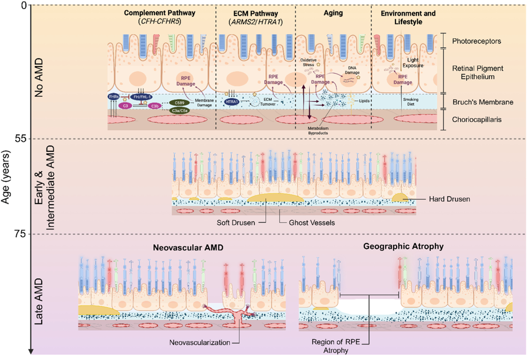
Modeling complex eye diseases like age-related macular degeneration (AMD) and glaucoma poses significant challenges, since these conditions depend highly on age-related changes that occur over several decades, with many contributing factors remaining unknown. Although both diseases exhibit a relatively high heritability of >50%, a large proportion of individuals carrying AMD- or glaucoma-associated genetic risk variants will never develop these diseases. Furthermore, several environmental and lifestyle factors contribute to and modulate the pathogenesis and progression of AMD and glaucoma. Several strategies replicate the impact of genetic risk variants, pathobiological pathways and environmental and lifestyle factors in AMD and glaucoma in mice and other species. In this review we will primarily discuss the most commonly available mouse models, which have and will likely continue to improve our understanding of the pathobiology of age-related eye diseases. Uncertainties persist whether small animal models can truly recapitulate disease progression and vision loss in patients, raising doubts regarding their usefulness when testing novel gene or drug therapies. We will elaborate on concerns that relate to shorter lifespan, body size and allometries, lack of macula and a true lamina cribrosa, as well as absence and sequence disparities of certain genes and differences in their chromosomal location in mice. Since biological, rather than chronological, age likely predisposes an organism for both glaucoma and AMD, more rapidly aging organisms like small rodents may open up possibilities that will make research of these diseases more timely and financially feasible. On the other hand, due to the above-mentioned anatomical and physiological features, as well as pharmacokinetic and -dynamic differences small animal models are not ideal to study the natural progression of vision loss or the efficacy and safety of novel therapies. In this context, we will also discuss the advantages and pitfalls of alternative models that include larger species, such as non-human primates and rabbits, patient-derived retinal organoids, and human organ donor eyes.

Keywords: Age-related disease; Age-related macular degeneration; Drusen; Eye disease; Geographic atrophy; Glaucoma; Mouse model; Non-human primate model; Optic nerve degeneration; Rabbit model; Retina.


Figures

Fig. 1.
Fig. 1.. Fundus and OCT images from early/intermediate AMD patient (A, B) and control subject (C, D).
Drusen can be seen as yellow spots in the fundus image (A) and as protrusions, as indicated by arrow heads, in the OCT image (B).
Fig. 2.
Fig. 2.. Natural History of age-related macular degeneration (AMD).
Schematic showing pathways driving AMD-associated pathology, which develops primarily at the interface between retinal pigment epithelium (RPE), Bruch’s membrane (BrM) and choriocapillaris. These pathways are at play prior to the clinical onset of early and intermediate AMD and subsequent progression to late AMD.
Fig. 3.
Fig. 3.. Altered dark adaptation (A) and dark-adapted ERG light responses in human AMD patients (B) and a mouse model of CFH variant (C).
(A) Recovery of visual sensitivity after bleach from control subjects (black) and from AMD patients (grey). (B) Leading edge of ERG light flash response from control (black) and dry (blue) and wet (red) AMD patients. (C) ERG light flash response amplitudes as a function of flash intensity in control (no high-fat diet, black; high-fat cholesterol-enriched diet, green) and in human CFH risk variant expressing mice (no high-fat diet, blue; high-fat cholesterol-enriched diet, pink). Adapted from (Murray et al., 2022), (A) (Dimopoulos et al., 2013), (B) (Landowski et al., 2019), (C).
Fig. 4.
Fig. 4.. Optic nerve head and retinal nerve fiber changes in glaucoma patients diagnosed using fundus photography.
Glaucomatous eye (A) has larger cup (bright center) to rim ratio compared to control eye (B). Images in C–F are from the same subject who developed glaucoma within 4-year period. In C the nerve fibers look normal but start to disappear from regions indicated in arrows in D-F. At the time when image F was photographed, patient had developed visual field deficits. (A, B) from (Abramoff et al., 2007), and (C–F) from (Quigley et al., 1992).
Fig. 5.
Fig. 5.
The major modeling strategies of AMD (A) and glaucoma (B) based on genetic variants (purple), environmental/lifestyle stressors (green) and pathological pathways (yellow).
Fig. 6.
Fig. 6.. Manhattan plot taken with permission from Fritsche et al., 2016 showing the fifty-two independent variants across 34 loci independently associated with AMD through genome-wide association studies.
The two most common contributors to disease are the CFH-CFHR5 region on chromosome 1q32 and the ARMS2/HTRA1 locus on chromosome 1q26. Other variants have a comparatively smaller effect on disease outcome.
Fig. 7.
Fig. 7.
A large part of the population-attributable risk for AMD can be traced to risk at the CFH-CFHR5 or ARMS2/HTRA1 loci, smoking and BMI. Additional factors including minor AMD-associated variants, hypertension, diet and light exposure have a comparatively smaller contribution to disease.
Fig. 8.
Fig. 8.. Autofluorescence deposits in Cfh−/− mouse retinas.
From (Coffey et al., 2007).
Fig. 9.
Fig. 9.. Drusen formation in old Sod1−/− mouse eyes.
Yellowish deposits in fundus photograph (A) and dome-shaped deposit between RPE (*) and Bruch’s membrane (**) (B). (Imamura et al., 2006).
Fig. 10.
Fig. 10.. Photopic negative ERG response (PhNR) in control (A) and glaucoma (B) patient, and in control (black, C) and OHT mouse model (grey, C).
A and B from (Preiser et al., 2013) and C from (Chrysostomou and Crowston, 2013).
Fig. 11.
Fig. 11.. Retinal electrophysiology from human donor eyes.
Eyes are collected from research donors or from organ donation between 0 and 4 h postmortem (A) and transported to laboratory in oxygenated Ames’ media (B). Ex vivo ERG responses (D) recorded using custom-build specimen holder (C) from macular punches obtained from macaque, research or organ donors as indicated in D. A, B and C adapted from (Abbas et al., 2022). Data in top and bottom left panel in D is from (Abbas et al., 2022).

References

    1. Abbas F, Becker S, Jones BW, Mure LS, Panda S, Hanneken A, Vinberg F, 2022. Revival of light signalling in the postmortem mouse and human retina. Nature 606, 351–357. - PMC - PubMed
    1. Abramoff MD, Alward WL, Greenlee EC, Shuba L, Kim CY, Fingert JH, Kwon YH, 2007. Automated segmentation of the optic disc from stereo color photographs using physiologically plausible features. Invest. Ophthalmol. Vis. Sci 48, 1665–1673. - PMC - PubMed
    1. Abu-Amero KK, Morales J, Bosley TM, 2006. Mitochondrial abnormalities in patients with primary open-angle glaucoma. Invest. Ophthalmol. Vis. Sci 47, 2533–2541. - PubMed
    1. Achberger K, Probst C, Haderspeck J, Bolz S, Rogal J, Chuchuy J, Nikolova M, Cora V, Antkowiak L, Haq W, Shen N, Schenke-Layland K, Ueffing M, Liebau S, Loskill P, 2019. Merging organoid and organ-on-a-chip technology to generate complex multi-layer tissue models in a human retina-on-a-chip platform. Elife 8. - PMC - PubMed
    1. Adam MF, Belmouden A, Binisti P, Brezin AP, Valtot F, Bechetoille A, Dascotte JC, Copin B, Gomez L, Chaventre A, Bach JF, Garchon HJ, 1997. Recurrent mutations in a single exon encoding the evolutionarily conserved olfactomedin-homology domain of TIGR in familial open-angle glaucoma. Hum. Mol. Genet 6, 2091–2097. - PubMed

Publication types