

Maternal fiber-rich diet promotes early-life intestinal development in offspring through milk-derived extracellular vesicles carrying miR-146a-5p
- PMID: 38365722
- PMCID: PMC10870446
- DOI: 10.1186/s12951-024-02344-4
Maternal fiber-rich diet promotes early-life intestinal development in offspring through milk-derived extracellular vesicles carrying miR-146a-5p
Abstract
Backgrounds: The intestinal development in early life is profoundly influenced by multiple biological components of breast milk, in which milk-derived extracellular vesicles (mEVs) contain a large amount of vertically transmitted signal from the mother. However, little is known about how maternal fiber-rich diet regulates offspring intestinal development by influencing the mEVs.
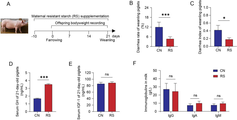
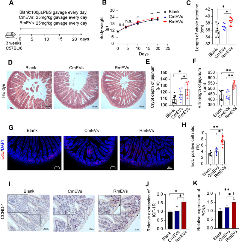
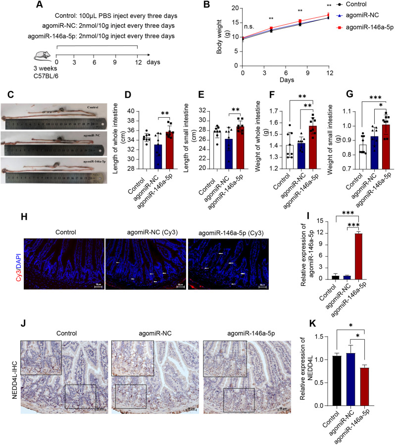
Results: In this study, we found that maternal resistant starch (RS) consumption during late gestation and lactation improved the growth and intestinal health of offspring. The mEVs in breast milk are the primary factor driving these beneficial effects, especially enhancing intestinal cell proliferation and migration. To be specific, administration of mEVs after maternal RS intake enhanced intestinal cell proliferation and migration in vivo (performed in mice model and indicated by intestinal histological observation, EdU assay, and the quantification of cyclin proteins) and in vitro (indicated by CCK8, MTT, EdU, and wound healing experiments). Noteworthily, miR-146a-5p was found to be highly expressed in the mEVs from maternal RS group, which also promotes intestinal cell proliferation in cells and mice models. Mechanically, miR-146a-5p target to silence the expression of ubiquitin ligase 3 gene NEDD4L, thereby inhibiting DVL2 ubiquitination, activating the Wnt pathway, and promoting intestinal development.
Conclusion: These findings demonstrated the beneficial role of mEVs in the connection between maternal fiber rich diet and offspring intestinal growth. In addition, we identified a novel miRNA-146a-5p-NEDD4L-β-catenin/Wnt signaling axis in regulating early intestinal development. This work provided a new perspective for studying the influence of maternal diet on offspring development.
Keywords: Intestinal development; Maternal diet; Milk-derived extracellular vesicles; Offspring; Resistant starch; miR-146a-5p.
© 2024. The Author(s).
Conflict of interest statement
The authors declare no competing interests.
Figures


Similar articles
-
Extracellular vesicles derived from M1 macrophages deliver miR-146a-5p and miR-146b-5p to suppress trophoblast migration and invasion by targeting TRAF6 in recurrent spontaneous abortion.Theranostics. 2021 Mar 31;11(12):5813-5830. doi: 10.7150/thno.58731. eCollection 2021. Theranostics. 2021. PMID: 33897883 Free PMC article.
-
Milk-derived extracellular vesicles alleviate ulcerative colitis by regulating the gut immunity and reshaping the gut microbiota.Theranostics. 2021 Jul 25;11(17):8570-8586. doi: 10.7150/thno.62046. eCollection 2021. Theranostics. 2021. PMID: 34373759 Free PMC article.
-
Breast cancer cell-derived extracellular vesicles transfer miR-182-5p and promote breast carcinogenesis via the CMTM7/EGFR/AKT axis.Mol Med. 2021 Jul 16;27(1):78. doi: 10.1186/s10020-021-00338-8. Mol Med. 2021. PMID: 34294040 Free PMC article.
-
The biological functions of maternal-derived extracellular vesicles during pregnancy and lactation and its impact on offspring health.Clin Nutr. 2023 Apr;42(4):493-504. doi: 10.1016/j.clnu.2023.02.007. Epub 2023 Feb 16. Clin Nutr. 2023. PMID: 36857958 Review.
-
Non-Coding RNAs in Human Breast Milk: A Systematic Review.Front Immunol. 2021 Sep 1;12:725323. doi: 10.3389/fimmu.2021.725323. eCollection 2021. Front Immunol. 2021. PMID: 34539664 Free PMC article.
Cited by
-
Maternal Diet Quality in Pregnancy and Human Milk Extracellular Vesicle and Particle microRNA.Epigenet Rep. 2025;3(1):1-10. doi: 10.1080/28361512.2025.2508883. Epub 2025 May 30. Epigenet Rep. 2025. PMID: 40631350
-
Milk-Derived Extracellular Vesicles and microRNAs: Potential Modulators of Intestinal Homeostasis.FASEB J. 2025 Aug 31;39(16):e70947. doi: 10.1096/fj.202501630R. FASEB J. 2025. PMID: 40838537 Free PMC article. Review.
-
White Adipocyte Stem Cell Expansion Through Infant Formula Feeding: New Insights into Epigenetic Programming Explaining the Early Protein Hypothesis of Obesity.Int J Mol Sci. 2025 May 8;26(10):4493. doi: 10.3390/ijms26104493. Int J Mol Sci. 2025. PMID: 40429638 Free PMC article. Review.
-
Microscopic messengers: Extracellular vesicles shaping gastrointestinal health and disease.Physiol Rep. 2025 Apr;13(7):e70292. doi: 10.14814/phy2.70292. Physiol Rep. 2025. PMID: 40165585 Free PMC article. Review.
-
Circ-0000197 derived from porcine milk small extracellular vesicles promotes intestinal barrier function by sponging miR-429.J Anim Sci Biotechnol. 2025 Jun 25;16(1):89. doi: 10.1186/s40104-025-01218-5. J Anim Sci Biotechnol. 2025. PMID: 40556020 Free PMC article.
References
-
- Zonneveld MI, van Herwijnen MJC, Fernandez-Gutierrez MM, Giovanazzi A, de Groot AM, Kleinjan M, van Capel TMM, Sijts A, Taams LS, Garssen J, et al. Human milk extracellular vesicles target nodes in interconnected signalling pathways that enhance oral epithelial barrier function and dampen immune responses. J Extracell Vesicles. 2021;10:e12071. doi: 10.1002/jev2.12071. - DOI - PMC - PubMed
MeSH terms
Substances
Grants and funding
LinkOut - more resources
Full Text Sources
