

Calcific bursitis of the Gruberi bursa: a case report
- PMID: 38365754
- PMCID: PMC10873953
- DOI: 10.1186/s13256-024-04377-7
Calcific bursitis of the Gruberi bursa: a case report
Abstract
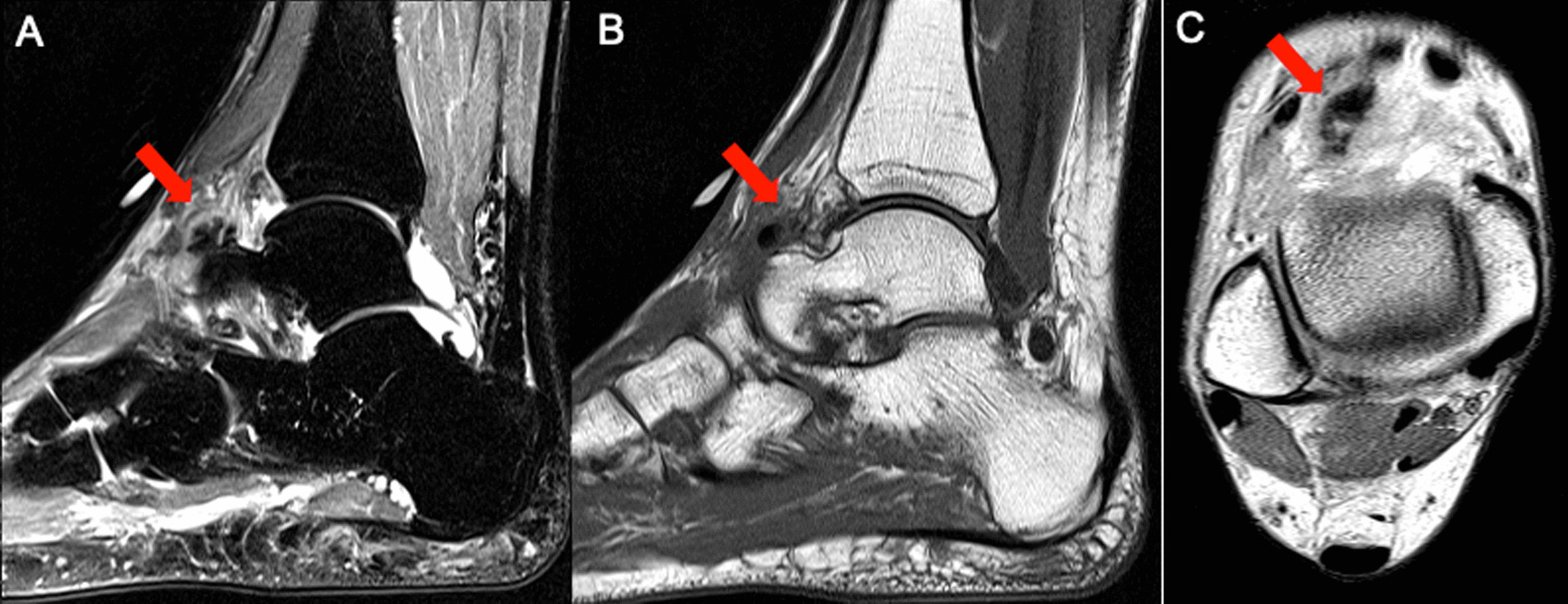
Background: Bursitis is the inflammation of a synovial bursa, a small synovial fluid-filled sac that acts as a cushion between muscles, tendons, and bones. Further, calcific bursitis results from calcium deposits on the synovial joint that exacerbates pain and swelling. The Gruberi bursa is located dorsolaterally in the ankle, between the extensor digitorium longus and the talus. Despite limited literature on its pathophysiology, the aim of this case is to discuss the bursa's association with calcific bursitis and its management via a case presented to our clinic.
Case presentation: A 47-year-old Caucasian female with no past medical or family history presents with acute right ankle pain following a minor injury 3 months prior with no improvement on analgesic or steroid therapy. Imaging demonstrated incidental calcium deposits. The day prior to presentation, the patient stated she used 1-pound ankle weights that resulted in mild swelling and gradual pain to the right dorsoanterior ankle. Physical exam findings displayed a significant reduction in the range of motion limited by pain. Imaging confirmed calcification within the capsule of the talonavicular joint, consistent with Gruberi bursitis. Initial management with prednisone yielded minimal improvement, requiring an interventional approach with ultrasound-guided barbotage that elicited immediate improvement.
Conclusion: The presented case report highlights a rare and unique instance of acute ankle pain and swelling caused by calcific Gruberi bursitis in a young female. Although the Gruberi bursa is a relatively new discovery, it contains inflammatory components that may predispose it to calcification and should be considered in the differential of ankle swelling. Therefore, utilizing a systematic approach to a clinical presentation and considering all differential diagnoses is essential.
Keywords: Ankle swelling; Calcific bursitis; Case report; Gruberi bursa; Non-operative.
© 2024. The Author(s).
Conflict of interest statement
The authors declare no competing interests.
Figures


References
-
- Ragab Y, Emad Y, Saad MA, et al. Contrast-enhanced magnetic resonance imaging (MRI) features of Gruberi bursitis as a very rare cause of dorsolateral ankle pain and swelling: case report and review of the literature. Radiol Case Rep. 2022;17(8):2612–2615. doi: 10.1016/j.radcr.2022.04.061. - DOI - PMC - PubMed
-
- Hochberg MC, Silman AJ, Smolen JS, et al. Rheumatology. Philadelphia: Elsevier Health Sciences; 2010.
Publication types
MeSH terms
Substances
LinkOut - more resources
Full Text Sources
Medical
Miscellaneous
