

Mosquito midgut stem cell cellular defense response limits Plasmodium parasite infection
- PMID: 38365823
- PMCID: PMC10873411
- DOI: 10.1038/s41467-024-45550-2
Mosquito midgut stem cell cellular defense response limits Plasmodium parasite infection
Abstract
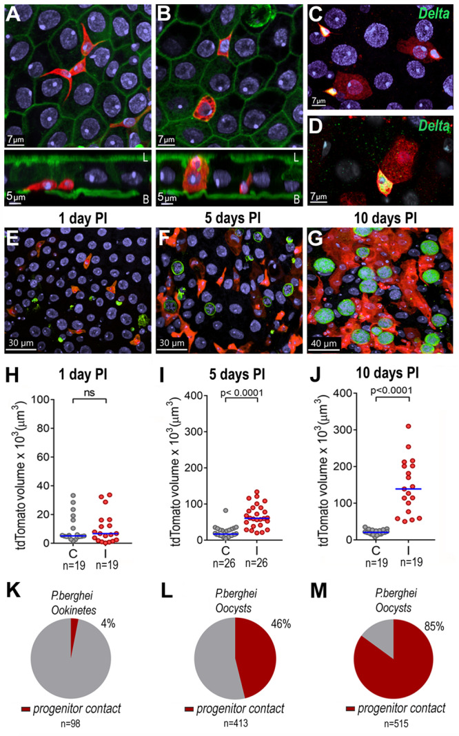
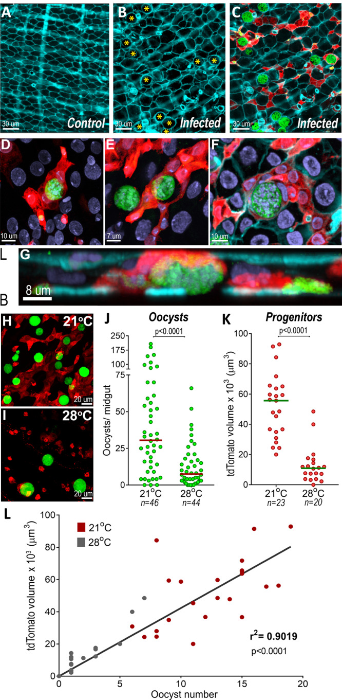
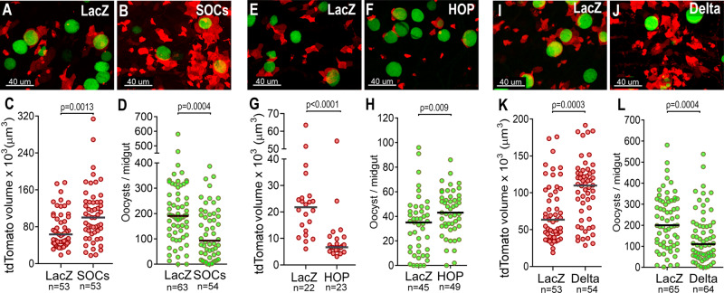
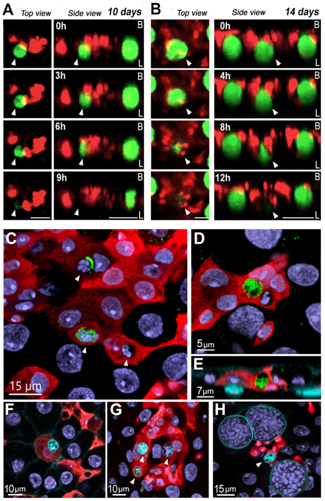
A novel cellular response of midgut progenitors (stem cells and enteroblasts) to Plasmodium berghei infection was investigated in Anopheles stephensi. The presence of developing oocysts triggers proliferation of midgut progenitors that is modulated by the Jak/STAT pathway and is proportional to the number of oocysts on individual midguts. The percentage of parasites in direct contact with enteroblasts increases over time, as progenitors proliferate. Silencing components of key signaling pathways through RNA interference (RNAi) that enhance proliferation of progenitor cells significantly decreased oocyst numbers, while limiting proliferation of progenitors increased oocyst survival. Live imaging revealed that enteroblasts interact directly with oocysts and eliminate them. Midgut progenitors sense the presence of Plasmodium oocysts and mount a cellular defense response that involves extensive proliferation and tissue remodeling, followed by oocysts lysis and phagocytosis of parasite remnants by enteroblasts.
© 2024. This is a U.S. Government work and not under copyright protection in the US; foreign copyright protection may apply.
Conflict of interest statement
The authors declare no competing interests.
Figures
Update of
-
Mosquito midgut stem cell cellular defense response limits Plasmodium parasite infection.bioRxiv [Preprint]. 2023 Aug 2:2023.08.02.551669. doi: 10.1101/2023.08.02.551669. bioRxiv. 2023. Update in: Nat Commun. 2024 Feb 16;15(1):1422. doi: 10.1038/s41467-024-45550-2. PMID: 37577486 Free PMC article. Updated. Preprint.
Similar articles
-
Late sporogonic stages of Plasmodium parasites are susceptible to the melanization response in Anopheles gambiae mosquitoes.Front Cell Infect Microbiol. 2024 Aug 1;14:1438019. doi: 10.3389/fcimb.2024.1438019. eCollection 2024. Front Cell Infect Microbiol. 2024. PMID: 39149419 Free PMC article.
-
Mosquito midgut stem cell cellular defense response limits Plasmodium parasite infection.bioRxiv [Preprint]. 2023 Aug 2:2023.08.02.551669. doi: 10.1101/2023.08.02.551669. bioRxiv. 2023. Update in: Nat Commun. 2024 Feb 16;15(1):1422. doi: 10.1038/s41467-024-45550-2. PMID: 37577486 Free PMC article. Updated. Preprint.
-
Additional Feeding Reveals Differences in Immune Recognition and Growth of Plasmodium Parasites in the Mosquito Host.mSphere. 2021 Mar 31;6(2):e00136-21. doi: 10.1128/mSphere.00136-21. mSphere. 2021. PMID: 33789941 Free PMC article.
-
Effect of a Blood Meal on Plasmodium Oocyst Growth Using the Enema Injection Method.Vector Borne Zoonotic Dis. 2025 Jun;25(6):408-415. doi: 10.1089/vbz.2024.0099. Epub 2025 May 7. Vector Borne Zoonotic Dis. 2025. PMID: 40329887
-
When a mean can be meaningless: evaluating mosquito infections with Plasmodium parasites.Parasitology. 2025 Jul 14:1-9. doi: 10.1017/S0031182025100541. Online ahead of print. Parasitology. 2025. PMID: 40659029 Review. No abstract available.
Cited by
-
A novel ATP-binding cassette protein (NoboABCG1.3) plays a role in the proliferation of Nosema bombycis.Parasitol Res. 2024 Dec 19;123(12):413. doi: 10.1007/s00436-024-08440-6. Parasitol Res. 2024. PMID: 39699667
-
Late sporogonic stages of Plasmodium parasites are susceptible to the melanization response in Anopheles gambiae mosquitoes.bioRxiv [Preprint]. 2024 May 31:2024.05.31.596773. doi: 10.1101/2024.05.31.596773. bioRxiv. 2024. Update in: Front Cell Infect Microbiol. 2024 Aug 01;14:1438019. doi: 10.3389/fcimb.2024.1438019. PMID: 38853990 Free PMC article. Updated. Preprint.
-
Advances in the dissection of Anopheles-Plasmodium interactions.PLoS Pathog. 2025 Mar 31;21(3):e1012965. doi: 10.1371/journal.ppat.1012965. eCollection 2025 Mar. PLoS Pathog. 2025. PMID: 40163471 Free PMC article. Review.
-
A single-cell atlas of the Culex tarsalis midgut during West Nile virus infection.bioRxiv [Preprint]. 2024 Nov 24:2024.07.23.603613. doi: 10.1101/2024.07.23.603613. bioRxiv. 2024. Update in: PLoS Pathog. 2025 Jan 27;21(1):e1012855. doi: 10.1371/journal.ppat.1012855. PMID: 39091762 Free PMC article. Updated. Preprint.
-
Late sporogonic stages of Plasmodium parasites are susceptible to the melanization response in Anopheles gambiae mosquitoes.Front Cell Infect Microbiol. 2024 Aug 1;14:1438019. doi: 10.3389/fcimb.2024.1438019. eCollection 2024. Front Cell Infect Microbiol. 2024. PMID: 39149419 Free PMC article.
References
MeSH terms
Substances
Grants and funding
LinkOut - more resources
Full Text Sources
Medical
