. 2024 Mar 12;11(3):ENEURO.0501-23.2024.
doi: 10.1523/ENEURO.0501-23.2024. Print 2024 Mar.

Synaptotagmin-7 Counteracts Short-Term Depression during Phasic Dopamine Release

Affiliations

Synaptotagmin-7 Counteracts Short-Term Depression during Phasic Dopamine Release

Joseph J Lebowitz et al. eNeuro. .

Abstract

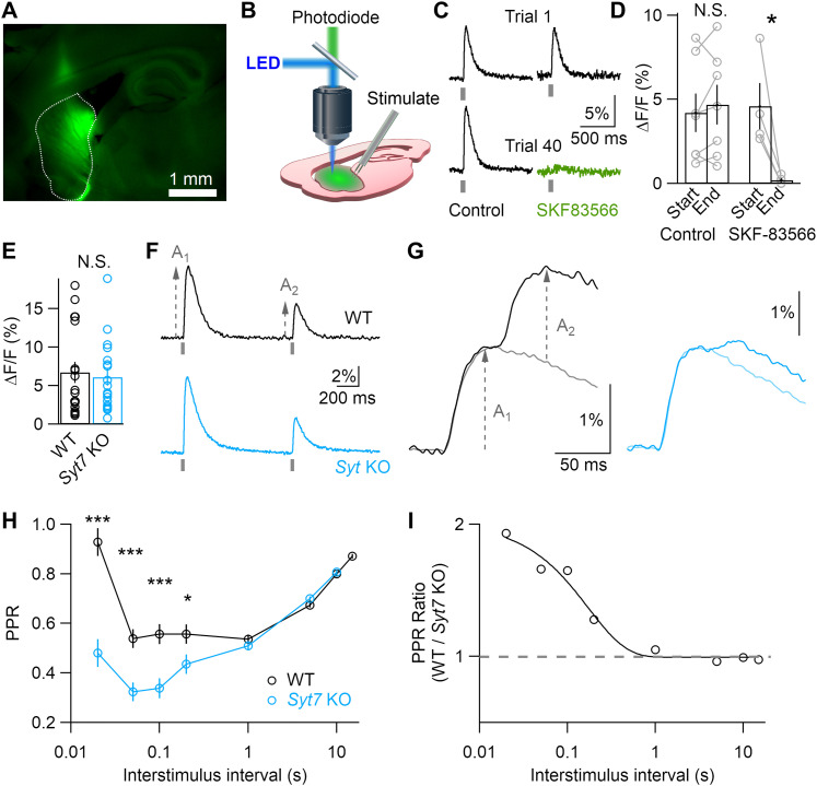
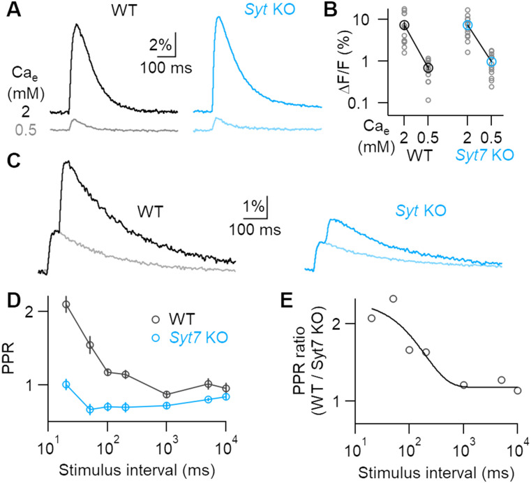
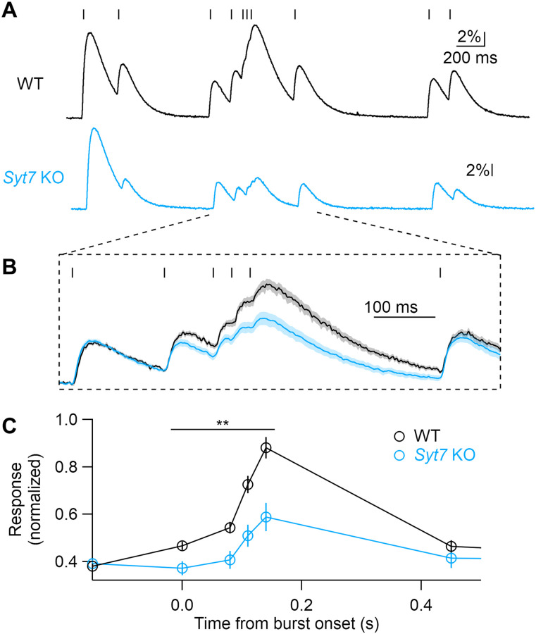
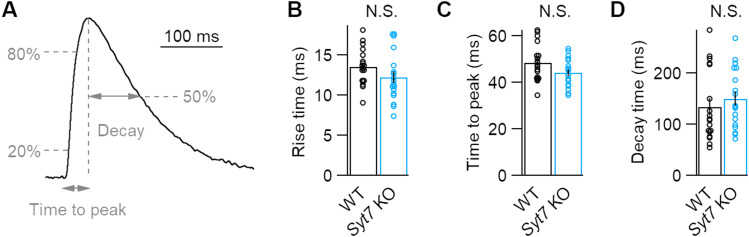
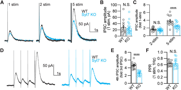
Dopamine neurons switch from tonic pacemaker activity to high-frequency bursts in response to salient stimuli. These bursts lead to superlinear increases in dopamine release, and the degree of this increase is highly dependent on firing frequency. The superlinearity and frequency dependence of dopamine release implicate short-term plasticity processes. The presynaptic Ca2+-sensor synaptotagmin-7 (SYT7) has suitable properties to mediate such short-term plasticity and has been implicated in regulating dopamine release from somatodendritic compartments. Here, we use a genetically encoded dopamine sensor and whole-cell electrophysiology in Syt7 KO mice to determine how SYT7 contributes to both axonal and somatodendritic dopamine release. We find that SYT7 mediates a hidden component of facilitation of release from dopamine terminals that can be unmasked by lowering initial release probability or by predepressing synapses with low-frequency stimulation. Depletion of SYT7 increased short-term depression and reduced release during stimulations that mimic in vivo firing. Recordings of D2-mediated inhibitory postsynaptic currents in the substantia nigra pars compacta (SNc) confirmed a similar role for SYT7 in somatodendritic release. Our results indicate that SYT7 drives short-term facilitation of dopamine release, which may explain the frequency dependence of dopamine signaling seen in vivo.

Keywords: dopamine; short-term plasticity; synaptotagmin.


Conflict of interest statement

The authors declare no competing financial interests.

Figures

Figure 1.
Figure 1.
Syt7 reduces short-term depression of striatal dopamine release. A, Representative fluorescent image of a sagittal brain slice with adeno-associated virus (AAV)-driven dLight expression in the dorsal striatum. B, Experimental method used to capture fluorescence with an amplified photodiode detector mounted on an upright microscope. C, Representative dLight transients recorded on the first and fortieth trials during an experiment with (right) or without (left) wash-in of the D1 antagonist SKF83566. D, Average response amplitudes for slices kept in control ACSF or superfused with the D1R antagonist SKF83566. E, Response amplitudes recorded from WT (N = 18) and Syt7 KO animals (N = 18). F, Representative responses to paired-pulse stimulation at 1 s intervals; each trace represents the average of five trials. G, As in F, but for 50 ms stimulus interval, illustrating how the response to a single stimulus (A1) was used to determine the amplitude of the second response (A2). H, Paired-pulse ratios (PPR = A2/A1) at varying interstimulus intervals. I, Ratio of WT and Syt7 KO PPR values, showing single exponential fit with 170 ms decay. Data in all figures expressed as mean ± SEM. Statistical significances assessed by Kruskal–Wallis (E) or unpaired Student's t test (D,H) are shown as *p < 0.05 and ***p < 0.001.
Figure 2.
Figure 2.
Lowering release probability reveals SYT7-mediated facilitation at striatal terminals. A, Representative dLight transients in WT and Syt7 KOs recorded from the same brain slice in high and low Cae. B, Response amplitudes recorded from slices where Cae was lowered from 2 to 0.5 mM (WT, N = 14; Syt7 KO, N = 13). C, Representative fluorescent transients evoked by paired-pulse stimulation at 50 ms intervals (dark traces) and single stimuli (light traces) in 0.5 mM Cae. D, Average paired-pulse ratios in 0.5 mM Cae at varying interstimulus intervals in WT (N = 15) and Syt7 KOs (N = 15). E, Ratio of average WT and Syt7 KO PPR, showing a single exponential fit with 196 ms decay.
Figure 3.
Figure 3.
SYT7 promotes dopamine release during phasic bursts. A, Stimulus train modeled on in vivo recordings from SNc dopamine neurons and representative dLight responses from WT and Syt7 KO animals. B, Averaged responses from WT (N = 9) and KO (N = 6), normalized to the initial response amplitude. C, Average normalized peak amplitudes for each stimulus, relative to the onset of the phasic burst. Statistical significance assessed by Student's t test, **p < 0.01.
Figure 4.
Figure 4.
The kinetics of dLight responses is unaffected by Syt7 depletion. A, Representative striatal dLight transients showing methods used to calculate kinetics. B–D, Averaged kinetics of dLight transients for both genotypes, as measured by the 20–80% rise time (B), time to peak (C), and 50% decay time (D). N = 18 for both genotypes. No significant differences were determined by unpaired Student's t tests (B,C) or Kruskal–Wallis test (D).
Figure 5.
Figure 5.
Syt7 mediates the facilitation of somatodendritic dopamine release. A, Representative D2-IPSCs induced by a single electrical stimulus (1 stim), a pair of stimuli at 40 Hz (2 stim), or a train of five stimuli at 40 Hz (5 stim). B, Amplitude of D2-IPSCs elicited by single stimuli in WT and Syt7 KO animals (WT, 28.4 pA ± 6.2 pA, N = 13 cells from four animals); KO, 25.3 ± 4.2 pA (N = 19 cells from four animals); p = 0.79, Mann–Whitney U test). C, Amplitude of D2-IPSCs induced by two or five stimuli at 40 Hz, normalized to the response elicited with one stimulus (2 stim—WT, 1.69 ± 0.13-fold 1 stim; KO, 1.45 ± 0.08-fold 1 stim, p = 0.64; 5 stim—WT, 3.84 ± 0.36-fold 1 stim, Syt7 KO, 2.52 ± 0.18-fold 1-stim, p < 0.0001; Sidak test following two-way ANOVA, same N as in A). D, Unmasking of facilitation protocol using three stimuli at 1 Hz, followed by a five-stimulus train at 100 Hz. The facilitated release is measured as the ratio of the five-stimulus IPSC to the third IPSC at 1 Hz. E, Quantification of facilitation in WT and Syt7 KO animals (WT, 4.61 ± 0.21, N = 14 cells, three animals; KO, 2.59 ± 0.15, N = 15 cells, three animals; p < 0.0001, unpaired t test). F, PPR measured for the first two 1 Hz stimuli (WT, 0.65 ± 0.02; KO, 0.64 ± 0.04, p = 0.90 unpaired t test, same N as in E). All data are presented as mean ± SEM with individual data points displayed. Statistical significances are shown as ****p < 0.0001.

Similar articles

Cited by

References

    1. Bacaj T, Wu D, Yang X, Morishita W, Zhou P, Xu W, Malenka RC, Südhof TC (2013) Synaptotagmin-1 and synaptotagmin-7 trigger synchronous and asynchronous phases of neurotransmitter release. Neuron 80:947–959. 10.1016/j.neuron.2013.10.026 - DOI - PMC - PubMed
    1. Banerjee A, Lee J, Nemcova P, Liu C, Kaeser PS (2020) Synaptotagmin-1 is the Ca2+ sensor for fast striatal dopamine release. Elife 9:e58359. 10.7554/eLife.58359 - DOI - PMC - PubMed
    1. Beckstead MJ, Ford CP, Phillips PE, Williams JT (2007) Presynaptic regulation of dendrodendritic dopamine transmission. Eur J Neurosci 26:1479–1488. 10.1111/j.1460-9568.2007.05775.x - DOI - PMC - PubMed
    1. Beckstead MJ, Grandy DK, Wickman K, Williams JT (2004) Vesicular dopamine release elicits an inhibitory postsynaptic current in midbrain dopamine neurons. Neuron 42:939–946. 10.1016/j.neuron.2004.05.019 - DOI - PubMed
    1. Caballero-Florán RN, Bendahmane M, Gupta JP, Chen X, Wu X, Morales A, Anantharam A, Jenkins PM (2023) Synaptotagmin-7 facilitates acetylcholine release in splanchnic nerve-chromaffin cell synapses during nerve activity. Neurosci Lett 800:137129. 10.1016/j.neulet.2023.137129 - DOI - PMC - PubMed