

HIF-2α-dependent TGFBI promotes ovarian cancer chemoresistance by activating PI3K/Akt pathway to inhibit apoptosis and facilitate DNA repair process
- PMID: 38365849
- PMCID: PMC10873328
- DOI: 10.1038/s41598-024-53854-y
HIF-2α-dependent TGFBI promotes ovarian cancer chemoresistance by activating PI3K/Akt pathway to inhibit apoptosis and facilitate DNA repair process
Abstract
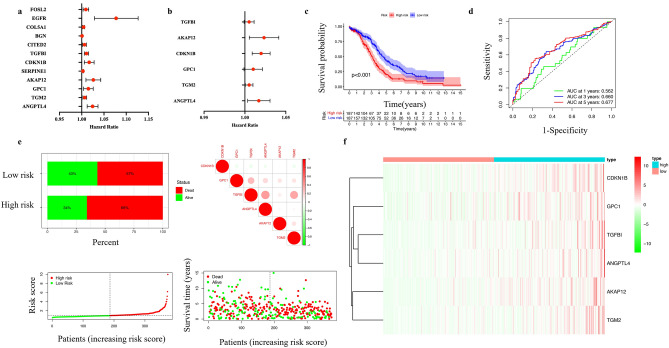
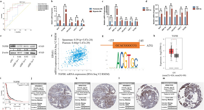
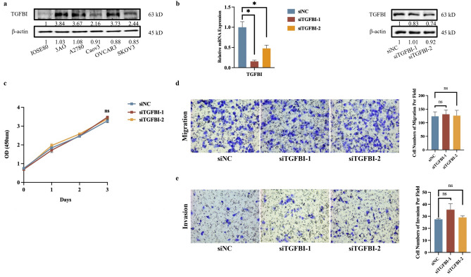
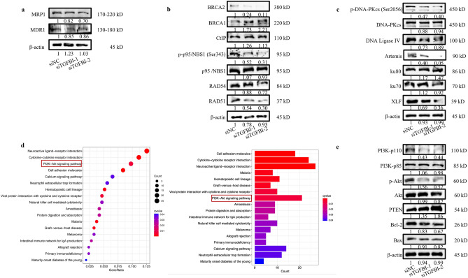
Hypoxia-mediated chemoresistance plays a crucial role in the development of ovarian cancer (OC). However, the roles of hypoxia-related genes (HRGs) in chemoresistance and prognosis prediction and theirs underlying mechanisms remain to be further elucidated. We intended to identify and validate classifiers of hub HRGs for chemoresistance, diagnosis, prognosis as well as immune microenvironment of OC, and to explore the function of the most crucial HRG in the development of the malignant phenotypes. The RNA expression and clinical data of HRGs were systematically evaluated in OC training group. Univariate and multivariate Cox regression analysis were applied to construct hub HRGs classifiers for prognosis and diagnosis assessment. The relationship between classifiers and chemotherapy response and underlying pathways were detected by GSEA, CellMiner and CIBERSORT algorithm, respectively. OC cells were cultured under hypoxia or transfected with HIF-1α or HIF-2α plasmids, and the transcription levels of TGFBI were assessed by quantitative PCR. TGFBI was knocked down by siRNAs in OC cells, CCK8 and in vitro migration and invasion assays were performed to examine the changes in cell proliferation, motility and metastasis. The difference in TGFBI expression was examined between cisplatin-sensitive and -resistant cells, and the effects of TGFBI interference on cell apoptosis, DNA repair and key signaling molecules of cisplatin-resistant OC cells were explored. A total of 179 candidate HRGs were extracted and enrolled into univariate and multivariate Cox regression analysis. Six hub genes (TGFBI, CDKN1B, AKAP12, GPC1, TGM2 and ANGPTL4) were selected to create a HRGs prognosis classifier and four genes (TGFBI, AKAP12, GPC1 and TGM2) were selected to construct diagnosis classifiers. The HRGs prognosis classifier could precisely distinguish OC patients into high-risk and low-risk groups and estimate their clinical outcomes. Furthermore, the high-risk group had higher percentage of Macrophages M2 and exhibited higher expression of immunecheckpoints such as PD-L2. Additionally, the diagnosis classifiers could accurately distinguish OC from normal samples. TGFBI was further verified as a specific key target and demonstrated that its high expression was closely correlated with poor prognosis and chemoresistance of OC. Hypoxia upregulated the expression level of TGFBI. The hypoxia-induced factor HIF-2α but not HIF-1α could directly bind to the promoter region of TGFBI, and facilitate its transcription level. TGFBI was upregulated in cisplatin-sensitive and resistant ovarian cancer cells in a cisplatin time-dependent manner. TGFBI interference downregulated DNA repair-related markers (p-p95/NBS1, RAD51, p-DNA-PKcs, DNA Ligase IV and Artemis), apoptosis-related marker (BCL2) and PI3K/Akt pathway-related markers (PI3K-p110 and p-Akt) in cisplatin-resistant OC cells. In summary, the HRGs prognosis risk classifier could be served as a predictor for OC prognosis and efficacy evaluation. TGFBI, upregulated by HIF-2α as an HRG, promoted OC chemoresistance through activating PI3K/Akt pathway to reduce apoptosis and enhance DNA damage repair pathway.
Keywords: Akt; Chemoresistance; DNA repair; Hypoxia; Ovarian cancer.
© 2024. The Author(s).
Conflict of interest statement
The authors declare no competing interests.
Figures


Similar articles
-
Ursolic acid inhibits proliferation and reverses drug resistance of ovarian cancer stem cells by downregulating ABCG2 through suppressing the expression of hypoxia-inducible factor-1α in vitro.Oncol Rep. 2016 Jul;36(1):428-40. doi: 10.3892/or.2016.4813. Epub 2016 May 18. Oncol Rep. 2016. PMID: 27221674
-
Activation of PI3K/AKT/mTOR signaling axis by UBE2S inhibits autophagy leading to cisplatin resistance in ovarian cancer.J Ovarian Res. 2023 Dec 19;16(1):240. doi: 10.1186/s13048-023-01314-y. J Ovarian Res. 2023. PMID: 38115063 Free PMC article.
-
Knockdown of KLF5 suppresses hypoxia-induced resistance to cisplatin in NSCLC cells by regulating HIF-1α-dependent glycolysis through inactivation of the PI3K/Akt/mTOR pathway.J Transl Med. 2018 Jun 14;16(1):164. doi: 10.1186/s12967-018-1543-2. J Transl Med. 2018. PMID: 29898734 Free PMC article.
-
Significance of flavonoids targeting PI3K/Akt/HIF-1α signaling pathway in therapy-resistant cancer cells - A potential contribution to the predictive, preventive, and personalized medicine.J Adv Res. 2024 Jan;55:103-118. doi: 10.1016/j.jare.2023.02.015. Epub 2023 Mar 4. J Adv Res. 2024. PMID: 36871616 Free PMC article. Review.
-
HER2 puzzle pieces: Non-Coding RNAs as keys to mechanisms, chemoresistance, and clinical outcomes in Ovarian cancer.Pathol Res Pract. 2024 Jun;258:155335. doi: 10.1016/j.prp.2024.155335. Epub 2024 Apr 29. Pathol Res Pract. 2024. PMID: 38723327 Review.
Cited by
-
Overexpression of HIF2α Enhances the Angiogenesis-Promoting Effect of hUC-MSC-Derived Extracellular Vesicles by Stimulating miR-146a.Protein Pept Lett. 2025;32(1):62-74. doi: 10.2174/0109298665347753241028072130. Protein Pept Lett. 2025. PMID: 39592897
-
KLC3 activates PI3K/AKT signaling and promotes ovarian cancer cell proliferation and migration through COL3A1.Oncol Rep. 2025 Jun;53(6):67. doi: 10.3892/or.2025.8900. Epub 2025 Apr 17. Oncol Rep. 2025. PMID: 40242978 Free PMC article.
-
Knockdown TNF family prognosis index crucial gene PDE4B promoted PANoptosis of ovarian carcinoma cell:Based in vitro and in vivo experiments.Transl Oncol. 2025 Jun;56:102333. doi: 10.1016/j.tranon.2025.102333. Epub 2025 Apr 16. Transl Oncol. 2025. PMID: 40245751 Free PMC article.
-
STAT3/TGFBI signaling promotes the temozolomide resistance of glioblastoma through upregulating glycolysis by inducing cellular senescence.Cancer Cell Int. 2025 Apr 3;25(1):127. doi: 10.1186/s12935-025-03770-6. Cancer Cell Int. 2025. PMID: 40181415 Free PMC article.
-
TGFBI promotes EMT and perineural invasion of pancreatic cancer via PI3K/AKT pathway.Med Oncol. 2025 Apr 26;42(6):181. doi: 10.1007/s12032-025-02736-y. Med Oncol. 2025. PMID: 40286004
References
-
- Dorayappan KDP, Wanner R, Wallbillich JJ, Saini U, Zingarelli R, Suarez AA, et al. Hypoxia-induced exosomes contribute to a more aggressive and chemoresistant ovarian cancer phenotype: A novel mechanism linking STAT3/Rab proteins. Oncogene. 2018;37(28):3806–3821. doi: 10.1038/s41388-018-0189-0. - DOI - PMC - PubMed
MeSH terms
Substances
Grants and funding
LinkOut - more resources
Full Text Sources
Medical
Molecular Biology Databases
Research Materials
Miscellaneous
