. 2024 Feb 16;15(1):1444.
doi: 10.1038/s41467-024-45273-4.

An ultrasensitive and broadband transparent ultrasound transducer for ultrasound and photoacoustic imaging in-vivo

Affiliations

An ultrasensitive and broadband transparent ultrasound transducer for ultrasound and photoacoustic imaging in-vivo

Seonghee Cho et al. Nat Commun. .

Abstract

Transparent ultrasound transducers (TUTs) can seamlessly integrate optical and ultrasound components, but acoustic impedance mismatch prohibits existing TUTs from being practical substitutes for conventional opaque ultrasound transducers. Here, we propose a transparent adhesive based on a silicon dioxide-epoxy composite to fabricate matching and backing layers with acoustic impedances of 7.5 and 4-6 MRayl, respectively. By employing these layers, we develop an ultrasensitive, broadband TUT with 63% bandwidth at a single resonance frequency and high optical transparency ( > 80%), comparable to conventional opaque ultrasound transducers. Our TUT maximises both acoustic power and transfer efficiency with maximal spectrum flatness while minimising ringdowns. This enables high contrast and high-definition dual-modal ultrasound and photoacoustic imaging in live animals and humans. Both modalities reach an imaging depth of > 15 mm, with depth-to-resolution ratios exceeding 500 and 370, respectively. This development sets a new standard for TUTs, advancing the possibilities of sensor fusion.


Conflict of interest statement

Chulhong Kim, Seonghee Cho and Joongho Ahn have financial interests in OPTICHO, which, however, did not support this work. The remaining authors declare no other competing interests.

Figures

Fig. 1
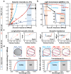
Fig. 1. Computed performance comparison of the proposed transparent ultrasound transducer (TUT), a conventional opaque ultrasound transducer (OUT), and a conventional TUT.
a Schematics of the proposed TUT, conventional OUT, and conventional TUT. b Comparison of the simulated acoustic impedance magnitudes (MRayl) and phase (rad) at a normalized frequency with a 100% bandwidth. c Comparison of the simulated power transmission efficiency and pulse-echo impulse responses. R, ringdown; FWHM, full-width-at-half-maximum; Bandwidth refers to the −6 dB width expressed as a fraction of the center frequency.
Fig. 2
Fig. 2. Acoustical, rheological, and optical properties of ceramic-epoxy composites.
a Experimentally measured and theoretically estimated viscosity vs maximum acoustic impedance for various ceramic-epoxy composites. Markers are experimental results, and solid lines are theoretically fitted. b Simulated light transmittance at 589 nm of 30 MHz acoustic quarter-wave plates made of various ceramic-epoxy composites. Only simulates up to 100 McPs. c Simulated and experimentally measured acoustic impedance and acoustic velocity vs volume fraction for various ceramic-epoxy composites (n = 12). d Photographs and experimentally measured optical transmittances of matching layers made of various transparent ceramic-epoxy composites. NIR, near infrared. Error bar denotes standard deviation.
Fig. 3
Fig. 3. Structure of the proposed transparent ultrasound transducer (TUT) and its optical transmittance.
a Structural schematic of the proposed TUT. b Demonstration of the optical clarity of the TUT. c Measured optical transmittance of the proposed TUT. d Measured pulse-echo responses and spectrum. ITO, indium tin oxide; LNO, lithium niobate; NIR, near infrared; OUT, opaque ultrasound transducer; R, ringdown; FWHM, full-width-at-half-maximum; Bandwidth refers to the -6 dB width expressed as a fraction of the center frequency.
Fig. 4
Fig. 4. Schematic of an integrated ultrasound (US)/photoacoustic (PA) microscopy system using a high-performance transparent ultrasound transducer (TUT), and its imaging performance.
a An integrated US/PA microscopy system. b US/PA full-width-at-half-maximum resolutions. c Signal-noise-ratio (SNR) vs penetration depth in chicken breast tissue (n = 140). Norm. amp, normalized amplitude; arb. units, arbitrary-units. Error bar denotes standard deviation.
Fig. 5
Fig. 5. Volumetric ultrasound (US) and photoacoustic (PA) images of a live mouse.
a US average intensity projection (AIP) and PA average amplitude projection (AAP) images of the mouse in the ventral (thorax and abdomen) and sagittal planes. b Depth-sliced US and depth-encoded PA maximum amplitude projection (MAP) images at various depths. 1, sternum; 2, mammary vessel; 3, epigastric vessel; 4, liver; 5, common carotid vessel; 6, subclavian vessel; 7, internal thoracic vessel; 8, heart; 9, vertebra; 10, tibia; 11, duodenum; 12, stomach; 13, jejunum; 14, cecum; 15, bladder; 16, preputial gland; 17, pelvis; 18, rib; 19, lateral thoracic vessel; 20, spleen; 21, kidney; 22, circumflex iliac vessel; 23, femur; 24, scapula; *, reverberation artifact.
Fig. 6
Fig. 6. 2D cross-sectional ultrasound (US)/ photoacoustic (PA) images cut along lines L1-5 in Fig. 5.
1, sternum; 2, heart; 3, lung; 4, internal thoracic vessels; 5, thoracic vessel; 6, liver; 7, gallbladder; 8, stomach; 9, vertebra; 10, jejunum; 11, abdominal vessel; 12, rib; 13, spleen; 14, kidney; *, reverberation artifact.
Fig. 7
Fig. 7. Volumetric ultrasound (US) and photoacoustic (PA) images of a human palm.
a US average intensity projection (AIP) and PA average amplitude projection (AAP) images of a human palm. b Depth-sliced US and depth-encoded PA maximum amplitude projection (MAP) images at various depths. 1, subcutaneous vessels; 2, palmar veins; 3, palmar digital arteries; 4, adductor pollicis muscles; 5, adductor minimi muscle.
Fig. 8
Fig. 8. 2D cross-sectional ultrasound (US)/ photoacoustic (PA) images cut along the line in Fig. 7a US.
1, palmar vein; 2, common palmar digital artery; 3, adductor pollicis; 4, subcutaneous vessels.
Fig. 9
Fig. 9. Perfusion kinematics monitoring of IR-1048 dye in live animals.
a Photoacoustic (PA) maximum amplitude projection (MAP) images of a live mouse from before injection to 9 hours after injection. b 2D cross-sectional PA images cut along the line on the liver. c 2D cross-sectional PA images cut along the line on the bladder. d In-vivo normalized PA amplitude profile in the liver and bladder regions over time after injection (n = 3). e Ex-vivo validation of IR-1048 accumulation in the liver at 30 minutes after injection. f Ex-vivo normalized PA amplitude profile in the extracted liver (n = 3). Norm. amp, normalized amplitude; arb. units, arbitrary-units. Error bar denotes standard error of three experiments.

Similar articles

Cited by

References

    1. Kruizinga P, et al. Compressive 3D ultrasound imaging using a single sensor. Sci. Adv. 2017;3:e1701423. doi: 10.1126/sciadv.1701423. - DOI - PMC - PubMed
    1. Ma T, Zhou B, Hsiai TK, Shung KK. A review of intravascular ultrasound-based multimodal intravascular imaging: the synergistic approach to characterizing vulnerable plaques. Ultrasonic imaging. 2016;38:314–331. doi: 10.1177/0161734615604829. - DOI - PMC - PubMed
    1. Park J, et al. Quadruple ultrasound, photoacoustic, optical coherence, and fluorescence fusion imaging with a transparent ultrasound transducer. P Natl. Acad. Sci. USA. 2021;118:e1920879118. doi: 10.1073/pnas.1920879118. - DOI - PMC - PubMed
    1. Choi, W. et al. Three-dimensional multistructural quantitative photoacoustic and US imaging of human feet in vivo. Radiology, 211029 (2022). - PubMed
    1. Kim J, et al. Multiparametric photoacoustic analysis of human thyroid cancers in vivo. Cancer Res. 2021;81:4849–4860. doi: 10.1158/0008-5472.CAN-20-3334. - DOI - PubMed