. 2024 Mar;31(3):360-377.
doi: 10.1038/s41418-024-01267-9. Epub 2024 Feb 16.

Tropomyosin1 isoforms underlie epithelial to mesenchymal plasticity, metastatic dissemination, and resistance to chemotherapy in high-grade serous ovarian cancer

Affiliations

Tropomyosin1 isoforms underlie epithelial to mesenchymal plasticity, metastatic dissemination, and resistance to chemotherapy in high-grade serous ovarian cancer

Tong Xu et al. Cell Death Differ. 2024 Mar.

Abstract

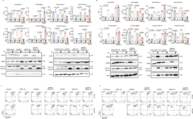
Phenotypic plasticity, defined as the ability of individual cells with stable genotypes to exert different phenotypes upon exposure to specific environmental cues, represent the quintessential hallmark of the cancer cell en route from the primary lesion to distant organ sites where metastatic colonization will occur. Phenotypic plasticity is driven by a broad spectrum of epigenetic mechanisms that allow for the reversibility of epithelial-to-mesenchymal and mesenchymal-to-epithelial transitions (EMT/MET). By taking advantage of the co-existence of epithelial and quasi-mesenchymal cells within immortalized cancer cell lines, we have analyzed the role of EMT-related gene isoforms in the regulation of epithelial mesenchymal plasticity (EMP) in high grade serous ovarian cancer. When compared with colon cancer, a distinct spectrum of downstream targets characterizes quasi-mesenchymal ovarian cancer cells, likely to reflect the different modalities of metastasis formation between these two types of malignancy, i.e. hematogenous in colon and transcoelomic in ovarian cancer. Moreover, upstream RNA-binding proteins differentially expressed between epithelial and quasi-mesenchymal subpopulations of ovarian cancer cells were identified that underlie differential regulation of EMT-related isoforms. In particular, the up- and down-regulation of RBM24 and ESRP1, respectively, represent a main regulator of EMT in ovarian cancer cells. To validate the functional and clinical relevance of our approach, we selected and functionally analyzed the Tropomyosin 1 gene (TPM1), encoding for a protein that specifies the functional characteristics of individual actin filaments in contractile cells, among the ovarian-specific downstream AS targets. The low-molecular weight Tpm1.8/9 isoforms are specifically expressed in patient-derived ascites and promote invasion through activation of EMT and Wnt signaling, together with a broad spectrum of inflammation-related pathways. Moreover, Tpm1.8/9 expression confers resistance to taxane- and platinum-based chemotherapy. Small molecule inhibitors that target the Tpm1 isoforms support targeting Tpm1.8/9 as therapeutic targets for the development of future tailor-made clinical interventions.


Conflict of interest statement

PWG and ECH receive funding from and are Directors of TroBio Therapeutics, a company commercializing anti-tropomyosin drugs. The remaining authors declare no competing interests.

Figures

Fig. 1
Fig. 1. A subpopulation of quasi-mesenchymal cells co-exists with epithelial cells in HGS ovarian cancer cell lines.
A FACS analysis of the ovarian cancer cell lines OV90, CAOV3, SKOV3, and COV504 with antibodies directed against CD44 and EpCAM. EpCAM/CD44 positive and negative areas were defined as previously described [17, 21] using multiple isotype controls and are shown by the quadrants in the plots. Using specific gates, cells were separated in CD44hiEpCAMhi and CD44hiEpCAMlo subpopulations. The percentages of CD44hiEpCAMlo and CD44hiEpCAMhi cells within each cell line are depicted in each quadrant. Notably, as previously observed for SW480 and HCT116, the ovarian cancer cell lines revealed a continuum of different EpCAM and CD44 expression levels with a large EpCAMhi (or EpCAMlo as in the case of SKOV3) cluster followed by a tail of gradually decreasing (increasing for SKOV3) EpCAM levels. By applying the indicated gates, cells were sorted into EpCAMhi and EpCAMlo subpopulations. Graphs show representative analysis from an individual experiment. B Phase contrast microscopy images of sorted EpCAMhi and EpCAMlo cells from EpCAMhi and EpCAMlo OV90 and CAOV3 sorted cells. Scale bar: 100 μm. While EpCAMhi cells show characteristic epithelial morphology, EpCAMlo cells showed a more spindle- and mesenchymal-like appearance. Scale bar: 100 µm. C Transwell migration assay (upper graph) and invasion assay (lower graph) of EpCAMhi (blue bar) and EpCAMlo (red bar) OV90 and CAOV3 cells. Each bar symbolizes the mean ± SD. D Principal component analysis (PCA) of the RNAseq profiles of EpCAMhi and EpCAMlo cells from the OV90 and CAOV3 lines. E Heatmap of common differentially expressed gene among EpCAMhi/lo and bulk subpopulations from the OV90 and CAOV3 cell lines (abs LFC > 1.5, P value < 0.01). Complete-linkage hierarchical clustering with split by k-means (k = 2) clustering was used. F Hallmark pathways based on the Gene Set Enrichment Analysis (GSEA) of OV90 and CAOV3 EpCAMlo expression profiles compared with EpCAMhi. Plots show only significantly expressed pathways, with a normalized enrichment score (NES) > 1 and P value < 0.05. G RT-qPCR expression analysis of ZEB1 in OV90, SKOV3 and COV504 transduced with an inducible control (shCT) and with a ZEB1-shRNA (shZEB1) construct. shRNA expression was induced with 1 μg/mL of doxycycline. GAPDH expression was used as control. Each bar represents the mean ± SD. P value is indicated. H FACS analysis of the OV90, SKOV3 and COV504 cell lines transfected with the shZEB1 and control constructs using antibodies against CD44 and EpCAM. Cells were induced with 1 μg/mL doxycycline for 72 h prior to the FACS analysis. The percentages of EpCAMlo and EpCAMhi cells within each cell line are depicted in each quadrant.
Fig. 2
Fig. 2. EpCAMlo cells co-exist with epithelial cells in high-grade serous ovarian cancer.
A Density plot shows the distribution of patient-derived ovarian cancer cells (scRNAseq data from Vazquez-Garcia et al. [22]) earmarked by expression of the EpCAMlo signature. The threshold was set as ≥0.1 for the subsequent analyses. The EpCAMlo signature was defined by genes that were identified as upregulated EpCAMlo cells in both CAOV3 and OV90. EpCAMlo upregulated genes were selected after differential expression analysis with EpCAMhi/bulk cells using the cut-offs log2FC > 1.5 and padj <0.05. B UMAP plot of patient-derived ovarian cancer cells. Cells positive for the EpCAMlo signature are highlighted in red and show enrichment within three clusters (#2, #4, and #6) labeled as EMT-like in the Vazquez-Garcia et al. study [22]. Please note that a substantial fraction of the EpCAMlo-like cells appears to fall outside these clusters and is distributed throughout the UMAP. C Violin plots showing the distribution of EpCAMlo-like cells (according to z-score) in different anatomical localization of ovarian cancers (adnexa, ascites, and non-adnexa). D Violin plots showing the expression levels of EMT-like signature of single cell cluster 2, 4 and 6. E Heatmap of hallmark gene sets across the EpCAMlo-like clusters in primary ovarian cancers (adnexa), ascites, and metastases (non-adnexa) based on the GSEA of three EpCAMlo-like clusters and bulk (p val <0.05). Complete-linkage hierarchical clustering was used.
Fig. 3
Fig. 3. Differential expression of RBPs ESRP1 and RBM24 regulates TPM1 isoforms in HGS ovarian cancer.
A Fold change analysis of RBPs [original list from reference [14]] differentially expressed between EpCAMlo and EpCAMhi cells in OV90 and CAOV3. Red bar indicates RBPs with significant differential expression (log2-fold change >2, and P value < 0.05). The dotted line represents the 1.5 absolute fold change cut-off. B RT-qPCR analysis of ESRP1 and RBM24 expression in OV90, CAOV3, and COV504 EpCAMhi/lo and bulk cells. GAPDH expression was employed as control (means ± SD, n = 3). P values are indicated. C Western blot analysis of ESRP1 and RBM24 expression in OV90, CAOV3, and COV504 EpCAMhi/lo and bulk cells. β-actin was used as loading control for western blots. D. TPM1 exon peak plots relative to the AS analysis of RNAseq data obtained from OV90 EpCAMhi/lo and CAOV3 bulk/EpCAMlo analysis. Each peak indicates the expression of specific exons; the height of each peak is indicative of the expression level of the specific exons. The exon-intron structure of the corresponding TPM1 isoforms is depicted below the exon peak plot. Exon 1a and 2b are specific to Tpm1.6/7 isoforms (brown), while exon 1b is specific to Tpm1.8/9 (green). Exon 6a (red) is only present in the Tpm1.7 and Tpm1.9 isoforms; whereas, exon 6b (yellow) earmarks the Tpm1.6 and Tpm1.8 isoforms. With the exception of exon 6a and 6b, exons 3 to 9d are present in all TPM1 isoforms. E RT-qPCR (histogram panels) and western analysis of TPM1 isoform expression in OV90, CAOV3, and COV504 EpCAMhi/lo and bulk cells. GAPDH expression was employed as control. (Means ± SD, n = 3). P value is indicated. β-actin was used as loading control for western blots. F CD44/EpCAM FACS analysis of the ovarian cancer cell lines PEA1 and PEA2. EpCAM/CD44 positive and negative areas were identified using multiple isotype controls. The percentages of cells within each quadrant are indicated. G RT-qPCR (left histogram panels) and western (right panel) analysis of ESRP1, RBM24, and TPM1 isoform expression in PEA1 and PEA2 ovarian cancer cell line; GAPDH expression was employed as control. (Means ± SD, n = 3). P values are indicated. β-actin was used as loading control for western blots.
Fig. 4
Fig. 4. The RBPs ESRP1 and RBM24 synergistically regulate TPM1 isoforms and the relative proportion of EpCAMhi/lo cells.
A RT-qPCR (histogram panels) and western (lower panel) analysis of ESRP1, RBM24, and TPM1 isoform expression in RBM24-OE (overexpressing) and shESRP1-KD (knockdown) OV90 and COV504 ovarian cancer cell line; GAPDH expression was employed as control (Means ± SD, n = 3). β-actin was used as loading control for western blots. B RT-qPCR (histogram panels) and western (lower panel) analysis of ESRP1, RBM24, and TPM1 isoform expression in ESRP1-OE (overexpressing) and shRBM24-KD (knockdown) OV90 and COV504 ovarian cancer cell line; GAPDH expression was employed as control (Means ± SD, n = 3). β-actin was used as loading control for western blots. C CD44/EpCAM FACS analysis of RBM24-OE/KD and ESRP1-OE/KD OV90 cells. Cells were induced with 1 μg/mL doxycycline for 72 h before analysis. The relative percentages of EpCAMlo and EpCAMhi cells are indicated in each quadrant. D CD44/EpCAM FACS analysis of RBM24-OE/KD and ESRP1-OE/KD COV504 cells. Cells were induced with 1 μg/mL doxycycline for 72 h before analysis. The relative percentages of EpCAMlo and EpCAMhi cells are indicated in each quadrant.
Fig. 5
Fig. 5. Ectopic expression of Tpm1.6/7 and Tpm1.8/9 isoforms results in increased migration and invasion, and decreased cell proliferation.
A RT-qPCR analysis of OV90, COV504, PEA1, and PEA2 ovarian cancer cell lines transduced to ectopically express the Tpm1.6/7-OE and Tpm1.8/9-OE isoforms; GAPDH expression was employed as control (Means ± SD, n = 3). P values are relative to the comparison with the parental cell lines. B Western analysis of OV90, COV504, PEA1, and PEA2 ovarian cancer cell lines transduced to ectopically express the Tpm1.6/7-OE and Tpm1.8/9-OE isoforms. β-actin was employed as loading control. C Proliferation assays of OV90, COV504, PEA1, and PEA2 ovarian cancer cell lines transduced to ectopically express the Tpm1.6/7-OE and Tpm1.8/9-OE isoforms. O.D. values are shown from day 1 to 6 (Means ± SD, n = 3). P values are relative to the comparison with the parental cell lines. D Transwell migration assay of OV90, COV504, PEA1, and PEA2 ovarian cancer cell lines transduced to ectopically express the Tpm1.6/7-OE and Tpm1.8/9-OE isoforms. 5×10⁴ cells were plated on TC-coated membranes and left O/N. The number of cells that migrated to the lower side of the membrane were counted and plotted (Means ± SD, n = 3). P values are relative to the comparison with the parental cell lines. E Confocal images of OV90 and COV504 parental and Tpm1.6/7/8/9-OE cells seeded on collagen layers and incubated for 6 days. As indicated by the arrows, Tpm1.8/9-OE cells appear to invade the collagen layer collectively as narrow linear strands with “leader” and “follower” cells. Scale bar: 250 μm. The number of cells invading the collagen was quantified and plotted (Means ± SD, n = 3). P values are relative to the comparison with the parental cell lines. Plots relative to the PEA1 and PEA2 ovarian cancer cell lines were also calculated (bottom). F Immunofluorescence analysis of OV90, COV504, PEA1 and PEA2 parental cells with antibodies directed against ARP2, Tpm1.6/7 and Tpm1.8/9. Nuclei were visualized by DAPI staining of DNA. Scale bar: 5 μm.
Fig. 6
Fig. 6. RNAseq analysis revealed TPM1 isoforms function in Wnt pathway and contribute to metastasis in vivo.
A Hierarchical clustering of the RNAseq data relative to Tpm1.6/7/8/9-OE OV90 cells. Complete-linkage hierarchical clustering was used. B Principal component analysis (PCA) of RNAseq profiles from parental and Tpm1.6/7/8/9-OE OV90 cells. C Hallmarks pathways based on the Gene Set Enrichment Analysis (GSEA) of parental and Tpm1.6/7/8/9-OE OV90 cells. The heatmap only includes significantly altered pathways, with NES > 1, and P value < 0.05. Complete-linkage hierarchical clustering was used. D Volcano plots showing differentially expressed genes between Tpm1.6/7-OE (left, green) and Tpm1.8/9-OE (pink, right) OV90 cells (abs LFC > 1.5, P value < 0.01). E TOP-Flash luciferase reporter analysis of Wnt signaling activity in Tpm1.6/7/8/9-OE (upper histogram) and upon knockdown by siRNA of Tpm1.6/7 and Tpm1.8/9 in OV90 and CAOV3 cells. P values are relative to the comparison with the parental cell lines (Means ± SD, n = 3-4). F Quantification of IVIS bioluminescence signals obtained from NSG mice injected IP with Tpm1.6/7/8/9-OE OV90 cells. Recipient animals were sacrificed 5 wk after injection. Four mice were analyzed for each type of transplanted cells. Data are presented as mean values ± SD. Y-axis meaning ROI (regions of interests) are user-defined areas within IVIS optical imaging. G Examples of IVIS bioluminescence signals from Tpm1.6/7/8/9-OE cell-injected mice at 5 wk after injection. The Spectrum in vivo imaging system was employed. For in vivo imaging purposes, mice were injected IP with D-luciferin (150 mg kg−1). H IVIS bioluminescence signals relative to specific organs from mice transplanted with the TPM1 isoform-OE cells. Y-axis meaning ROI (regions of interests) are user-defined areas within IVIS optical imaging. I Tpm1.6/7 and Tpm1.8/9 IHC (upper panels) and ISH (BaseScope; lower panels) analyses of tumoroids derived from ascetic fluids from mice transplanted with the TPM1 isoform-OE cells. Scale bars: 100 μm (large panels) and 15 μm (inlets) for IHC; 20 μm and 5 μm (inlets) for ISH. J Colony formation assay relative to cells derived from tumoroids obtained from mice transplanted with the TPM1 isoform-OE cells (Means ± SD, n = 3). Y-axis means the percentage of tumoroids single cells form into colonies.
Fig. 7
Fig. 7. Tpm1.8/9 isoforms are enriched in malignant ascites from ovarian cancer cells and confer resistance to platinum- and taxane-based therapies.
A Examples of IHC (left panels) and ISH (BaseScope; right panels) analyses of patient-derived ovarian cancers with antibodies (IHC) and oligonucleotides probes (ISH) specific for the Tpm1.6/7 and Tpm1.8/9 isoforms. Ovarian cancer tissues were obtained from a primary tumor and a metastasis (without chemotherapy). Scale bar: 100 μm and 15 μm (inlets) for IHC; 20 μm and 5 μm (inlets) for ISH. B Schematic flowchart of the analysis of ascites from late-stage ovarian cancer patients. C FACS analysis and sorting strategy of CD45CD90+ and CD45CD90 cells from patient-derived ascites. From left to right: FSC-A/SSC–A, FSC-W/FSC-A, SSC-W/SSC-A, FSC-A/CD45 and CD45-CD90+/− single cell gates. D RT-qPCR analysis of Tpm1.6/7 and Tpm1.8/9 expression in sorted CD45CD90+ and CD45CD90 cells from patient-derived ascites (n = 13) sorted by FACS; GAPDH expression was employed as control (Means ± SD). E RT-qPCR analysis of Tpm1.6/7 and Tpm1.8/9 expression in OV90 and COV504 cells exposed to cisplatin and paclitaxel cells; GAPDH expression was employed as control (Means ± SD, n = 3-4). F Dose-response curves relative to Tpm1.6/7/8/9-OE cells grown in the presence of different concentrations of paclitaxel and cisplatin (log scale and cell viability on the x and y axis, respectively). IC50 values were calculated from biological triplicates to quintuplicates for each experiment (Means ± SD, n = 3–5). G RT-qPCR (left histogram panels) and western (right) analysis of TPM1 isoform expression in siTpm1.6/7 and siTpm1.8/9 knockdown OV90 and COV504 cells; GAPDH expression was employed as control (Means ± SD, n = 3-4). β-actin was employed as loading control for the western blots. H Dose-response curves of siTpm1.6/7 and siTpm1.8/9 knockdown OV90 and COV504 cells cultured in the presence of different concentrations of paclitaxel (left) and cisplatin (right) concentrations (log scale and cell viability on the x and y axis, respectively). IC50 values were calculated from biological triplicates to quintuplicates for each experiment (Means ± SD, n = 3–5).
Fig. 8
Fig. 8. Small molecule inhibitors directed against Tpm1.8/9 isoforms antagonize their effects on EMT, Wnt signaling, and resistance to chemotherapy.
A Upper panels: RT-qPCR analysis of TPM1 isoforms and EMT-related gene expression in OV90 parental cells cultured for 24 h in the presence of compound #1 or #3 at 0, 2, 5, and 10 μM. The values were calculated by normalizing with the untreated cells. P values < 0.05 are shown by red bars while gray bars indicate lower values; GAPDH expression was employed as control (Means ± SD, n = 3). Lower panels: western analysis of TPM1 isoform expression in OV90 parental cells cultured for 24 h in the presence of compound #1 or #3 at 0, 2, 5, and 10 μM. Β-actin was used as loading control for western blots. B RT-qPCR analysis of TPM1 isoforms and EMT-related gene expression in OV90 EpCAMlo cells cultured for 24 h in the presence of compound #1 or #3 at 0, 2, 5, and 10 μM. The values were calculated by normalizing with the untreated cells. P values < 0.05 are shown by red bars while gray bars indicate lower values; GAPDH expression was employed as control (Means ± SD, n = 3). Lower panels: western analysis of TPM1 isoform expression in OV90 EpCAMlo cells cultured for 24 h in the presence of compound #1 and #3 at 0, 2, 5, and 10 μM. β-actin was used as loading control for western blots. C Dose-response curves of parental OV90 cells treated with compound #1 or #3 in the presence of different paclitaxel and cisplatin concentrations. IC50 values were calculated from biological triplicates for each experiment (Means ± SD, n = 3). D Dose-response curves of OV90 EpCAMlo cells treated with compound #1 or #3 in the presence of different paclitaxel and cisplatin concentrations. IC50 values were calculated from biological triplicates for each experiment (Means ± SD, n = 3). E Dose-response curves of parental CAOV3 cells treated with compound #1 or #3 in the presence of different paclitaxel and cisplatin concentrations. IC50 values were calculated from biological quadruplicates for each experiment (Means ± SD, n = 4). F Dose-response curves of CAOV3 EpCAMlo cells treated with compound #1 or #3 in the presence of different paclitaxel and cisplatin concentrations. IC50 values were calculated from biological quadruplicates for each experiment (Means ± SD, n = 4). G TOP-Flash luciferase reporter analysis of Wnt signaling activity in OV90, COV504 and CAOV3 parental (upper panel) and EpCAMlo (lower panel) cells treated with compound #1 or #3 (Means ± SD, n = 3–5).

References

    1. Jayson GC, Kohn EC, Kitchener HC, Ledermann JA. Ovarian cancer. Lancet. 2014;384:1376–88. doi: 10.1016/S0140-6736(13)62146-7. - DOI - PubMed
    1. Siegel RL, Miller KD, Jemal A. Cancer statistics, 2018. CA Cancer J Clin. 2018;68:7–30. doi: 10.3322/caac.21442. - DOI - PubMed
    1. Bast RC, Jr, Hennessy B, Mills GB. The biology of ovarian cancer: new opportunities for translation. Nat Rev Cancer. 2009;9:415–28. doi: 10.1038/nrc2644. - DOI - PMC - PubMed
    1. Kurman RJ, Shih IeM. Pathogenesis of ovarian cancer: lessons from morphology and molecular biology and their clinical implications. Int J Gynecol Pathol. 2008;27:151–60. - PMC - PubMed
    1. Bowtell DD, Böhm S, Ahmed AA, Aspuria P-J, Bast RC, Beral V, et al. Rethinking ovarian cancer II: reducing mortality from high-grade serous ovarian cancer. Nat Rev Cancer. 2015;15:668–79. doi: 10.1038/nrc4019. - DOI - PMC - PubMed

Publication types