

Improving the accuracy of bone mineral density using a multisource CBCT
- PMID: 38366012
- PMCID: PMC10873385
- DOI: 10.1038/s41598-024-54529-4
Improving the accuracy of bone mineral density using a multisource CBCT
Abstract
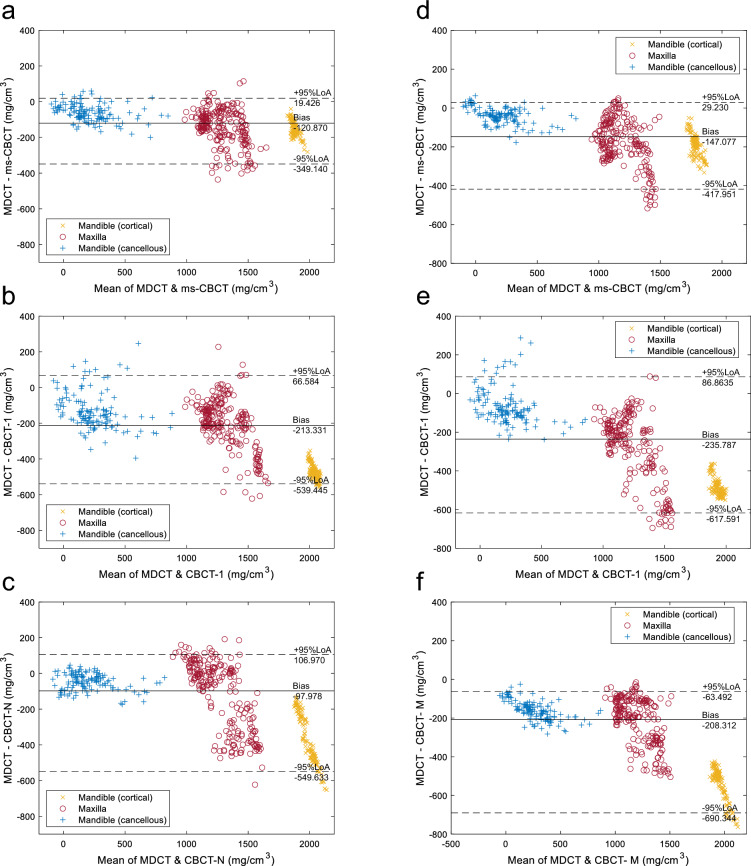
Multisource cone beam computed tomography CBCT (ms-CBCT) has been shown to overcome some of the inherent limitations of a conventional CBCT. The purpose of this study was to evaluate the accuracy of ms-CBCT for measuring the bone mineral density (BMD) of mandible and maxilla compared to the conventional CBCT. The values measured from a multi-detector CT (MDCT) were used as substitutes for the ground truth. An anthropomorphic adult skull and tissue equivalent head phantom and a homemade calibration phantom containing inserts with varying densities of calcium hydroxyapatite were imaged using the ms-CBCT, the ms-CBCT operating in the conventional single source CBCT mode, and two clinical CBCT scanners at similar imaging doses; and a clinical MDCT. The images of the anthropomorphic head phantom were reconstructed and registered, and the cortical and cancellous bones of the mandible and the maxilla were segmented. The measured CT Hounsfield Unit (HU) and Greyscale Value (GV) at multiple region-of-interests were converted to the BMD using scanner-specific calibration functions. The results from the various CBCT scanners were compared to that from the MDCT. Statistical analysis showed a significant improvement in the agreement between the ms-CBCT and MDCT compared to that between the CBCT and MDCT.
© 2024. The Author(s).
Conflict of interest statement
The authors declare the following competing interests: O.Z. has equity ownership and serves on the board of directors of XinVisio, LLC., to which the technologies used or evaluated in this project have been licensed, and NuRay Co., Ltd, which manufactures the x-ray sources used in this study. J.L. has equity ownership in XinVisio, LLC. and NuRay Co. Ltd. O.Z., J.L., C.R.I., Y.Z.L., and B.L. are co-inventors of licensed technology evaluated in this study. All activities have been approved by the institutional COI committee. All other authors declare no competing interests.
Figures


Similar articles
-
Development and validation of a 3D anthropomorphic phantom for dental CBCT imaging research.Med Phys. 2023 Nov;50(11):6714-6736. doi: 10.1002/mp.16661. Epub 2023 Aug 21. Med Phys. 2023. PMID: 37602774
-
Evaluation of the feasibility of a multisource CBCT for maxillofacial imaging.Phys Med Biol. 2023 Aug 14;68(17):10.1088/1361-6560/acea17. doi: 10.1088/1361-6560/acea17. Phys Med Biol. 2023. PMID: 37487498 Free PMC article.
-
The impact of CBCT reconstruction and calibration for radiotherapy planning in the head and neck region - a phantom study.Acta Oncol. 2014 Aug;53(8):1114-24. doi: 10.3109/0284186X.2014.927073. Epub 2014 Jun 30. Acta Oncol. 2014. PMID: 24975372
-
Accuracy of MDCT and CBCT in three-dimensional evaluation of the oropharynx morphology.Eur J Orthod. 2018 Jan 23;40(1):58-64. doi: 10.1093/ejo/cjx030. Eur J Orthod. 2018. PMID: 28453722
-
A dose-neutral image quality comparison of different CBCT and CT systems using paranasal sinus imaging protocols and phantoms.Eur Arch Otorhinolaryngol. 2022 Sep;279(9):4407-4414. doi: 10.1007/s00405-022-07271-4. Epub 2022 Jan 27. Eur Arch Otorhinolaryngol. 2022. PMID: 35084532 Free PMC article.
Cited by
-
The Value of Mandibular Indices on Cone Beam Computed Tomography in Secondary Causes of Low Bone Mass.J Clin Med. 2024 Aug 16;13(16):4854. doi: 10.3390/jcm13164854. J Clin Med. 2024. PMID: 39200996 Free PMC article.
-
Accuracy of the Hounsfield Unit Values Measured by Implant Planning Software.Dent J (Basel). 2024 Dec 17;12(12):413. doi: 10.3390/dj12120413. Dent J (Basel). 2024. PMID: 39727470 Free PMC article.
-
Prediction of Dental Implants Primary Stability With Cone Beam Computed Tomography-Based Homogenized Finite Element Analysis.Clin Implant Dent Relat Res. 2025 Apr;27(2):e70016. doi: 10.1111/cid.70016. Clin Implant Dent Relat Res. 2025. PMID: 40033523 Free PMC article.
-
The impact of jawbone regions (molar area, premolar area, anterior area) and bone density on the accuracy of robot-assisted dental implantation: a preliminary study.Front Bioeng Biotechnol. 2025 Feb 25;13:1536957. doi: 10.3389/fbioe.2025.1536957. eCollection 2025. Front Bioeng Biotechnol. 2025. PMID: 40070547 Free PMC article.
-
Bone Densitometric Analysis using Cone Beam Computed Tomography (Cone Beam CT) and Computed Tomography (CT): Establishing the Correlation between Predicted and Actual Values.Adv Biomed Res. 2025 Jan 30;14:8. doi: 10.4103/abr.abr_313_24. eCollection 2025. Adv Biomed Res. 2025. PMID: 40213598 Free PMC article.
References
MeSH terms
Grants and funding
LinkOut - more resources
Full Text Sources
Medical
