Review
. 2024 Jun;25(6):424-442.
doi: 10.1038/s41580-024-00703-5. Epub 2024 Feb 16.

The cell biology of ferroptosis

Affiliations
Review

The cell biology of ferroptosis

Scott J Dixon et al. Nat Rev Mol Cell Biol. 2024 Jun.

Abstract

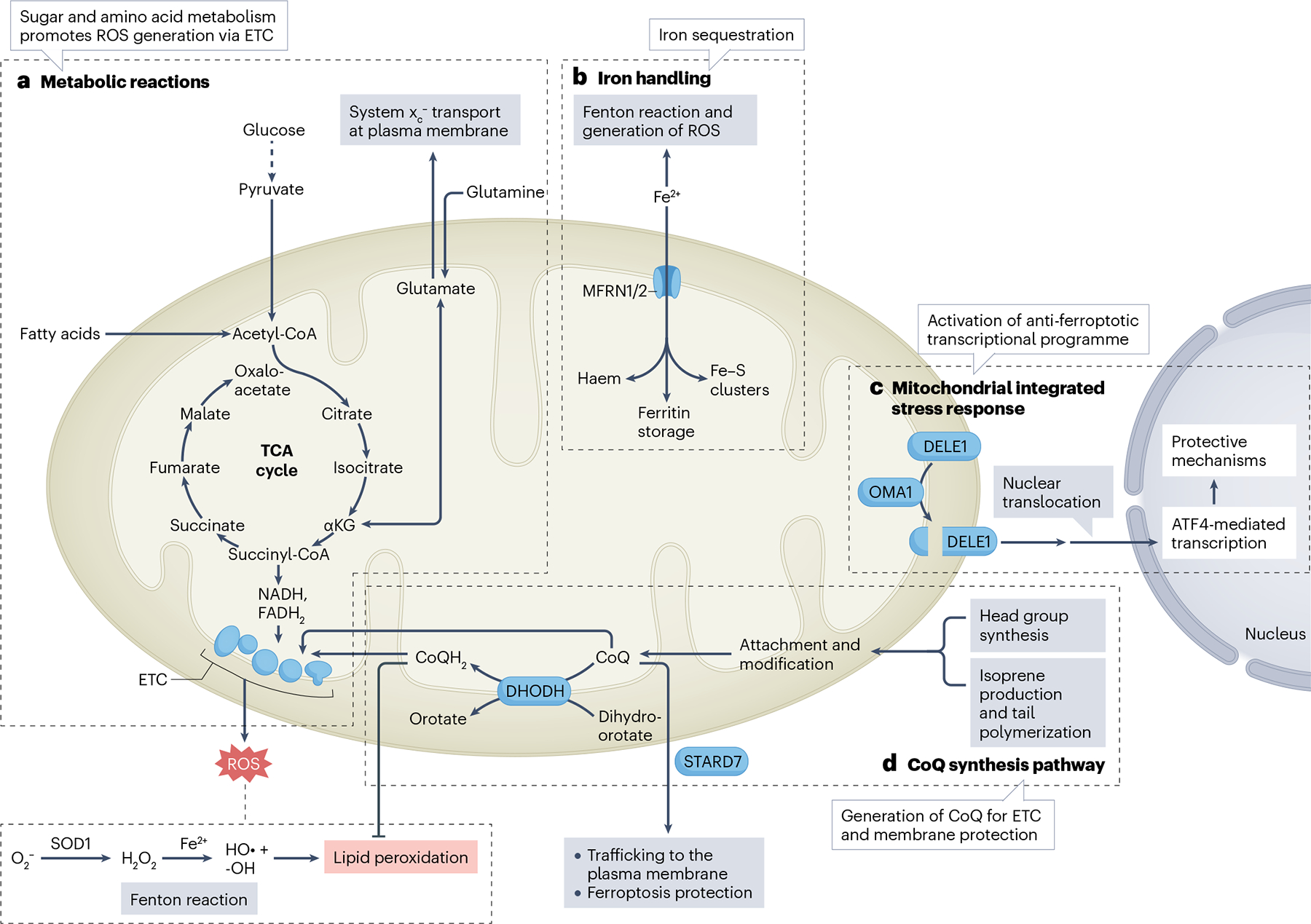
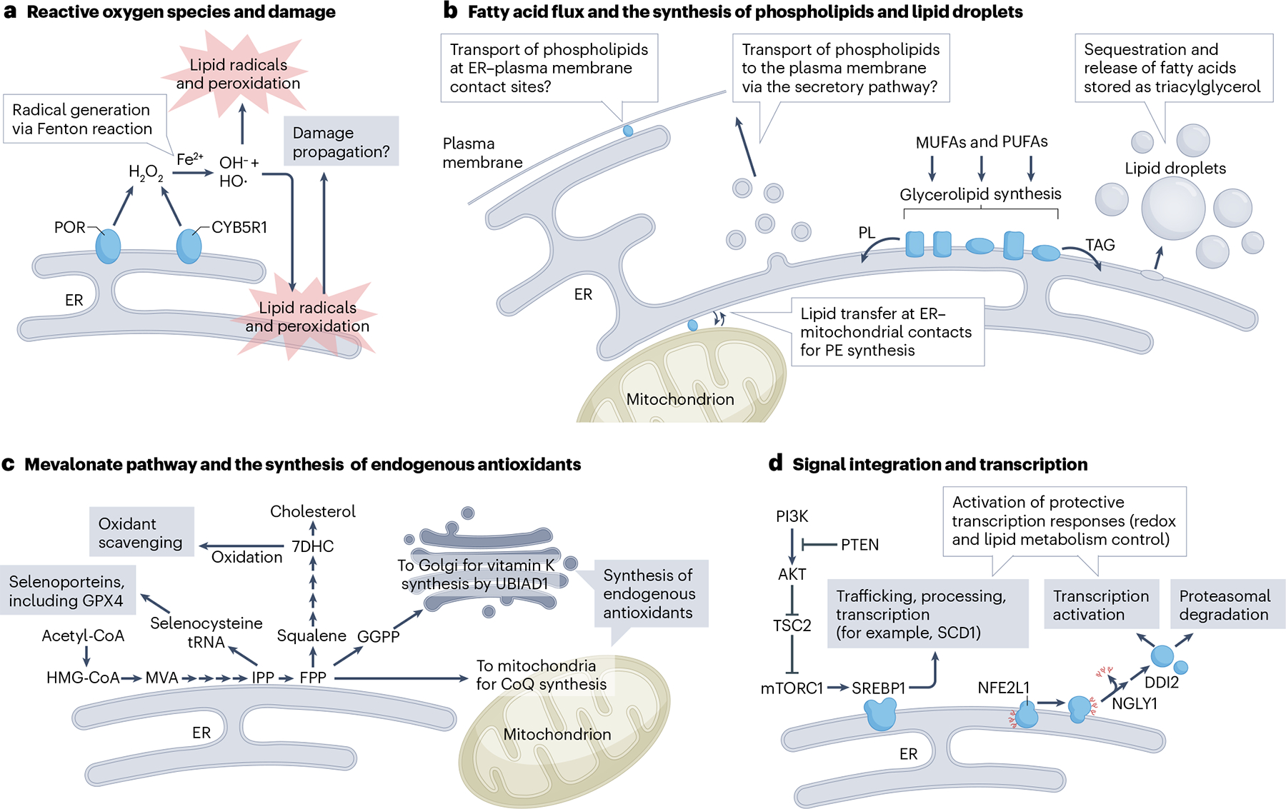
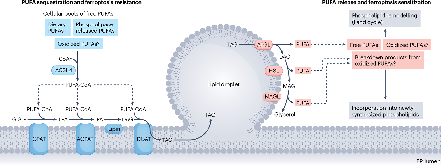
Ferroptosis is a non-apoptotic cell death mechanism characterized by iron-dependent membrane lipid peroxidation. Here, we review what is known about the cellular mechanisms mediating the execution and regulation of ferroptosis. We first consider how the accumulation of membrane lipid peroxides leads to the execution of ferroptosis by altering ion transport across the plasma membrane. We then discuss how metabolites and enzymes that are distributed in different compartments and organelles throughout the cell can regulate sensitivity to ferroptosis by impinging upon iron, lipid and redox metabolism. Indeed, metabolic pathways that reside in the mitochondria, endoplasmic reticulum, lipid droplets, peroxisomes and other organelles all contribute to the regulation of ferroptosis sensitivity. We note how the regulation of ferroptosis sensitivity by these different organelles and pathways seems to vary between different cells and death-inducing conditions. We also highlight transcriptional master regulators that integrate the functions of different pathways and organelles to modulate ferroptosis sensitivity globally. Throughout this Review, we highlight open questions and areas in which progress is needed to better understand the cell biology of ferroptosis.


Conflict of interest statement

Competing interests

S.J.D. is a co-founder of Prothegen and a member of the scientific advisory board for Hillstream BioPharma. S.J.D. holds patents related to ferroptosis. J.A.O. is a member of the scientific advisory board for Vicinitas Therapeutics and holds patents related to ferroptosis.

Figures

Fig. 1 |
Fig. 1 |. The general mechanism of ferroptosis.
a, Ferroptosis execution can be distinguished from the regulation of ferroptosis sensitivity. Ferroptosis execution involves membrane lipid peroxidation, leading to plasma membrane rupture and cell death. The glutathione (GSH)–glutathione peroxidase 4 (GPX4) and NADPH–ferroptosis suppressor protein 1 (FSP1) systems limit membrane lipid peroxidation and inhibit ferroptosis. Ferroptosis sensitivity is dictated by the propensity of the cell to accumulate high levels of membrane lipid peroxides. Whether a cell will accumulate high levels of lipid peroxides relates to the amount of iron and oxidizable lipids in the cell, and to the status of the cellular oxidant-producing and antioxidant systems. The regulation of ferroptosis sensitivity likely encompasses hundreds of distinct metabolites, enzymes and other biomolecules that impinge upon the membrane lipid peroxidation and membrane-rupturing mechanisms. b, Plasma membrane rupture is the key event that results in cell death during ferroptosis. It involves the accumulation of phospholipid hydroperoxides, which may be generated enzymatically (for example, via lipoxygenases) and non-enzymatically through radical-mediated reactions. Phospholipid peroxidation has several consequences for the cell, including altered ion fluxes (for example, increased Piezo-mediated Ca2+ uptake, increased transient receptor potential (TRP)-mediated Ca2+ and Na+ uptake, and reduced Na+/K+ ATPase-mediated Na+ export and K+ uptake), water ingress and biophysical effects on the membrane. Cell swelling and increased membrane stiffness can further activate ion channels (for example, Piezo and TRP) in a feedforward manner, further enhancing ion fluxes and accelerating plasma membrane rupture. c, Important ferroptosis defence mechanisms localize to the plasma membrane. The system xc antiporter imports cystine in exchange for glutamate. In the cytosol, cystine is rapidly reduced to cysteine. Cysteine can be used to synthesize (reduced) GSH. GSH is the cofactor for GPX4, an enzyme that can convert toxic phospholipid peroxides (L-OOH) into benign lipid alcohols (L-OH). Parallel to GPX4, the oxidoreductase FSP1 uses NAD(P)H to regenerate the reduced form of radical-trapping antioxidants (for example, coenzyme Q10 (CoQ) or vitamin K), which in turn terminate the lipid peroxidation process by donating electrons to phospholipid peroxyl radicals (LOO). Receptor-mediated endocytosis influences ferroptosis sensitivity. For example, the uptake of iron in complex with transferrin by the transferrin receptor enhances ferroptosis sensitivity while uptake of the selenium-rich protein SEPP1 by its cognate receptor LDL receptor-related protein 8 (LRP8) suppresses ferroptosis sensitivity. Iron, and presumably selenium in the form of selenocysteine, are subsequently released from the lysosome. Iron can react with soluble and lipid peroxides to generate hydroxyl and lipid alkoxyl radicals that promote lipid peroxidation. Cysteine and selenium released from the lysosome can be used to synthesize GSH and the selenoprotein GPX4, respectively. ACSL4, acyl-CoA synthetase long-chain family member 4; CHMP, charged multivesicular body protein; CoA, coenzyme A; GR, GSH reductase; GSSG, oxidized GSH; PKCβII, protein kinase Cβ; PUFA, polyunsaturated fatty acid.
Fig. 2 |
Fig. 2 |. Ferroptosis regulation in the mitochondria.
Mitochondria play several context-dependent roles in the regulation of ferroptosis sensitivity. a, Metabolic reactions — the breakdown of sugar and amino acids via glycolysis and the tricarboxylic acid (TCA) cycle yields NADH and FADH2, which pass electrons to the mitochondrial electron transport chain (ETC). The ETC can partially reduce oxygen to generate reactive oxygen species (ROS) such as superoxide (O2•−) at complexes I and III. Dismutation of O2•− yields hydrogen peroxide, which can result in the formation of hydroxyl radicals (HO•) via reactions with Fe2+ in the Fenton reaction. Glutamine can be catabolized to glutamate (a substrate for the system xc antiporter) and then to the TCA cycle intermediate α-ketoglutarate (αKG). αKG synthesis promotes ferroptosis, possibly by enhancing mitochondrial ROS production. b, Iron handling — mitoferrin 1 (MFRN1) and MFRN2 mediate iron transport across the inner mitochondrial membrane. The synthesis of Fe–S clusters and haem prosthetic groups in the mitochondria consumes labile iron that otherwise may accumulate to promote ROS accumulation. c, Mitochondrial integrated stress response — OMA1-dependent proteolytic processing of DAP3 binding cell death enhancer 1 (DELE1) releases a protein fragment that initiates the integrated stress response, culminating in activating transcription factor 4 (ATF4)-dependent transcription of a programme that enhances glutathione metabolism and protects against ferroptosis. d, Coenzyme Q10 (CoQ) synthesis — the final steps in de novo CoQ synthesis occur in the mitochondria. CoQ is employed as a key electron carrier in the ETC. CoQ can be reduced by dihydroorotate dehydrogenase (DHODH) to CoQH2, which can either transfer electrons to complex III in the ETC or function as a local radical-trapping antioxidant to prevent peroxidation of mitochondrial lipids and limit ferroptosis. In addition, CoQ is trafficked by a processed form of StAR-related lipid transfer protein 7 (STARD7) from the mitochondria to the plasma membrane, where it can contribute to ferroptosis protection mediated by ferroptosis suppressor protein 1. CoA, coenzyme A.
Fig. 3 |
Fig. 3 |. Ferroptosis regulation in the ER.
a, Cytochrome P450 oxidoreductase (POR) and cytochrome b5 reductase 1 (CYB5R1) are endoplasmic reticulum (ER) enzymes that promote ferroptosis by generating H2O2, which can further react with iron to produce reactive oxygen species species that initiate lipid peroxidation, leading to ferroptosis. Lipid peroxidation in the ER is an early event in ferroptosis. Whether damage propagates from the ER or accumulates at a slower rate in other membranes is an open question (see also Box 2). b, The ER is the primary site of lipid synthesis, including the synthesis of glycerolipids, namely phospholipids (PLs) and triacylglycerols (TAGs). The incorporation of monounsaturated fatty acids (MUFAs) and polyunsaturated fatty acids (PUFAs) into PLs or TAGs helps establish the overall sensitivity of the cell to lipid peroxidation and ferroptosis. TAGs are stored within lipid droplets that emerge from the outer leaflet of the ER. PLs are trafficked to the plasma membrane via the secretory pathway or transferred at ER–plasma membrane contact sites. Phosphatidylethanolamine (PE) is often oxidized during ferroptosis. The synthesis of PE involves the transfer of phosphatidylserine from the ER to mitochondria, where it is enzymatically converted to PE prior to transport back to the ER. The relative contributions of these PL trafficking pathways to ferroptosis remain to be defined. c, A complex series of enzymatic steps in the ER mediate the synthesis of cholesterol via the mevalonate pathway. Several intermediates in this pathway protect against ferroptosis. Farnesyl pyrophosphate (FPP) is needed to synthesize the endogenous radical-trapping antioxidants coenzyme Q10 (CoQ) and vitamin K in the mitochondria and Golgi, respectively. Isopentenyl pyrophosphate (IPP) is attached to the selenocysteine tRNA as an isopentenyl moiety, a key modification for its function, promoting the synthesis of selenoproteins, including the anti-ferroptotic enzyme glutathione peroxidase 4 (GPX4). 7-Dehydrocholesterol (7DHC) is highly prone to oxidation and can suppress ferroptosis by competing with PL for oxidation. Squalene also suppresses ferroptosis, but the mechanism is not understood. d, Proteolytic processing of the membrane-tethered transcription factors sterol regulatory element-binding protein 1 (SREBP1) and nuclear factor erythroid 2-related factor 1 (NFE2L1) releases soluble, active transcription factors that traffic to the nucleus and initiate transcriptional programmes to control lipid metabolism and the cellular oxidative stress response. The phosphoinositide 3-kinase (PI3K)–mechanistic target of rapamycin complex 1 (mTORC1) pathway promotes ferroptosis resistance by increasing SREBP1 activation and expression of stearoyl-CoA desaturase 1 (SCD1), which generates ferroptosis-suppressive MUFAs. NFE2L1 is dislocated from the ER into the cytoplasm, where it is deglycosylated by N-glycanase 1 (NGLY1) and proteolytically processed by DNA-damage inducible 1 homologue 2 (DDI2). The amount of NFE2L1 that escapes proteasomal clearance is a determinant of NFE2L1 transcriptional signalling. HMG-CoA, 3-hydroxy-3-methylglutaryl coenzyme A; MVA, mevalonate; GGPP, geranylgeranyl pyrophosphate; UBIAD1, UBIA prenyltransferase domain-containing protein 1.
Fig. 4 |
Fig. 4 |. PUFA flux and sequestration in lipid droplets.
Polyunsaturated fatty acids (PUFAs), which may be derived from dietary sources or released from the phospholipids by phospholipases, are conjugated with coenzyme A (CoA) by acyl-CoA synthetase enzymes such as acyl-CoA synthetase long-chain family member 4 (ACSL4), a process known as fatty acid activation, and are then incorporated into triacylglycerol (TAG) that is stored in lipid droplets. Sequestration of PUFAs within TAGs can reduce the amount of PUFAs available to be incorporated into phospholipids and may also protect PUFAs from oxidation. PUFAs esterified within TAG can be released from lipid droplets via lipolysis, which involves a series of lipid droplet-associated lipases, or via lipophagy. The released PUFAs may be incorporated into newly synthesized phospholipids or existing phospholipids (via the Lands cycle), thereby sensitizing membranes to oxidative damage. Lipid droplets may sequester and/or release oxidized PUFAs or their breakdown products to influence ferroptosis. AGPAT, acyl-CoA:acylglycerol phosphate acyltransferase; ATGL, adipose triglyceride lipase; DAG, diacylglycerol; DGAT, acyl-CoA:diacylglycerol acyltransferase; ER, endoplasmic reticulum; G-3-P, glycerol-3-phosphate; GPAT, glycerol phosphate acyltransferase; HSL, hormone-sensitive lipase; LPA, lysophosphatidic acid; MAG, monoacylglycerol; MAGL, monoacylglycerol lipase; PA, phosphatidic acid.
Fig. 5 |
Fig. 5 |. A cellular map of ferroptosis regulation.
Cellular sensitivity to ferroptosis is influenced by spatially segregated processes that occur in many organelles. These include processes that regulate fatty acid availability and the incorporation of oxidizable fatty acids into phospholipids, the synthesis and recycling of endogenous antioxidants, the production and metabolism of reactive oxygen species (ROS), and iron metabolism and sequestration. CoQ, coenzyme Q10; MUFA, monounsaturated fatty acid; NFE2L1, nuclear factor erythroid 2-related factor 1; PUFA, polyunsaturated fatty acid; VLCFA, very-long-chain fatty acid; SREBP, sterol regulatory element-binding protein.

References

    1. Dixon SJ et al. Ferroptosis: an iron-dependent form of nonapoptotic cell death. Cell 149, 1060–1072 (2012). - PMC - PubMed
    1. Galy B, Conrad M & Muckenthaler M Mechanisms controlling cellular and systemic iron homeostasis.Nat. Rev. Mol. Cell Biol 10.1038/s41580-023-00648-1 (2023). - DOI - PubMed
    1. Conrad M & Pratt DA The chemical basis of ferroptosis. Nat. Chem. Biol 15, 1137–1147 (2019). - PubMed
    1. Ursini F & Maiorino M Lipid peroxidation and ferroptosis: the role of GSH and GPx4. Free Radic. Biol. Med 152, 175–185 (2020). - PubMed
    1. Conrad M et al. Regulation of lipid peroxidation and ferroptosis in diverse species. Genes Dev 32, 602–619 (2018). - PMC - PubMed

LinkOut - more resources