. 2024 Apr;37(2):257-272.
doi: 10.1007/s10334-024-01149-8. Epub 2024 Feb 17.

Fast online spectral-spatial pulse design for subject-specific fat saturation in cervical spine and foot imaging at 1.5 T

Affiliations

Fast online spectral-spatial pulse design for subject-specific fat saturation in cervical spine and foot imaging at 1.5 T

Christian Karl Eisen et al. MAGMA. 2024 Apr.

Abstract

Objective: To compensate subject-specific field inhomogeneities and enhance fat pre-saturation with a fast online individual spectral-spatial (SPSP) single-channel pulse design.

Methods: The RF shape is calculated online using subject-specific field maps and a predefined excitation k-space trajectory. Calculation acceleration options are explored to increase clinical viability. Four optimization configurations are compared to a standard Gaussian spectral selective pre-saturation pulse and to a Dixon acquisition using phantom and volunteer (N = 5) data at 1.5 T with a turbo spin echo (TSE) sequence. Measurements and simulations are conducted across various body parts and image orientations.

Results: Phantom measurements demonstrate up to a 3.5-fold reduction in residual fat signal compared to Gaussian fat saturation. In vivo evaluations show improvements up to sixfold for dorsal subcutaneous fat in sagittal cervical spine acquisitions. The versatility of the tailored trajectory is confirmed through sagittal foot/ankle, coronal, and transversal cervical spine experiments. Additional measurements indicate that excitation field (B1) information can be disregarded at 1.5 T. Acceleration methods reduce computation time to a few seconds.

Discussion: An individual pulse design that primarily compensates for main field (B0) inhomogeneities in fat pre-saturation is successfully implemented within an online "push-button" workflow. Both fat saturation homogeneity and the level of suppression are improved.

Keywords: 1.5 T MRI; Dynamic RF pulses; Dynamic transmission; Fat saturation; Pulse design.


Conflict of interest statement

CKE and AMN receive research support from Siemens Healthcare GmbH. PL, JH, DR and DG are employees of Siemens Healthcare GmbH. SL is an employee of Siemens Healthcare Pty Ltd.

Figures
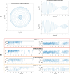
Fig. 1
Fig. 1
a Excitation k-space trajectory and gradients for the final parameter set. These parameters are optimized with six volunteer data sets (sagittal cervical spine) and ten initial points following MATLABs pattern search algorithm (part of the Global Optimization Toolbox in R2019b) and individual pulse calculations with subsequent Bloch simulations. The shown trajectory is used for all further experiments. b Complex RF pulse shapes for the proposed fat saturation method (SPSP) in standard and accelerated configurations for an exemplary volunteer measurement. The first column shows the RF magnitude and the second column represents the corresponding phase. As sampling compression factors (sc) increase, the RF pulse shape becomes more discretized
Fig. 2
Fig. 2
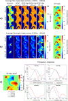
Multi-slice examples of phantom (1 slice block) (a), sagittal cervical spine (3 slice blocks) (b) and foot/ankle (4 slice blocks) (c) measurements with no fat saturation, Dixon, Gaussian fat sat and different SPSP configurations. The fat saturation quality achieved with Dixon is targeted with the pre-saturation approaches. a Fat ROI is highlighted with yellow arrows. Gaussian fat sat is not capable of saturating fat in presence of large B0 offsets, leading to insufficient fat saturation (red arrow). SPSP fat saturation improves fat signal suppression for all slices (green arrows). B0 inhomogeneities along slice direction do not show strong variations. b In vivo measurements comparing the same methods and settings used for phantom acquisitions. The SPSP fat pre-saturation shows a lower residual fat signal, especially in subcutaneous fat (green arrows). The configuration "SPSP bfgs100 sc6" differs from the less accelerated settings. This is mainly visible in the dorsal subcutaneous fat (red arrows). c In vivo foot/ankle measurements demonstrating applicability of the trajectory to other body parts. The fat saturation towards the toes (red arrow in Gaussian fat saturation) can be improved with the proposed method (green arrows). Inaccuracies in the B0 maps lead to incorrect fat saturation in higher slices (represented by slice 27, red arrows)
Fig. 3
Fig. 3
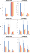
“Remaining signal” and “inhomogeneity” results for a phantom, b volunteer sagittal cervical spine and c volunteer sagittal foot/ankle measurements showing a clear improvement of fat saturation using the proposed SPSP approach compared to Gaussian fat sat in both metrics. Dixon indicates a state, where fat is saturated and water is not excited. For a Dixon has almost no fat signal left for the entire fat ROI. Water signal is only slightly excited for all pre-saturation approaches. Dixon shows some water excitation caused by the implementation of the vendor. Quantitatively no great differences between the SPSP approaches are visible. For b SPSP performs better than Gaussian fat sat in all considered ROIs, especially in subcutaneous fat. The analysis of the sagittal foot/ankle measurements (c) shows a clear improvement with the SPSP approach compared to the conventional Gaussian pulse in dorsal subcutaneous fat and at the metatarsal bones, which confirms the visual impression. Dixon outperforms the pre-saturation approaches in terms of fat saturation quality and homogeneity
Fig. 4
Fig. 4
Simulated frequency response of Gaussian fat sat and different SPSP configurations at a fat and b water frequencies with a frequency range of ± 20 Hz for an exemplary volunteer data set. Strong deviations from the target FAs (110°/0°) are noticeable for Gaussian fat sat as a result of the corresponding B0 inhomogeneities. Therefore, unsatisfactory fat saturation and undesired water excitation are expected. Saturation quality of SPSP fat saturation decreases with stronger accelerated configurations for all slices, but still remains better than Gaussian fat sat in all settings. c Illustration of the specific frequency responses of four exemplary voxels showing that the Gaussian pulse is shifted along the frequency axis due to B0 inhomogeneities, while the SPSP pulses have location-dependent response curves responding to the field shifts. Yellow points mark the targets of the pulse optimization for fat and water, respectively
Fig. 5
Fig. 5
Flip angle error (average and standard deviation) of simulated FAs compared to target FAs (110°/0°) at fat (− 3.4 ppm) and water frequencies (0.0 ppm) for cervical spine sagittal, coronal, transversal and foot/ankle sagittal simulations based on five volunteer data sets. A universal application of the offline determined trajectory parameters based on sagittal cervical spine data sets to the presented body regions and orientations seems feasible
Fig. 6
Fig. 6
Exemplary multi-slice volunteer measurement of “SPSP standard” configuration considering the actual B1 map (1st column) and the nominal B1 value (2nd column) based on the adjust transmitter voltage. Results in dorsal subcutaneous fat and spine are similar, whereas ventral subcutaneous fat (especially in the chest area) shows slightly higher residual signal when the nominal B1 values is used. A similar B1 pattern is observed for all B1 maps, with lower values towards the abdomen, especially in the chest area

Similar articles

Cited by

References

    1. Bley TA, Wieben O, Francois CJ, Brittain JH, Reeder SB. Fat and water magnetic resonance imaging. J Magn Reson Imaging. 2010;31(1):4–18. doi: 10.1002/jmri.21895. - DOI - PubMed
    1. Delfaut EM, Beltran J, Johnson G, Rousseau J, Marchandise X, Cotten A. Fat suppression in MR imaging: techniques and pitfalls. Radiographics. 1999;19(2):373–382. doi: 10.1148/radiographics.19.2.g99mr03373. - DOI - PubMed
    1. de Kerviler E, Leroy-Willig A, Clement O, Frija J. Fat suppression techniques in MRI: an update. Biomed Pharmacother. 1998;52(2):69–75. doi: 10.1016/S0753-3322(98)80006-1. - DOI - PubMed
    1. Hu HH, Bornert P, Hernando D, Kellman P, Ma J, Reeder S, Sirlin C. ISMRM workshop on fat-water separation: insights, applications and progress in MRI. Magn Reson Med. 2012;68(2):378–388. doi: 10.1002/mrm.24369. - DOI - PMC - PubMed
    1. Haase A, Frahm J, Hanicke W, Matthaei D. 1H NMR chemical shift selective (CHESS) imaging. Phys Med Biol. 1985;30(4):341–344. doi: 10.1088/0031-9155/30/4/008. - DOI - PubMed