. 2024 Apr 23;133(4):621-642.
doi: 10.1093/aob/mcae019.

Histochemical, metabolic and ultrastructural changes in leaf patelliform nectaries explain extrafloral nectar synthesis and secretion in Clerodendrum chinense

Affiliations

Histochemical, metabolic and ultrastructural changes in leaf patelliform nectaries explain extrafloral nectar synthesis and secretion in Clerodendrum chinense

Shobhon Paul et al. Ann Bot. .

Abstract

Background and aims: Extrafloral nectaries are nectar-secreting structures present on vegetative parts of plants which provide indirect defences against herbivore attack. Extrafloral nectaries in Clerodendrum chinense are patelliform-shaped specialized trichomatous structures. However, a complete understanding of patelliform extrafloral nectaries in general, and of C. chinense in particular, has not yet been established to provide fundamental insight into the cellular physiological machinery involved in nectar biosynthesis and secretory processes.

Methods: We studied temporal changes in the morphological, anatomical and ultrastructural features in the architectures of extrafloral nectaries. We also compared metabolite profiles of extrafloral nectar, nectary tissue, non-nectary tissue and phloem sap. Further, both in situ histolocalization and normal in vitro activities of enzymes related to sugar metabolism were examined.

Key results: Four distinct tissue regions in the nectar gland were revealed from histochemical characterization, among which the middle nectariferous tissue was found to be the metabolically active region, while the intermediate layer was found to be lipid-rich. Ultrastructural study showed the presence of a large number of mitochondria along with starch-bearing chloroplasts in the nectariferous region. However, starch depletion was noted with progressive maturation of nectaries. Metabolite analysis revealed compositional differences among nectar, phloem sap, nectary and non-nectary tissue. Invertase activity was higher in secretory stages and localized in nectariferous tissue and adjacent region.

Conclusions: Our study suggests extrafloral nectar secretion in C. chinense to be both eccrine and merocrine in nature. A distinct intermediate lipid-rich layer that separates the epidermis from nectary parenchyma was revealed, which possibly acts as a barrier to water flow in nectar. This study also revealed a distinction between nectar and phloem sap, and starch could act as a nectar precursor, as evidenced from enzymatic and ultrastructural studies. Thus, our findings on changing architecture of extrafloral nectaries with temporal secretion revealed a cell physiological process involved in nectar biosynthesis and secretion.

Keywords: Clerodendrum chinense; in situ histolocalization; Extrafloral nectary; nectar; nectariferous tissue; patelliform nectaries; phloem sap.


Figures

Fig. 1.
Fig. 1.
Clerodendrum chinense extrafloral nectary. A. Single plant under glasshouse conditions. B. EFNs grouped on the junction between the petiole and leaf blade (arrow). Inset showing stereo-microscopic view. C. Camponotus compressus ant visiting extrafloral nectary; accumulated nectar drops at the lamina base (inset). D. Nectar secretion rates in different stages of extrafloral nectary. E. Total chlorophyll content. Bar graph represents mean ± CI (95 %), n = 5 (D), n = 4 (E). Different letters represent differences among the different stages (P < 0.05).
Fig. 2.
Fig. 2.
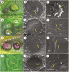
Macrostructure and surface ultrastructure of C. chinense EFNs at different secretory stages visualized via stereo- and scanning electron microscopy. Pre-secretory stage: A. EFN macrostructures without any visible nectar droplets. B, C. Surface ultrastructures showing bulging sacs in the subcuticular space depicting nectar accumulation. Secretory stage: D. Nectar droplet visible on the EFN. E, F. Openings for nectar release at the post-secretory stage. G. Brown spots indicating closing of the nectar release path. H, I. Clearly visible collapsed bulging sacs. J. A distinct type of smaller concave EFN is visible on the lamina surface. K, L. Openings for nectar release on the smaller nectaries.
Fig. 3.
Fig. 3.
Histological overview of EFNs in C. chinense. A. EFN after TBO staining showing three histologically distinct regions. B. Intermediate layer (arrow). C. Thickened cell walls between cells of the intermediate layer (arrow). D. EFNs showing dense nectariferous parenchyma (unstained). E. Intermediate layer showing blue fluorescence under UV light. F. Unstained intermediate layer after ruthenium red staining. G. EFN at the pre-secretory stage showing vascular patches after TBO staining. H. EFN at the secretory stage after TBO staining. I. EFN at the post-secretory stage showing less dense nectariferous cells and vascular connections after TBO staining. Scale bars represent 100 µm for A, D, E, G–I; 25 µm for B; 50 µm for F; 10 µm for C. Abbreviations: s.e, secretory epidermis; n.p, nectariferous parenchyma; s.n.p, sub-nectariferous parenchyma; i.l, intermediate layer.
Fig. 4.
Fig. 4.
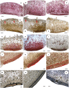
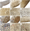
Histology of extrafloral nectary tissues at pre-secretory, secretory and post-secretory stages. A, B. PAS staining showing the presence of neutral carbohydrates in the nectariferous tissues of pre-secretory and secretory stages, respectively. C. Post-secretory stage showing faintly stained regions indicating depletion of neutral carbohydrates. D, E. Lugol’s iodine staining showing the presence of starch grains in the sub-nectariferous tissues of pre-secretory and secretory stages, respectively. F. No starch grains are visible at the post-secretory stage. G, H. Xylidine Ponceau staining showing the presence of total proteins in the secretory epidermis and nectariferous tissue of pre-secretory and secretory stages, respectively. I. Post-secretory stage showing depletion of total proteins in the nectariferous tissue. J, K, L. Oil Red O staining showing the presence of lipid-rich cuticular and intermediate layers at all three stages. M, N, O. Sudan Black B staining showing the presence of lipid-rich intermediate layers at all three stages. A clear blockage is noted in the secretory epidermis at the post-secretory stage (O). Scale bars represents 100 µm for A–I; 50 µm for J–O.
Fig. 5.
Fig. 5.
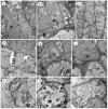
Transmission electron micrographs of the secretory epidermis, intermediate layer and nectariferous parenchyma at pre-secretory and secretory stages. A, B, C. Palisade-like secretory epidermal cells at secretory stage. D. Intermediate cell layer with thickened wall and plasmodesmatal connections (inset) at secretory stage. E. Junction between the secretory epidermis and intermediate cell at secretory stage. F. Junction between the intermediate cell and nectariferous parenchyma cell at secretory stage. G. Nectariferous parenchyma cells with several starch-containing chloroplasts and mitochondria at pre-secretory stage. H. Nectariferous parenchyma cells showing plasmodesmatal connection with adjacent fusing small vacuoles cells at secretory stage. I. Nectariferous parenchyma cells showing numerous small vacuoles, mitochondria and a few chloroplasts at pre-secretory stages. Abbreviations: se, secretory epidermis; ic, intermediate cell; np, nectariferous parenchyma; tw, thickened wall; m, mitochondria; cp, chloroplast; st, starch grain, v, vacuole, n, nucleus.
Fig. 6.
Fig. 6.
Transmission electron micrographs of nectariferous and sub-nectariferous parenchyma at different stages. Pre-secretory stage: A. Peripheral arrangement of chloroplasts in the sub-nectariferous parenchyma cell. B. Chloroplasts in the nectariferous cell with numerous starch grains. C, D. Plasmodesmatal connection between adjacent nectariferous cells. Secretory stage: E. Nectariferous parenchyma cell with several mitochondria. F, G. Vesicles tending toward plasma membrane (arrows) for their subsequent coalescence with the membrane, an important part in merocrine secretion. Post-secretory stage: H. Reduction of starch grains in the post-secretory stage along with the movement of chloroplasts towards the periphery in the nectariferous cell due to the formation of large vacuoles. I, J. Chloroplasts with reduced starch grains. Abbreviations: m, mitochondria; cp, chloroplast; st, starch grain, v, vacuole.
Fig. 7.
Fig. 7.
Variable composition of metabolites in different nectar and nectary samples. A. Heatmap representation of the variable metabolite composition in extrafloral nectar, nectary tissue, non-nectary tissue and phloem sap. Relative abundance is shown in the heatmap where dark blue colour indicates the lowest value and red indicates the highest value, with other shades in between corresponding to the gradient between extremes. The corresponding PLS-DA shows a clear separation of different samples. B. Scores plot showing 85.1 % variation between the samples as collectively shown in components 1 and 2.
Fig. 8.
Fig. 8.
Variation in relative abundances of metabolites from various stages of nectary and non-nectary tissue samples. A. Heatmap representation of the variable metabolite abundances in different stages of nectary and non-nectary tissues. Relative abundance is shown in the heatmap where dark blue indicates the lowest value and red indicates the highest value, with other shades corresponding to the gradient between extremes. The corresponding PLS-DA shows a separation of different samples. B. Scores plot showing 55.6 % of the variation between the samples as collectively shown in components 1 and 2.
Fig. 9.
Fig. 9.
In vitro activities of carbohydrate-metabolizing enzymes at different nectary developmental stages. A. Invertase activities. B. Amylase activities. C. Sucrose phosphate synthase activities. D. Sucrose synthase activities. Bars represent the mean of three biological replicates ± CI (95 %). Different letters represent differences among the different secretory stages (P < 0.05).
Fig. 10.
Fig. 10.
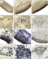
Histo-enzymatic localization of invertase on EFN tissues by the formation of blue formazan precipitate. A, B, C. EFN sections under control conditions with no reactions performed at pre-secretory, secretory and post-secretory stages, respectively. D, E, F. Visualization of vascular bundles of EFNs at pre-secretory, secretory and post-secretory stages, respectively, under control conditions. G, H, I. Localization of invertase in the EFN tissues at pre-secretory, secretory and post-secretory stages, respectively. Note: invertase activity was higher in the secretory epidermis, intermediate layer and nectariferous parenchyma in pre-secretory and secretory stages compared to post-secretory stage. J, K, L. In vascular bundles of EFNs, invertase activity was localized in the phloem elements at the pre-secretory, secretory and post-secretory stages, respectively. Scale bars represent 100 µm for A–C, G–I; 50 µm for D–F, J–L.
Fig. 11.
Fig. 11.
Histo-enzymatic localization of sucrose synthase on EFN tissues by the formation of blue formazan precipitate. A, B, C. EFN sections under control conditions with no reactions performed at pre-secretory, secretory and post-secretory stages, respectively. D, E, F. Visualization of vascular bundles of EFNs at pre-secretory, secretory and post-secretory stages, respectively, under control conditions. G, H, I. Localization of sucrose synthase in the EFN tissues at pre-secretory, secretory and post-secretory stages, respectively Note: sucrose synthase activity was higher in the sub-nectariferous parenchyma in pre-secretory and secretory stages compared to post-secretory stage. J, K, L. In vascular bundles of EFNs, sucrose synthase activity was localized in the phloem elements at the pre-secretory, secretory and post-secretory stages, respectively. Scale bars represent 100 µm for A–C, G–I; 50 µm for D–F, J–L.
Fig. 12.
Fig. 12.
A. A conceptual model of nectar secretion from the patelliform foliar extrafloral nectary glands in C. chinense. This diagram is based on the results of the present study combined with previous observations (Lin et al., 2014; Heil, 2015; Chatt et al., 2021). In foliar secretory tissue, nectar precursors are the primary photosynthates that have three possible fates. First, they can undergo phloem loading where sucrose serves as the major transport sugar. During active nectar secretion, phloem unloading drives phloem sap into nectary tissue via the sucrose transporter (SUT), where it is cleaved by sucrose synthase (SuSy) to create a hexose pool that can directly participate in nectar chemistry after being cleaved by cell wall invertase (CWIN4) and transported by the hexose transporter (HT). Second, primary photosynthates (i.e. triose-phosphates) undergo alteration to form hexose phosphates leading to the sucrose biosynthesis pathway, with sucrose phosphate synthase (SPS) catalysing the crucial regulatory step. The sucrose efflux transporter SWEET9 secretes sucrose into the apoplastic space, and cell wall invertase (CWIN4) cleaves it into nectar hexoses. Osmotic gradients cause water accumulation and exudation of nectar via nectary microchannels. Third, photosynthates can remain stored as starch in the chloroplasts and this is eventually broken down by amylase and directed towards the sucrose biosynthesis pathway leading to nectar secretion. B. Schematic diagram highlighting the major changes that occur at the cellular level during the process of nectar secretion in the nectariferous parenchyma. Numerous small vacuoles present during the pre-secretory stages coalesce to form large vacuoles in the secretory and post-secretory stages leading to the peripheral orientation of cell organelles. Starch granules present in the chloroplasts of pre-secretory stages undergo degradation in the subsequent stages and may serve as the precursor of nectar components. The large number of mitochondria provides energy for the secretory processes.

Similar articles

Cited by

References

    1. Afzal S, Singh NK, Singh N, Chaudhury N.. 2021. Structural analysis of extrafloral nectaries of Senna occidentalis L.: insights on diversity and evolution. Planta 254: 125. - PubMed
    1. Arnon DI. 1949. Copper enzymes in isolated chloroplasts, polyphenolxidase in Beta vulgaris. Plant Physiology 24: 1–15. - PMC - PubMed
    1. Beccari N, Mazzi V.. 1966. Manuale di tecnica microscopica. Guida pratica alla ricercar istologica e istochimica. Torino: Società Editrice Libraria.
    1. Bentley BL. 1977. Extrafloral nectaries and protection by pugnacious bodyguards. Annual Review of Ecology and Systematics 8: 407–427.
    1. Bhattacharya R, Saha S, Kostina O, Muravnik L, Mitra A.. 2020. Replacing critical point drying with a low-cost chemical drying provides comparable surface image quality of glandular trichomes from leaves of Millingtonia hortensis L. f. in scanning electron micrograph. Applied Microscopy 50: 15. - PMC - PubMed

Publication types

MeSH terms

LinkOut - more resources