. 2024 Jul;245(1):109-136.
doi: 10.1111/joa.14024. Epub 2024 Feb 16.

The vomeronasal system of the wolf (Canis lupus signatus): The singularities of a wild canid

Affiliations

The vomeronasal system of the wolf (Canis lupus signatus): The singularities of a wild canid

Irene Ortiz-Leal et al. J Anat. 2024 Jul.

Abstract

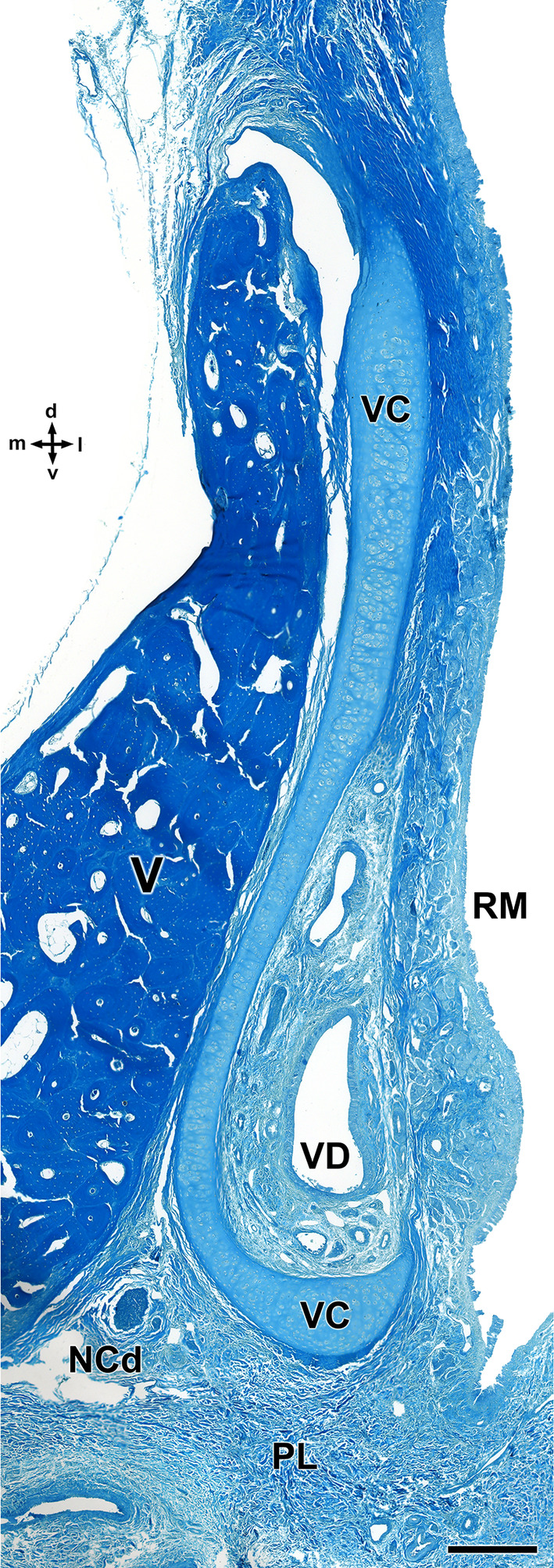
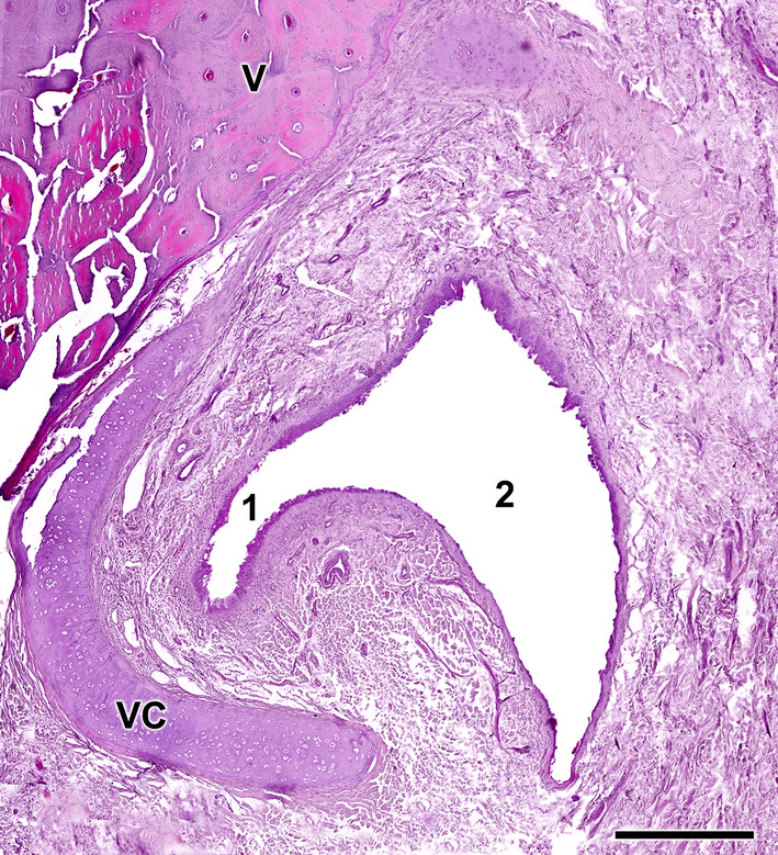
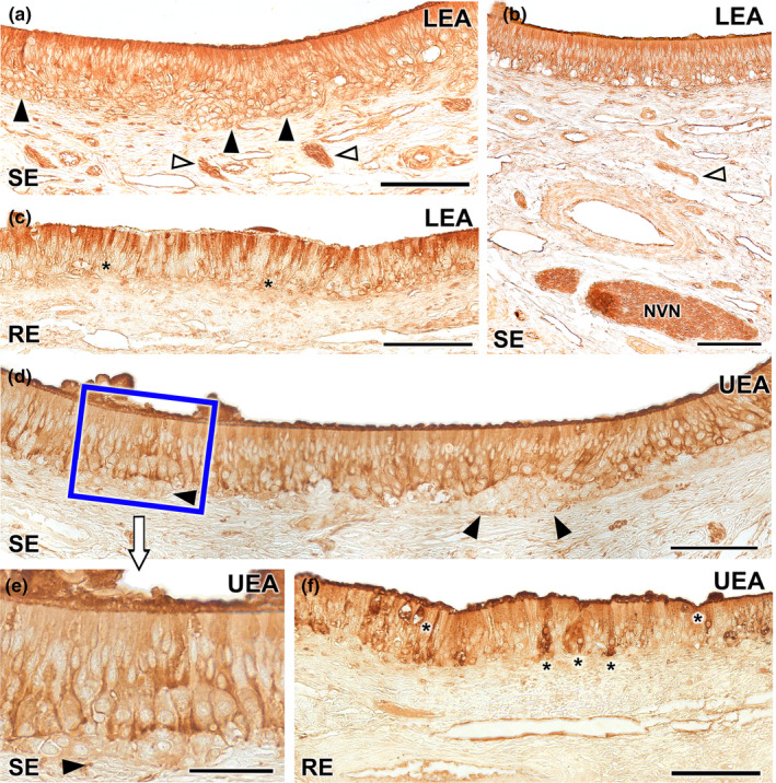
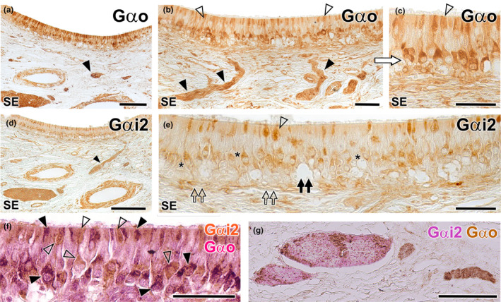
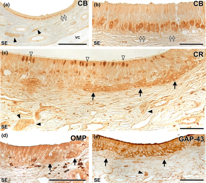
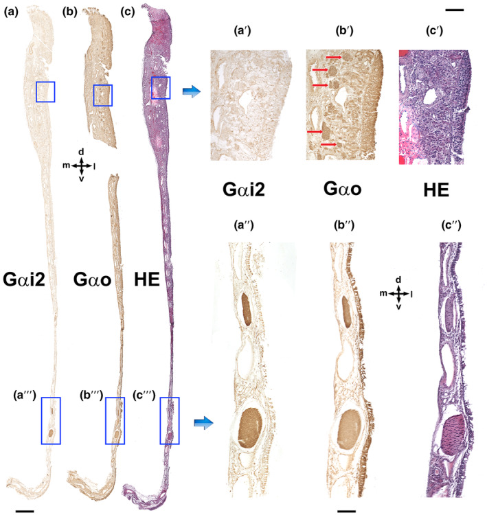
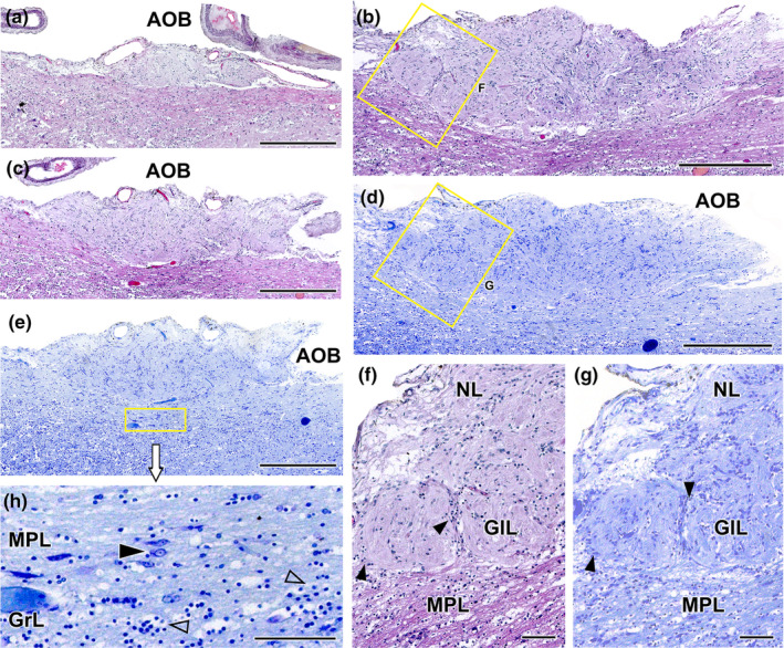
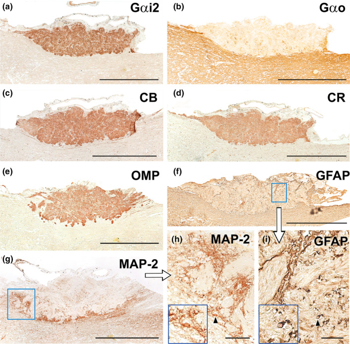
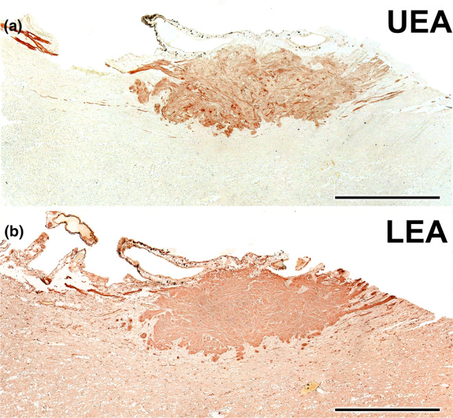
Wolves, akin to their fellow canids, extensively employ chemical signals for various aspects of communication, including territory maintenance, reproductive synchronisation and social hierarchy signalling. Pheromone-mediated chemical communication operates unconsciously among individuals, serving as an innate sensory modality that regulates both their physiology and behaviour. Despite its crucial role in the life of the wolf, there is a lacuna in comprehensive research on the neuroanatomical and physiological underpinnings of chemical communication within this species. This study investigates the vomeronasal system (VNS) of the Iberian wolf, simultaneously probing potential alterations brought about by dog domestication. Our findings demonstrate the presence of a fully functional VNS, vital for pheromone-mediated communication, in the Iberian wolf. While macroscopic similarities between the VNS of the wolf and the domestic dog are discernible, notable microscopic differences emerge. These distinctions include the presence of neuronal clusters associated with the sensory epithelium of the vomeronasal organ (VNO) and a heightened degree of differentiation of the accessory olfactory bulb (AOB). Immunohistochemical analyses reveal the expression of the two primary families of vomeronasal receptors (V1R and V2R) within the VNO. However, only the V1R family is expressed in the AOB. These findings not only yield profound insights into the VNS of the wolf but also hint at how domestication might have altered neural configurations that underpin species-specific behaviours. This understanding holds implications for the development of innovative strategies, such as the application of semiochemicals for wolf population management, aligning with contemporary conservation goals.

Keywords: accessory olfactory bulb; chemical communication; immunohistochemistry; lectins; pheromones; vomeronasal organ; vomeronasal system; wolf.


Figures

FIGURE 1
FIGURE 1
Macroscopic cross sectional study of the nasal cavity of the wolf. (a) Lateral view of the head of the wolf showing the eight consecutive levels chosen for the macroscopic sectional study. (b) The central part of the vomeronasal organ (VNO) is located at level 6 (yellow line in “a”, enlarged section in “b and d”). The VNO corresponds to two tubular structures located in the ventral part of the nasal septum (black arrows), lateral to the vomer bone (asterisk) and ventral to the cartilage of the nasal septum (circle). (c) Transverse sections of the nasal cavity ordered from rostral (1) to caudal (8), corresponding to the levels represented in (a). (d) At higher magnification, it can be seen how the VNO is enveloped by a cartilaginous capsule (arrowheads). In the central part of the VNO, the vomeronasal ducts can be observed. CT, canine tooth; DT, dorsal turbinate; NS, nasal septum; VT, ventral turbinate.
FIGURE 2
FIGURE 2
Topographical relationship of the wolf VNO. (a) Fronto‐dorsal view of the skull skeleton showing the relationship of the lateral part of the vomer bone (V) to the palatine fissure. (b–d) Computerised tomography images of the head in the horizontal (b), parasagittal (c) and transverse (d) planes. Arrows indicate the location of the VNO. CT, canine tooth; IB, incisive bone; NB, nasal bone; VC, vomeronasal cartilage; VT, ventral turbinate.
FIGURE 3
FIGURE 3
Dissection of the nasal and cranial cavities of the wolf. (a). Dorsolateral view of the nasal cavity, showing the projection zone of the VNO (arrow) and the myelinated branches forming the caudal nasal nerve (arrowheads) entering the VNO. (b) Enlargement of the inset shown in (c) displaying the vomeronasal nerves leaving the VNO (arrowheads) in a caudodorsal direction towards the medial portion of the cribriform plate of the ethmoid. RM: Respiratory mucosa. OM: Olfactory mucosa. (c) Lateral view of the nasal and cranial cavities, showing in the latter the left‐brain hemisphere. The projection area of the VNO (arrow), the vomeronasal nerves in a caudodorsal direction (inset), the caudal nasal nerve (open arrowhead) and the olfactory bulb (arrowhead) are also indicated. FS, Frontal sinus. Scale bar = 1.5 cm.
FIGURE 4
FIGURE 4
Wolf VNO after its full extraction in association to the vomer bone. (a) Transverse cross section of the nasal septum corresponding to the level 9 shown in Figure 1. From a caudodorsal viewpoint, the VNOs (arrows) are visualised on both sides of the base of the nasal septum (NS) over the thin vertical projection of the vomer bone (V). (b) On a more rostral level, corresponding to level 3 of Figure 1, a ventral view of the vomer bone shows both VNOs with its distinctive rounded and elongated shape (arrows). The lancet points to the lifted respiratory mucosa of the nasal cavity that covers the VNO. (c) Dissected out and partially transversely sectioned VNO (black arrows) showing the crescent shape vomeronasal duct (white arrows). a and b, fresh samples; (c), BF‐fixed sample. Scale bars: (a–b) = 0.5 cm, (c) = 0.2 cm.
FIGURE 5
FIGURE 5
Ventral view of the palate of the wolf after removal of the mandible. (a) The mucosa of the roof of the oral cavity is observed, and in its most anterior part, the location of the incisive papilla (IP) and palatine rugae (PR) are indicated. (b) Skeleton of the corresponding area where the palatine fissures (PF) are observed on both sides of the midline. PB: Palatine bone. (c) Superposition of images analogous to “a and b” to show the exact location of the PF on the mucosa of the palate. The IP is also shown (arrow). Scale bar: (a) 1 cm, (b–c) 1.5 cm.
FIGURE 6
FIGURE 6
Encephalon and olfactory bulb of the wolf. (a–d) Dorsal, ventral, lateral and medial views of BF‐fixed encephalon. The white box highlights the dorsal, ventral, lateral and medial views of the olfactory bulb (OB) respectively. (e) Lateral view of the right olfactory bulb, separated from a formalin‐fixed brain. (f) Medial view of the left MOB separated from the BF‐fixed brain. (g) Dorsocaudal view of the left OB, BF fixed. The area where the accessory olfactory bulb is presumptively located is indicated by the broken red circle. Bs, brain stem; Cb, cerebellum; Cc, corpus callosum; Cd, caudal; d, dorsal; FL, frontal lobe; Ia, interthalamic adhesion; l, lateral; LOT, lateral olfactory tract; m, medial; nII, optical nerve; nIII, oculomotor nerve; OP, olfactory peduncle; Pi, piriform lobe; Po, Pons; R, rostral. Scale bar = 1 cm.
FIGURE 7
FIGURE 7
Histological transverse section of the wolf vomeronasal organ in its central portion, stained with haematoxylin–eosin. This central level exemplifies the major histological features of the VNO. VC, vomeronasal cartilage. In its central portion, it forms an incomplete ring which opens dorsally. Arrowhead, artery; Co, connective tissue; D, dorsal; l, lateral; LT, Lymphoid tissue; m, medial; NCd, caudal nasal nerve; NVN, vomeronasal nerves; RE, respiratory epithelium; RM, respiratory mucosa; SE, neurosensory epithelium; v, ventral; VD, vomeronasal duct; VG, vomeronasal glands; Vv, vomeronasal veins. Scale bar = 500 μm.
FIGURE 8
FIGURE 8
Transverse sections of the wolf VNO at two selected levels. (a) Rostral level. (b) Caudal level. Co, connective tissue; Gls. vomeronasal glands; NCd, caudal nasal nerve; NVN, vomeronasal nerve; RM, respiratory mucosa; SE, Sensory epithelium; VG, vomeronasal glands; VC, vomeronasal cartilage; VD, vomeronasal duct; and Vv, vomeronasal veins. Staining: Gallego's trichrome (a) and haematoxylin–eosin (b). d, dorsal; l, lateral; m, medial; and v, ventral. Scale bar = 500 μm.
FIGURE 9
FIGURE 9
Histological study of the epithelia of the wolf vomeronasal duct. (a) Medial parenchyma of the organ stained with Gallego's trichrome. The overlying sensory neuroepithelium (SE), enlarged in (b, c), shows a large lamina propria with veins (Vv), vomeronasal nerves (NVN) and connective tissue. (b) Two large clusters of cells similar in appearance to neuroreceptor cells (arrowheads) are observed in the lamina propria. (c) Cellular components of the neuroepithelium: 1, basal cells; 2, neuroreceptor cells (open arrowheads); 3, sustentacular cells; 4, cell processes layer; and 5, mucomicrovillar complex. (d) Neurosensory epithelium and lamina propria, stained with haematoxylin–eosin. (e) Enlargement of the area is shown in “d” showing the five components of the sensory neuroepithelium. Open arrowheads: neurosensory cells. (f) Enlargement of the area of respiratory epithelium (RE) is shown in (g), stained with Gallego's trichrome. Note the presence of cilia (white arrowhead), chemosensory cells (black arrowheads) and goblet cells (*). (g) Respiratory epithelium stained with Gallego's trichrome. (h) Enlargement of the Figure “c”, displaying the luminal surface of the SE, covered by the microvillar complex. (i) Enlargement of the luminal surface of the RE, showing the ciliated covering. Scale bars: (a, d, g) = 100 μm; (b, c, e, f) = 50 μm; (h, i) = 10 μm.
FIGURE 10
FIGURE 10
Vomeronasal glands of the wolf. The VNO parenchyma presents an abundant number of glands, which are more developed in its dorsal area. (a, b) The arrowheads delimit the glandular areas of interest. Both Gallego's trichrome (a) and haematoxylin–eosin (b) stainings show the serous tubuloalveolar nature of the wolf vomeronasal glands. (c) Alcian blue staining shows the positivity of these acini to this stain. VD, vomeronasal duct; Vv, vomeronasal veins; (*) Respiratory mucosa. Scale bar = 250 μm.
FIGURE 11
FIGURE 11
Vasculature of the wolf VNO. (a) Image of the anterior portion of the VNO is shown in Figure 8a exemplifies the presence of a profuse venous ring (Vv) surrounding the vomeronasal duct (VD). The arteries (arrowheads) are small and sparse. (b) Arteries indicated in A showed at higher magnification. (c) Caudal section of the VNO showing the glove‐fingered termination of the vomeronasal duct (VD). Numerous veins (Vv) and small arterial trunks (arrowhead) predominate at this level. (d) In a central section of the VNO, large venous sinuses (Vv) run along the lateral portion of the parenchyma. NCd, caudal nasal nerve. Staining: (a, b d) Gallego's trichrome; (c) haematoxylin–eosin. Scale bars: (a, d) = 250 μm; (c) = 100 μm; (b) = 50 μm.
FIGURE 12
FIGURE 12
Histological study of the wolf vomeronasal organ innervation. (a). Unmyelinated branches of the vomeronasal nerves (*). They are characterised by their homogeneous appearance, with densely packed nerve bundles. (b). The lateral part of the parenchyma contains myelinated nerve branches originating from the caudal nasal nerve (*). Their structure is looser and the void spaces corresponding to the myelin sheaths are visible. Staining: Haematoxylin–eosin. Scale bar = 100 μm.
FIGURE 13
FIGURE 13
Decalcified cross section of the anterior portion of the wolf nasal septum, stained with Gallego's trichrome, showing the topographical relationship of the VNO with the vomer bone. The vomeronasal cartilage (VC) presents an elongated morphology, accompanying the shape of the vomer bone (V) and opens laterally. d, dorsal; l, lateral; LP, lamina propria; m, medial; NCd, caudal nasal nerve; PL, palate; RM, respiratory mucosa; v, ventral; VD, vomeronasal duct. Scale bar = 250 μm.
FIGURE 14
FIGURE 14
Opening of the vomeronasal duct into the incisive duct of the wolf. The vomeronasal duct (1) establishes a direct communication with the incisive duct (2). Anatomically, the latter runs laterally to the organ, allowing a double direct communication between the oral and nasal cavities. Staining: haematoxylin–eosin. VC, vomeronasal cartilage; and V, vomer bone. Scale bar = 500 μm.
FIGURE 15
FIGURE 15
Lectin histochemical labelling of the vomeronasal epithelium. (a–c) LEA lectin labelling: The neuroreceptor epithelium (SE) shows intense labelling throughout all its components including neuroepithelial clusters (black arrowheads) and nerve bundles in the lamina propria (white arrowheads) (a). This labelling extends to the vomeronasal nerves (NVN) (b). (c) The respiratory epithelium shows a diffuse LEA positivity, concentrated mainly in the most apical part of the epithelium, while the basal cells remain unlabelled (asterisk). (d–f) UEA lectin: Positivity is demonstrated across the sensory epithelium of the VNO excluding the neuronal clusters (arrowheads) which are not labelled (d). At higher magnification, an intensity gradient increasing with depth can be discerned (e). The unlabelled neuronal clusters are shown (arrowhead). (f) The respiratory epithelium exhibits fainter labelling, with few positive cells scattered along the epithelium (*). UEA labelling is also markedly concentrated in the mucociliary layer. Scale bar: (a–d, f) = 100 μm; (e) = 50 μm.
FIGURE 16
FIGURE 16
Immunohistochemical study of the wolf VNO. Gαo (a–c): Immunolabelling with anti‐Gαo shows a pattern of neuronal labelling predominantly concentrated in the neuroreceptor cells present in the basal layers of the neuroepithelium (a and b) and extending to the adjacent vomeronasal nerves (arrowheads). At higher magnification (c: enlarged area of the blue box in image b), it is appreciated how the labelling extends along the entire length of the immunopositive neuroreceptor cells, from the apical dendrite (open arrowhead) to the soma. Immunopositive neuroreceptor cells embrace the intraepithelial capillaries of the VNO. Gαi2 (d, e): The labelling is predominantly concentrated in the neuroreceptor cells mainly located in the central zone of the epithelium (asterisk) and the vomeronasal nerves (arrowhead). Unlike Gαo, no immunopositive neurons are identified around the intraepithelial capillaries (black double arrow). The dendritic knobs are less numerous than in Gαo but more dilated (open arrowhead). The deep neuronal clusters are immunonegative (open double arrows). (f–g): f Double immunostaining for Gαi2 and Gαo confirms the presence of two subpopulations of vomeronasal neuroreceptor cells. Anti‐Gαi2 (brown) immunostains a subpopulation of cells predominantly located in the central zones, which have thick dendritic knobs (open arrowheads). Anti‐Gαo (red) stains a cell subpopulation mainly located in a more basal zone (black arrowheads). (g) In some cases, as the one shown, the vomeronasal nerves in the parenchyma are predominantly composed either of Gαi2 type fibres (red) or Gαo type fibres (brown). Scale bars: (a, d and g) = 100 μm; (b, c, e and f) = 50 μm.
FIGURE 17
FIGURE 17
Immunohistochemical study of the wolf VNO. Calbindin (CB) (a–b): At low magnification (f) uniform labelling is observed throughout the neuroreceptor cell layer, extending into the vomeronasal nerves (black arrowheads). No immunopositivity is observed in the clusters (double arrows). At higher magnification (g: magnification of the blue box in f) how the immunopositive cells correspond to a regularly aligned subpopulation in an intermediate zone between the basal and apical layers is appreciated. The terminal knobs are poorly labelled. Calretinin (CR) (c): A strong immunopositivity to the neuronal clusters is observed (arrows). In addition, a subpopulation of neuroreceptor cells whose dendrites show dilated terminal knobs can be identified (white arrowheads). Vomeronasal nerves (black arrowhead). OMP (d): Anti‐OMP produces a diffuse labelling throughout the epithelium, including the basal clusters (arrows). GAP‐43 (e): Pattern similar to (i) but without labelling the neuronal clusters (arrows). Arrowhead: vomeronasal nerves. SE: Neurosensory epithelium. VC: Vomeronasal cartilage. Scale bars: (a) = 250 μm; (d and e) = 100 μm; (b and c) = 50 μm.
FIGURE 18
FIGURE 18
Immunolabelling of the VNO nasal septum mucosa with anti‐G proteins subunits. (a) Anti‐Gαi2 exclusively stains the vomeronasal nerves as they run through the mucosa of the nasal septum (a"). Therefore, anti‐Gαi2 does not produce positive immunolabelling of the olfactory nerves, which run in the upper part of the olfactory mucosa (a'). (b) Anti‐Gαo (b) stains both the vomeronasal nerves (b″) and the olfactory nerves in the mucosa (b′, red arrows). (c) Haematoxylin–eosin adjacent section. d, Dorsal; l, lateral; m, medial; and v, ventral. Scale bars: (a–c) = 500 μm; (a'–c′ and a"–c″) = 100 μm.
FIGURE 19
FIGURE 19
Histological study of the wolf accessory olfactory bulb stained with haematoxylin–eosin and Nissl staining. (a–c). A general view of the AOB can be seen at three selected sagittal levels stained with HE. They show the elongated shape of this structure and the predominance of the nervous (NL) and glomerular layers (GlL). (d, e) Sagittal Nissl‐stained sections at low magnification allows to appreciate the development of the AOB. (f) At higher magnification (corresponding to box in b), two glomerular formations clearly defined by periglomerular cells (arrowheads) are appreciated. (g) The magnification of the superficial area of the AOB (box in d) allows to discriminate the presence of a mitral‐plexiform layer (MPL). (h) Enlargement of the deep area of the AOB (corresponding to box in e) shows mitral cells (black arrowhead) in the MPL as well as granular cells (open arrowhead) in the deeper granular layer (GrL). Scale bars: (a–e) = 500 μm; (f–h) = 100 μm.
FIGURE 20
FIGURE 20
Immunohistochemical study of wolf AOB. (a) Anti‐Gαi2 uniformly and intensely label the superficial layers of the AOB (nervous and glomerular layers). The entire surrounding neuropil is negative. (b) Anti‐Gαo produces a reverse pattern to the one shown in A, where the neuropil surrounding the superficial layers is strongly immunopositive, including the mitral‐plexiform and granular layers of the AOB. However, the superficial layer is clearly negative, although immunopositive punctae areas are observed. (c–e) The calcium binding proteins, calbindin (c) and calretinin (d), as well as OMP (e) show an identical labelling pattern to that obtained with anti‐Gαi2, concentrated in both the nerve and glomerular layers and immunonegative for the neuropil. (f) Anti‐GFAP (f and enlarged area in i) produces a trabecular labelling pattern in the nerve and glomerular layers, which corresponds to the ensheathing glia accompanying the vomeronasal nerve endings. Occasionally, cell bodies belonging to these glial cells are visible (arrowhead). The cell body is magnified in the bottom left‐hand box. (g) Anti‐MAP‐2 immunolabelling does not produce immunopositive labelling in the superficial layers (nervous and glomerular), but it strongly labels an irregular band corresponding to the MPL layer. (h) MAP‐2‐immunopositive prolongations originating from the MPL can be observed running between the glomeruli of the AOB. (h: enlargement of the box in g). The cell body of an immunopositive interneuron (arrowhead) is shown in an enlarged view in the box at the bottom left. Scale bars: (a–g) = 500 μm; (h–i) = 50 μm.
FIGURE 21
FIGURE 21
Lectin histochemical study of the wolf AOB. (a) UEA lectin labels uniformly and strongly the superficial layers of the AOB (nervous and glomerular layers). The entire surrounding neuropil is negative. (b) LEA‐lectin produces a similar labelling to UEA but with higher intensity in the AOB superficial layers and a diffuse pattern in the surrounding neuropil. Scale bar = 500 μm.

Similar articles

Cited by

References

    1. Abellán‐Álvaro, M. , Martínez‐García, F. , Lanuza, E. & Agustín‐Pavón, C. (2022) Inhibition of the medial amygdala disrupts escalated aggression in lactating female mice after repeated exposure to male intruders. Communications Biology, 5(1), 980. Available from: 10.1038/s42003-022-03928-2 - DOI - PMC - PubMed
    1. Adams, D.R. & Wiekamp, M.D. (1984) The canine vomeronasal organ. Journal of Anatomy, 138(Pt 4), 771–787. - PMC - PubMed
    1. Alonso, J.R. , Briñón, J.G. , Crespo, C. , Bravo, I.G. , Arévalo, R. & Aijón, J. (2001) Chemical organization of the macaque monkey olfactory bulb: II. Calretinin, calbindin D‐28k, parvalbumin, and neurocalcin immunoreactivity: calcium‐binding proteins in the monkey OB. Journal of Comparative Neurology, 432(3), 389–407. Available from: 10.1002/cne.1110 - DOI - PubMed
    1. Alsafy, M.A.M. , El‐gendy, S.A.A. & Abumandour, M.M.A. (2014) Computed tomography and gross anatomical studies on the head of one‐humped camel (Camelus dromedarius). The Anatomical Record, 297(4), 630–642. Available from: 10.1002/ar.22865 - DOI - PubMed
    1. Asa, C.S. , Mech, L.D. , Seal, U.S. & Plotka, E.D. (1990) The influence of social and endocrine factors on urine‐marking by captive wolves (Canis lupus). Hormones and Behavior, 24(4), 497–509. Available from: 10.1016/0018-506X(90)90038-Y - DOI - PubMed

Publication types