. 2024 Jul;61(4):541-549.
doi: 10.1177/03009858241231556. Epub 2024 Feb 17.

Eptesipox virus-associated lesions in naturally infected big brown bats

Affiliations

Eptesipox virus-associated lesions in naturally infected big brown bats

Ursula G Perdrizet et al. Vet Pathol. 2024 Jul.

Abstract

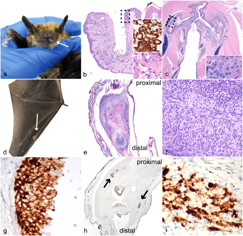
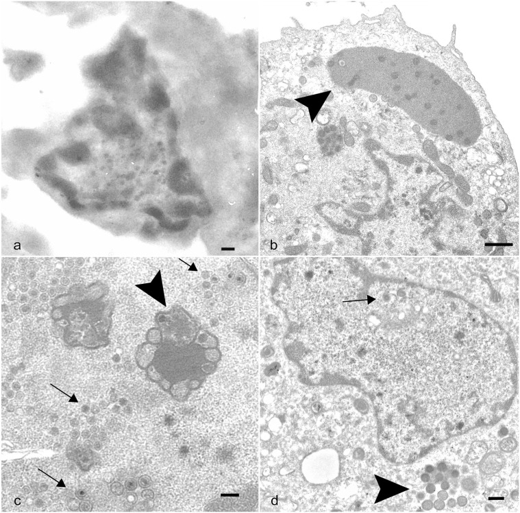
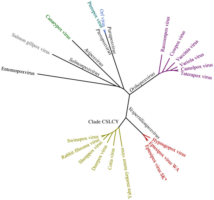
Bats have many unique qualities amongst mammals; one of particular importance is their reported tolerance to viruses without developing disease. Here, the authors present evidence to the contrary by describing and demonstrating viral nucleic acids within lesions from eptesipox virus (EfPV) infection in big brown bats. One hundred and thirty bats submitted for necropsy from Saskatchewan, Canada, between 2017 and 2021 were screened for EfPV by polymerase chain reaction (PCR); 2 had amplifiable poxvirus DNA. The lesions associated with infection were oral and pharyngeal ulcerations and joint swelling in 2/2 and 1/2 cases, respectively. These changes were nonspecific for poxvirus infection, although intracytoplasmic viral inclusion bodies within the epithelium, as observed in 2/2 bats, are diagnostic when present. Viral nucleic acids, detected by in situ hybridization (ISH), were observed in the epithelium adjacent to ulcerative lesions from both cases and within the joint proliferation of 1 case. A new isolate of EfPV was obtained from 1 case and its identity was confirmed with electron microscopy and whole genome sequencing. Juxtanuclear replication factories were observed in most cells; however, rare intranuclear virus particles were also observed. The significance of the presence of virus particles within the nucleus is uncertain. Whole genome assembly indicated that the nucleotide sequence of the genome of this EfPV isolate was 99.7% identical to a previous isolate from big brown bats in Washington, USA between 2009 and 2011. This work demonstrates that bats are not resistant to the development of disease with viral infections and raises questions about the dogma of poxvirus intracytoplasmic replication.

Keywords: big brown bat; chiroptera; eptesipox virus; joint; oral ulcer; poxviridae; ultrastructure; whole genome sequencing.


Conflict of interest statement

Declaration of Conflicting InterestsThe author(s) declared no potential conflicts of interest with respect to the research, authorship, and/or publication of this article.

Figures

Figure 1.
Figure 1.
Big brown bat eptesipox virus-associated lesions. (a, b, d–f, h, i) Index case from which virus was isolated. (a) Left lower lip. Gross image of swelling and ulceration (arrow) and swelling of right carpus. (b) Lower left lip and mandible. Ulcer of lip and gingiva, the dashed box represents the location of higher magnification inset. Hematoxylin and eosin (HE). Inset (bottom): higher magnification of keratinocytes with intracytoplasmic inclusion bodies. HE. Inset (top): eptesipox virus (EfPV) in situ hybridization (ISH) probe binding is predominantly restricted to the cytoplasm. (c) Pharynx. Extensive ulceration in the second big brown bat identified with EfPV infection, the dashed box represents the location of higher magnification inset. HE. Inset: higher magnification of intracytoplasmic pink viral inclusion bodies in the epithelium. HE (d) Metacarpophalangeal joint of the third digit of the left wing with joint swelling (arrow). (e) Left third metacarpophalangeal joint that is expanded by spindle cell proliferation. HE. (f) Left third metacarpophalangeal joint. Higher magnification showing large numbers of neutrophils amongst the spindle cells. HE. (g) Pharynx. Serial section of the ulcerated pharyngeal epithelium in (c) with probe binding restricted primarily to the cytoplasm. EfPV ISH. (h) Left third metacarpophalangeal joint. A serial section of the joint in (e) with multifocal probe binding to EfPV nucleic acids (arrows). EfPV ISH. (i) Left third metacarpophalangeal joint. Higher magnification demonstrating more intense binding in the nucleus than the cytoplasm of spindle cells. EfPV ISH.
Figure 2.
Figure 2.
Big brown bat eptesipox virus transmission electron micrographs of joint proliferation and cell culture. (a) Left third metacarpophalangeal joint. Transmission electron micrograph (TEM) of a spindle cell from an unstained serial section of the joint with a mottled nucleus containing round to oblong structures with an electron-lucent outer layer and an electron-dense core. Scale bar: 200 nm. (b–d) Eptesicus fuscus kidney cell line 3b cells infected with eptesipox virus, TEM. (b) Cell with large juxtanuclear replication factories in the cytoplasm (arrowhead) characteristic of poxviral infections. Scale bar: 1 µm. (c) Cell with intranuclear structures consistent with viral factories (arrowhead) and virions (arrows). Scale bar: 200 nm. (d) Cell with intranuclear virus core and capsids (arrow) and intracytoplasmic immature virions (arrowhead). Scale bar: 400 nm.
Figure 3.
Figure 3.
Maximum likelihood phylogenetic tree analysis of poxviruses using concatenated amino acid alignments of 13 genes rooted on an entomopoxvirus. All other viruses belong to the Chordopoxvirinae subfamily and genera of the branches are in black. The new isolate eptesipox virus/Saskatoon/01/2020 is indicated by an asterisk (*). CSCLY, Capripoxvirus, Suipoxvirus, Leporipoxvirus, Cervidpoxvirus, and Yatapoxvirus genera; WA, Washington; SK, Saskatoon.

Similar articles

Cited by

References

    1. Baker KS, Leggett RM, Bexfield NH, et al. Metagenomic study of the viruses of African straw-coloured fruit bats: detection of a chiropteran poxvirus and isolation of a novel adenovirus. Virology. 2013;441(2):95–106. - PMC - PubMed
    1. Blank H, Davis C, Collins C. Electron microscopy for the diagnosis of cutaneous viral infections. Br J Dermatol. 1970;83(1):69–80. - PubMed
    1. Bolger AM, Lohse M, Usadel B. Trimmomatic: a flexible trimmer for Illumina sequence data. Bioinformatics. 2014;30(15):2114–2120. - PMC - PubMed
    1. Bratke KA, McLysaght A. Identification of multiple independent horizontal gene transfers into poxviruses using a comparative genomics approach. BMC Evol Biol. 2008;8:67. - PMC - PubMed
    1. Calderón A, Guzmán C, Oviedo-Socarras T, et al. Two cases of natural infection of Dengue-2 virus in bats in the Colombian Caribbean. Trop Med Infect Dis. 2021;6(1):35. - PMC - PubMed

Publication types

LinkOut - more resources