. 2024 Feb 27;43(2):113795.
doi: 10.1016/j.celrep.2024.113795. Epub 2024 Feb 15.

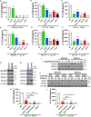
The IRAK1/IRF5 axis initiates IL-12 response by dendritic cells and control of Toxoplasma gondii infection

Affiliations

The IRAK1/IRF5 axis initiates IL-12 response by dendritic cells and control of Toxoplasma gondii infection

Milton Pereira et al. Cell Rep. .

Abstract

Activation of endosomal Toll-like receptor (TLR) 7, TLR9, and TLR11/12 is a key event in the resistance against the parasite Toxoplasma gondii. Endosomal TLR engagement leads to expression of interleukin (IL)-12 via the myddosome, a protein complex containing MyD88 and IL-1 receptor-associated kinase (IRAK) 4 in addition to IRAK1 or IRAK2. In murine macrophages, IRAK2 is essential for IL-12 production via endosomal TLRs but, surprisingly, Irak2-/- mice are only slightly susceptible to T. gondii infection, similar to Irak1-/- mice. Here, we report that upon T. gondii infection IL-12 production by different cell populations requires either IRAK1 or IRAK2, with conventional dendritic cells (DCs) requiring IRAK1 and monocyte-derived DCs (MO-DCs) requiring IRAK2. In both populations, we identify interferon regulatory factor 5 as the main transcription factor driving the myddosome-dependent IL-12 production during T. gondii infection. Consistent with a redundant role of DCs and MO-DCs, mutations that affect IL-12 production in both cell populations show high susceptibility to infection in vivo.

Keywords: CP: Immunology; CP: Microbiology; IL-12; IRAK; IRF5; Toll-like receptors; Toxoplasma gondii; cell signaling; dendritic cells; innate immunity; monocytes; myddosome.


Conflict of interest statement

Declaration of interests The authors declare no competing interests.

Figures

Figure 1.
Figure 1.. IRAKs control primary infection with T. gondii
(A and C) Survival data from C57BL/6 mice IP infected with T. gondii Me49 (25 cysts per mouse): WT (n = 35), Irak1−/− (n = 19), Irak2−/− (n = 19), Irak1−/−Irak2−/− (n = 27), Irak4 Ki (n = 22), Irak1−/−Irak4 Ki (n = 12), Irak2−/−Irak4 Ki (n = 22), and Irak4−/− (n = 8). (B) Cysts counts in the brain of WT (n = 15), Irak1−/− (n = 14), and Irak2−/− (n = 13) mice IP infected with T. gondii Me49 (25 cysts per mouse) for 40 days. (D–G) Quantification of IL-12 p40 and IFN-γ in the plasma (D and E) and peritoneal cavity (F and G) 5 days post IP infection with T. gondii Me49 (25 cysts per mouse) in WT (n = 13), Irak1−/− (n = 7), Irak2−/− (n = 14), Irak1−/−Irak2−/− (n = 6), Irak4 Ki (n = 12), Irak1−/−Irak4 Ki (n = 4), Irak2−/−Irak4 Ki (n = 14), and Irak4−/− (n = 5) mice or mice mock-infected with PBS (WT uninfected, n = 10). (H and I) IL-12 p40 production in splenocytes unprimed (H) or primed with IFN-γ (100 ng mL−1, 24 h) after in vitro infection with T. gondii Me49 tachyzoites at multiplicity of infection (MOI) 3 for 24 h (n = 2 for all strains). n represents the number of animals. In (A) and (C), survival data were pooled from five independent experiments. In (B) and (D)–(I), data were pooled from two (H and I), three (B), or four (D–G) independent experiments and are presented as mean ± standard error of the mean (SEM). Each independent experiment consisted of three technical replicates. *p < 0.05, **p < 0.01, ***p < 0.001, ****p < 0.0001 in relation to the WT (one-way analysis of variance [ANOVA] with Tukey’s multiple-comparisons test).
Figure 2.
Figure 2.. IRAK1 is required for IL-12 production in CD11c+ DCs
(A–F) IL-12 p40 production by BMDMs (A and D), splenic CD11b+ (B and E), and CD11c+ DCs (C and F) treated with R848 (1 μg mL−1, 24 h) (A–C) or exposed to T. gondii Me49 tachyzoites (MOI 3, 24 h) (D–F). (G and H) Immunoblot of whole-cell lysates from CD11c+ DCs, splenic CD11b+ cells, and BMDMs. (I) Immunoblot of phosphatase- and deubiquitinase-treated whole-cell lysates from splenic CD11b+ and CD11c+ DCs exposed to T. gondii Me49 tachyzoites (MOI 3) for up to 24 h. (J) Immunoblot of phosphatase- and deubiquitinase-treated whole-cell lysates from splenic CD11b+ and CD11c+ DCs from the indicated strains exposed to T. gondii Me49 tachyzoites (MOI 3) for up to 24 h. (K and L) IL-12 p40 production in CD11c+ DCs of the indicated strains exposed to T. gondii Me49 tachyzoites (MOI 3, 24 h). In (A)–(F), (K), and (L), data were pooled from three independent experiments and are presented as mean ± SEM; each independent experiment consisted of three technical replicates. Images are representative of three (G and H), four (J), or six (I) independent experiments. ns, not significant. *p < 0.05, **p < 0.01, ***p < 0.001, ****p < 0.0001 in relation to the WT (ANOVA with Tukey’s multiple-comparisons test).
Figure 3.
Figure 3.. IRAK1 and IRAK2 are redundant for NF-κB and MAPK activation, while IRAK4 kinase activity is essential
(A–F) Immunoblot analysis of CD11c+ DCs of the indicated strains. Images are representative of three independent experiments. (A and B) RelA phosphorylation, total RelA, IkB-a phosphorylation, and degradation in CD11c+ DCs treated with R848 (1 μg mL−1, 30 min) or exposed to T. gondii Me49 tachyzoytes (MOI 3, 30 min). (C and D) RelA and c-Rel in nuclear extracts of CD11c+ DCs exposed to T. gondii Me49 tachyzoites (MOI 3, 30 min). (E and F) Phosphorylation of ERK, JNK, and p38 in CD11c+ DCs exposed to T. gondii Me49 tachyzoites (MOI 3, 15 and 30 min).
Figure 4.
Figure 4.. IL-12 production by CD11c+ DCs is mediated by IRAK1 via IRF5
(A–C) IRF5 immunoblots from nuclear extracts of CD11c+ DCs (A and B) or splenic CD11b+ cells (C) infected with T. gondii Me49 tachyzoites (MOI 3, 2 h). (D–F) IL-12 p40 production in WT and Irf5−/− BMDMs (D), splenic CD11b+ (E), and CD11c+ DCs (F) treated with R848 (1 μg mL−1, 24 h) or exposed to T. gondii Me49 tachyzoites (MOI 3, 24 h). (G) ChIP-qPCR using control immunoglobulin G (IgG) or anti-IRF5 in WT and Irf5−/− CD11c+ DCs, uninfected or infected with T. gondii Me49 tachyzoites (MOI 3, 2 h). (H and I) IRF5 Immunoblot of whole-cell lysates from CD11c+ DCs, splenic CD11b+, and BMDMs (H) and densitometric quantifications (I). (J) IRF5 immunoblots from nuclear extracts of CD11c+ DCs infected with T. gondii Me49 tachyzoites (MOI 3, 2 h) with or without the IKKβ inhibitors BI605906 (10 μg mL−1) or TPCA-1 (10 μg mL−1). (K) IL-12 p40 production by CD11c+ DCs exposed to T. gondii Me49 tachyzoites (MOI 3, 24 h) with or without the IKKβ inhibitors BI605906 (10 μg mL−1) or TPCA-1 (10 μg mL−1). (L) Immunoblot of p-IKKβ in whole-cell lysates of CD11c+ DCs infected with T. gondii Me49 tachyzoites (MOI 3, 2 h). Images are representative of two (C and J) or three (A, B, H, and L) independent experiments. In (D)–(G), (I), and (K), data were pooled from three independent experiments and are presented as mean ± SEM; each independent experiment consisted of three technical replicates. ***p < 0.001, ****p < 0.0001 (ANOVA with Tukey’s multiple-comparisons test). #p < 0.05, ##p < 0.01, ###p < 0.001 (unpaired t test).
Figure 5.
Figure 5.. Production of inflammatory cytokines and chemokines by DCs requires IRAK1, IRAK4, and IRF5
(A) Volcano plot depicting T. gondii-infected versus uninfected CD11c+ DCs (WT). The horizontal dashed line represents a —log false discovery rate of 2, and the vertical dashed lines represent a log2 fold change of −1 and +1. (B) Heatmap showing the expression and hierarchical clustering of selected genes in CD11c+ DCs from the indicated strains, uninfected or infected with T. gondii Me49 tachyzoites (MOI 3, 4 h). (C–F) Quantification of IL-12 p40 (C), CCL5 (D), TNF (E), and CCL22 (F) in culture supernatants of CD11c+ DCs of the indicated strains infected with T. gondii Me49 (MOI 3) for 4 and 24 h. In (A) and (B), Data were pooled from four independent experiments. In (C)–(F), data were pooled from four independent experiments and are presented as mean ± SEM; each independent experiment consisted of one technical replicate. *p < 0.05, **p < 0.01, ***p < 0.001, ****p < 0.0001 (ANOVA with Tukey’s multiple-comparisons test).
Figure 6.
Figure 6.. IRAK1-mediated CNBP activation enhances IL-12 production in CD11c+ DCs
(A–C) CNBP nuclear translocation in CD11c+ DCs (A and B) and splenic CD11b+ cells (C) infected with T. gondii Me49 tachyzoites (MOI 3, 2 h). (D) CNBP nuclear translocation in CD11c+ DCs infected with T. gondii Me49 tachyzoites (MOI 3, 2 h) with or without the IKKβ inhibitors BI605906 (10 μg mL−1) or TPCA-1 (10 μg mL−1). (E and F) IL-12 p40 production in Cnbpfl/flVav-iCre−/− and Cnbpfl/flVav-iCre+/+ CD11c+ DCs stimulated with R848 (1 μg mL−1, 24 h) (E) or T. gondii Me49 tachyzoites (MOI 3, 24 h) (F). (G) IRF5 nuclear translocation in Cnbpfl/flVav-iCre−/− and Cnbpfl/flVav-iCre+/+ CD11c+ DCs exposed to T. gondii Me49 tachyzoites (MOI 3, 2 h). (H) IRF5 and CNBP co-immunoprecipitation in WT and Irf5−/− CD11c+ DCs exposed to T. gondii Me49 tachyzoites (MOI 3, 2 h). (I) ChIP-qPCR using control IgG or anti-IRF5 in Cnbpfl/flVav-iCre−/− and Cnbpfl/flVav-iCre+/+ CD11c+ DCs, uninfected or infected with T. gondii Me49 tachyzoites (MOI 3, 2 h). Images are representative of two (C and D) or three (A, B, G, and H) independent experiments. In (E), (F), and (I), data were pooled from three independent experiments and are presented as mean ± SEM; each independent experiment consisted of three technical replicates. ***p < 0.001, ****p < 0.0001 (ANOVA with Tukey’s multiple-comparisons test). #p < 0.05, ##p < 0.01, ###p < 0.001 (unpaired t test).
Figure 7.
Figure 7.. IRF5 controls primary infection with T. gondii
(A) Survival data from C57BL/6 WT (n = 16) and Irf5−/− (n = 15) mice IP infected with T. gondii Me49 (25 cysts per mouse). (B–E) Quantification of IL-12 p40 and IFN-γ in the plasma (B and C) and peritoneal cavity (D and E) in WT (n = 14) and Irf5−/− (n = 12) mice 5 days post IP infection with T. gondii Me49 (25 cysts per mouse) or mock infected with PBS WT (n = 4) and Irf5−/− (n = 3) (uninfected controls). (F and G) IL-12 p40 production in splenocytes unprimed (n = 5 for each strain) (F) or primed with IFN-γ (100 ng mL−1, 24 h) (n = 3 for each strain) (G) after in vitro infection with T. gondii Me49 tachyzoites at MOI 3 for 24 h n represents the number of animals. In (A), survival data were pooled from two independent experiments. In (B)–(G), data were pooled from three independent experiments and are presented as mean ± SEM; each independent experiment consisted of three technical replicates. *p < 0.05, **p < 0.01, ***p < 0.001, ****p < 0.0001 in relation to the WT (ANOVA with Tukey’s multiple-comparisons test). ###p < 0.001 (unpaired t test).

Similar articles

Cited by

References

    1. Akira S, Uematsu S, and Takeuchi O, (2006). Pathogen Recognition and Innate Immunity. Cell 124, 783–801. 10.1016/j.cell.2006.02.015. - DOI - PubMed
    1. Fitzgerald KA, and Kagan JC (2020). Toll-like Receptors and the Control of Immunity. Cell 180, 1044–1066. 10.1016/j.cell.2020.02.041. - DOI - PMC - PubMed
    1. Pereira M, and Gazzinelli RT (2023). Regulation of innate immune signaling by IRAK proteins. Front. Immunol. 14, 1133354. - PMC - PubMed
    1. Cao Z, Xiong J, Takeuchi M, Kurama T, and Goeddel DV (1996). TRAF6 is a signal transducer for interleukin-1. Nature 383, 443–446. 10.1038/383443a0. - DOI - PubMed
    1. Pauls E, Nanda SK, Smith H, Toth R, Arthur JSC, and Cohen P (2013). Two Phases of Inflammatory Mediator Production Defined by the Study of IRAK2 and IRAK1 Knock-in Mice. J.I. 191, 2717–2730. 10.4049/jimmunol.1203268. - DOI - PMC - PubMed

Publication types

Substances