. 2024 Apr;21(3):e00336.
doi: 10.1016/j.neurot.2024.e00336. Epub 2024 Feb 16.

Ultrahigh frequency transcutaneous electrical nerve stimulation for neuropathic pain alleviation and neuromodulation

Affiliations

Ultrahigh frequency transcutaneous electrical nerve stimulation for neuropathic pain alleviation and neuromodulation

Szu-Han Chen et al. Neurotherapeutics. 2024 Apr.

Abstract

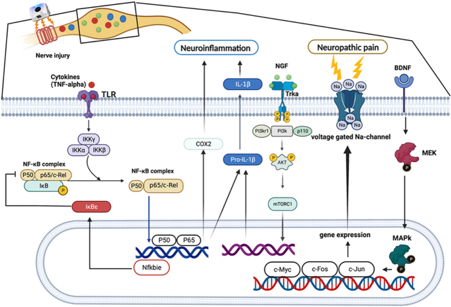
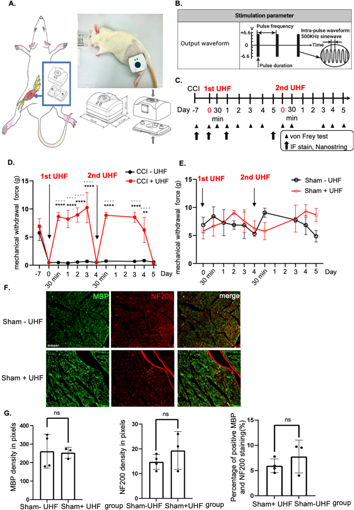
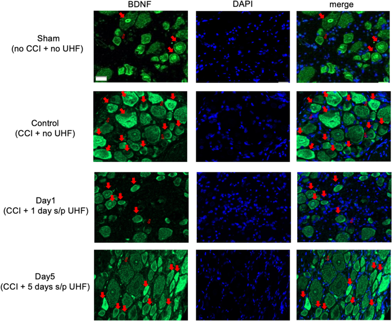
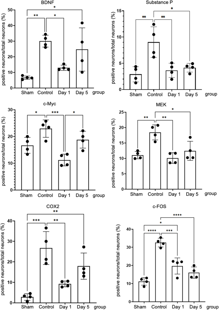
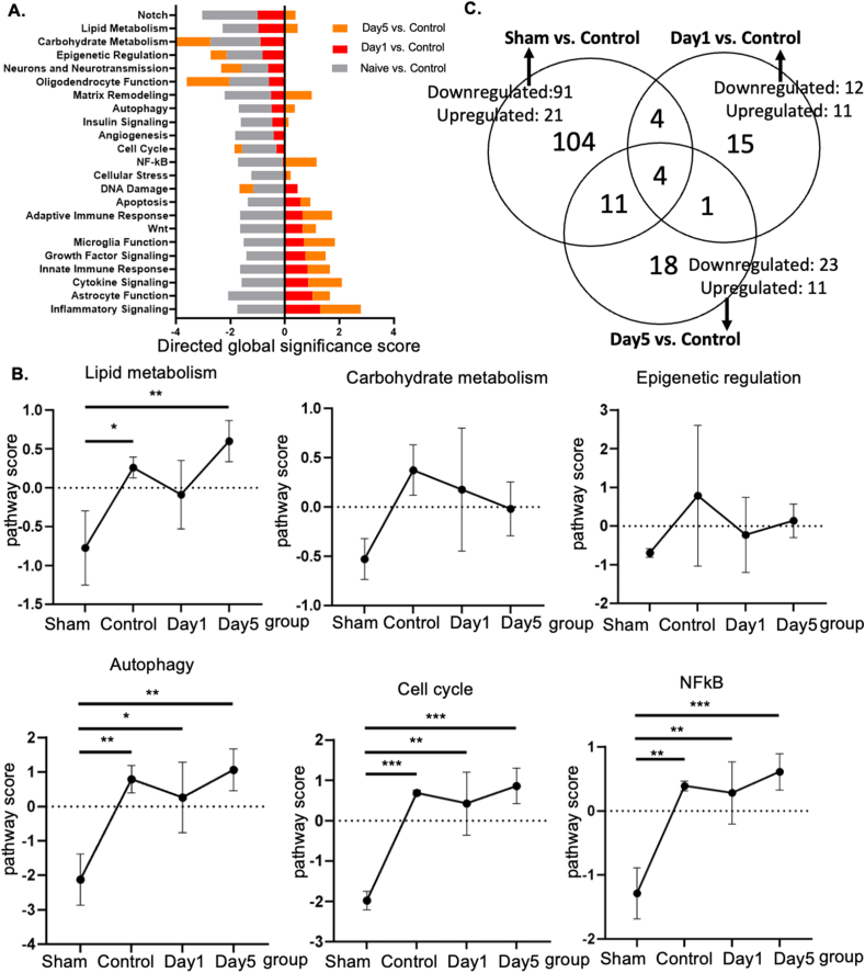
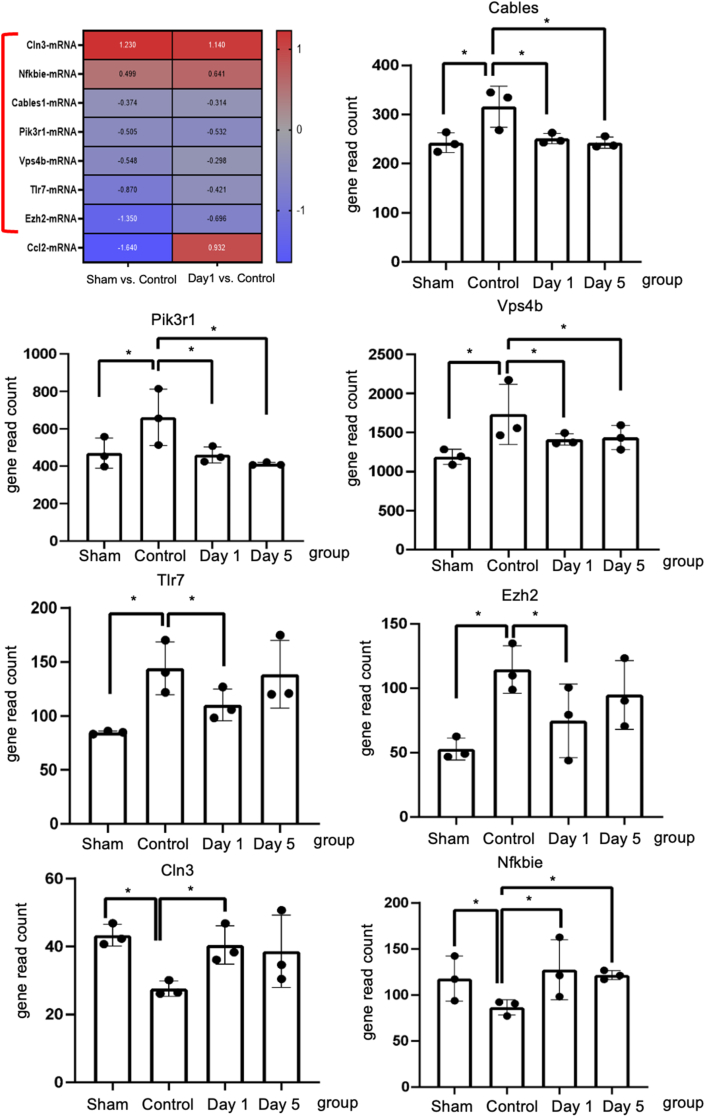
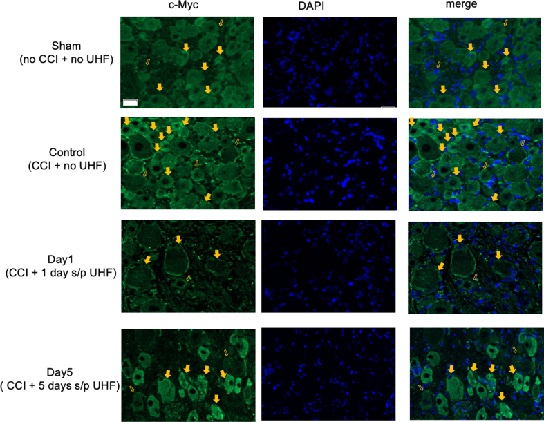
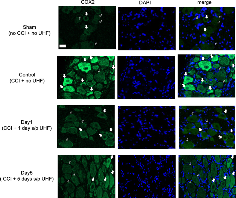
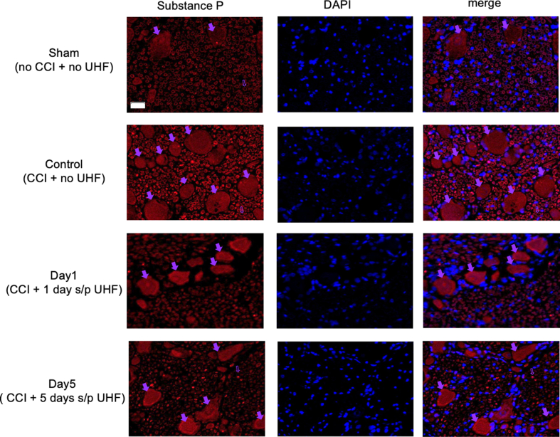
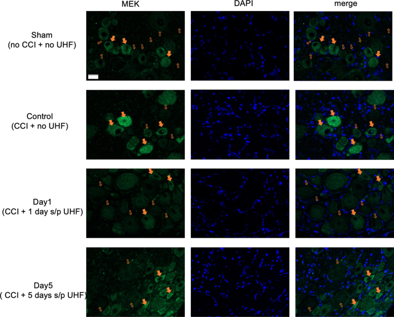
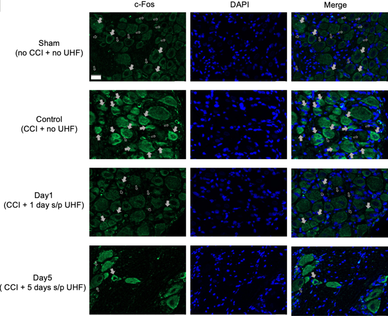
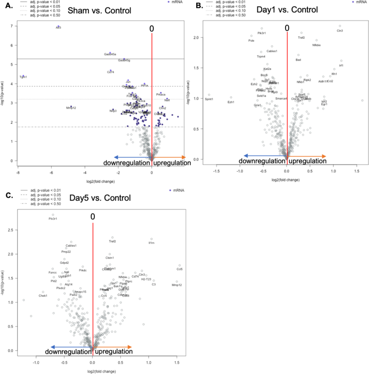
A challenging complication in patients with peripheral compressive neuropathy is neuropathic pain. Excessive neuroinflammation at the injury site worsens neuropathic pain and impairs function. Currently, non-invasive modulation techniques like transcutaneous electrical nerve stimulation (TENS) have shown therapeutic promise with positive results. However, the underlying regulatory molecular mechanism for pain relief remains complex and unexplored. This study aimed to validate the therapeutic effect of ultrahigh frequency (UHF)-TENS in chronic constriction injury of the rat sciatic nerve. Alleviation of mechanical allodynia was achieved through the application of UHF-TENS, lasting for 3 days after one session of therapy and 4 days after two sessions, without causing additional damage to the myelinated axon structure. The entire tissue collection schedule was divided into four time points: nerve exposure surgery, 7 days after nerve ligation, and 1 and 5 days after one session of UHF therapy. Significant reductions in pain-related neuropeptides, MEK, c-Myc, c-FOS, COX2, and substance P, were observed in the injured DRG neurons after UHF therapy. RNA sequencing of differential gene expression in sensory neurons revealed significant downregulation in Cables, Pik3r1, Vps4b, Tlr7, and Ezh2 after UHF therapy, while upregulation was observed in Nfkbie and Cln3. UHF-TENS effectively and safely relieved neuropathic pain without causing further nerve damage. The decreased production of pain-related neuropeptides within the DRG provided the therapeutic benefit. Possible molecular mechanisms behind UHF-TENS may result from the modulation of the NF-κB complex, toll-like receptor-7, and phosphoinositide 3-kinase/Akt signaling pathways. These results suggest the neuromodulatory effects of UHF-TENS in rat sciatic nerve chronic constriction injury, including alleviation of neuropathic pain, amelioration of pain-related neuropeptides, and regulation of neuroinflammatory gene expression. In combination with the regulation of related neuroinflammatory genes, UHF-TENS could become a new modality for enhancing the treatment of neuropathic pain in the future.

Keywords: Compression neuropathy; Neuroinflammation; Neuropathic pain; Transcutaneous electrical nerve stimulation; Ultrahigh frequency.


Conflict of interest statement

Declaration of competing interest The authors declare the following financial interests/personal relationships which may be considered as potential competing interests: Yuan-Yu Hsueh reports financial support was provided by National Science and Technology Council. Szu-Han Chen reports financial support was provided by National Science and Technology Council. Wei-Tso Lin reports a relationship with Gimer Medical Co., Ltd, New Taipei City, Taiwan that includes: employment. If there are other authors, they declare that they have no known competing financial interests or personal relationships that could have appeared to influence the work reported in this paper.

Figures

Image 1
Graphical abstract
Fig. 1
Fig. 1
Functional behavior outcome by ultrahigh frequency transcutaneous electrical stimulation therapy. (A) Animal model of chronic constriction injury over the ipsilateral sciatic nerve. The device of UHF-TENS was applied on the skin of the treated side sciatic nerve under stimulation protocol. (B) Stimulation parameter and output waveform in UHF (C) Timeline and study design of UHF-TENS treatment in this study. (Arrowhead: time point for von Frey test; Arrow: time point for tissue collection for IF and nanostring) (D, E) The mechanical withdrawal force (g) indicated the forces that induced paw withdrawal by the Von Frey test for injured or uninjured nerves with or without UHF treatment. (n ​= ​6 per group in injured group; n ​= ​3 per group in uninjured group) Data were presented with mean ​± ​standard deviation, ∗∗∗∗ indicated statistical significance in UHF group as compared to the no UHF group; p ​< ​0.0001; ++++ indicated statistical significance at different time point as compared to day 0 in UHF group). (F, G) Immunofluorescent staining of uninjured sciatic nerve group. Mature axon staining with NF200 revealed no significant difference between the uninjured group with and without electrical stimulation therapy. Similar pattern was also observed in Schwann cell staining with MBP alone and colocalization image. NF200/MBP colocalization revealed that the amount of myelinated axon after UHF in the uninjured nerve group was no significant difference in the uninjured nerve group without UHF stimulation. (n ​= ​3 in UHF group and n ​= ​4 in control group; Scale bar: 50 ​μm; ns indicated no significant difference).
Fig. 2
Fig. 2
Immunofluorescent staining of BDNF in the injured side of dorsal root ganglion under UHF-TENS treatment. A significant increase in signal intensity was induced by nerve constriction injury in the control group compared to sham group as the baseline. After UHF stimulation, the signal intensity of BDNF decreased significantly on Day 1 and returned to baseline level on Day 5. (n ​= ​4 per group; Scale bar: 20 ​μm; positive neuron: red solid arrowhead; negative neuron: red hollow arrowhead).
Fig. 3
Fig. 3
Quantitative analysis of immunofluorescent staining of neuropathic pain markers on the injured side of dorsal root ganglion. BDNF (brain-derived neurotrophic factor), cyclooxygenase 2 (COX2), and c-Myc significant increase in signal intensity were induced after nerve injury in the control group compared to the uninjured nerve as the baseline. After UHF stimulation on Day 1, the signal intensity decreased and returned to increase on day five after electrical stimulation therapy. Similarly, substance P (SP), MEK, and c-Fos over injured DRG significantly increased signal intensity after nerve injury in the control group compared to the uninjured nerve as the baseline. After electrical stimulation therapy on Day 1, the signal intensity decreased and still reduced compared to the injured nerve of the control group on day five after UHF stimulation. (n ​= ​4 per timepoint Data was presented with mean ​± ​standard deviation, ∗ indicated p ​< ​0.05; ∗∗ indicated p ​< ​0.01, ∗∗∗ indicated p ​< ​0.001, ∗∗∗∗ indicated p ​< ​0.00001, and ns indicated no significant difference).
Fig. 4
Fig. 4
Genetic profiling of injured nerve modified by UHF stimulation by Nanostring nCounter Neuroinflammation panel (A) Neuroinflammation pathway-specific changes in gene differential expression among sham group, control group, and UHF treated nerve at Day 1 and Day 5 by directed global significance score. (B) The pathway scores of lipid metabolism, carbohydrate metabolism, epigenetic regulation, autophagy, cell cycle, and NFkB were upregulated after peripheral nerve injury and downregulated after UHF treatment. (C) Venn diagram showing the overlap of the significant differential expression gene lists for the three pairwise comparison. The overlap of Sham vs. Control and Day1 vs. Control area identifies eight genes that are both modified by injury as well as UHF treatment after CCI. (n ​= ​3 for each group).
Fig. 5
Fig. 5
The Nanostring gene analysis of inflammatory genes in the dorsal root ganglion. The heatmap shows eight significant differential expression gene lists overlap genes between comparisons Day −7 (sham) vs. Day 0 (control) and Day 1 (UHF) vs. Day 0 (control). Among these, the gene expression of the below 7 genes, Cln3, Nfkbie, Cables1, Pik3r1, Vps4b, Tlr7, and Ezh2, indicate the potential UHF regulatory effects. The Nfkbie and Cln3 were upregulated after nerve injury in control group, but significantly downregulated by UHF treatment. The Tlr7, Pik3r1, Eh2, Vps4b, and Cables were downregulated after nerve injury in control group, while upregulated by UHF treatment. (n ​= ​3 for each group; ∗ indicated p ​< ​0.05).
Fig. 6
Fig. 6
Sketch map of the regulatory molecular mechanism of UHF-TENS for neuropathic pain in DRG neuron of CCI rats, in terms of neuropathic pain formation and neuroinflammation signal modulation.
Figure S1
Figure S1
Immunohistochemical staining of c-Myc in the injured side of dorsal root ganglion under UHF-TENS treatment. A significant increase in signal intensity was induced by nerve constriction injury in the control group compared to sham group as the baseline. After UHF stimulation, the signal intensity of c-Myc decreased significantly on Day 1 and returned to baseline level on Day 5. (n=3-4 per group; 20 μm; positive neuron: yellow solid arrowhead; negative neuron: yellow hollow arrowhead).
Figure S2
Figure S2
Immunohistochemical staining of COX2 in the injured side of dorsal root ganglion under UHF-TENS treatment. A significant increase in signal intensity was induced by nerve constriction injury in the control group compared to sham group as the baseline. After UHF stimulation, the signal intensity of COX2 decreased significantly on Day 1 and returned to baseline level on Day 5. (n=3-4 per group; Scale bar:20 μm; positive neuron: white solid arrowhead; negative neuron: white hollow arrowhead).
Figure S3
Figure S3
Immunohistochemical staining of Substance P in the injured side of dorsal root ganglion under UHF-TENS treatment. A significant increase in signal intensity was induced by nerve constriction injury in the control group compared to sham group as the baseline. After UHF stimulation, the signal intensity of Substance P decreased significantly on Day 1 through Day 5. (n=4 per group; Scale bar: 20 μm; positive neuron: purple solid arrowhead; negative neuron: purple hollow arrowhead).
Figure S4
Figure S4
Immunohistochemical staining of MEK in the injured side of dorsal root ganglion under UHF-TENS treatment. A significant increase in signal intensity was induced by nerve constriction injury in the control group compared to sham group as the baseline. After UHF stimulation, the signal intensity of MEK decreased significantly on Day 1through Day 5. (n=4 per group; Scale bar: 20 μm; positive neuron: orange solid arrowhead; negative neuron: orange hollow arrowhead).
Figure S5
Figure S5
Immunohistochemical staining of c-Fos in the injured side of dorsal root ganglion under UHF-TENS treatment. A significant increase in signal intensity was induced by nerve constriction injury in the control group compared to sham group as the baseline. After UHF stimulation, the signal intensity of -Fos decreased significantly on Day 1 through Day 5. (n=4 per group; Scale bar: 20 μm; positive neuron: grey solid arrowhead; negative neuron: grey hollow arrowhead).
Figure S6
Figure S6
(A–C) Volcano plot of differential gene expression at sham group, Day 1, and Day 5, comparing with control group. (n=4 for each group).

References

    1. Cavalli E., Mammana S., Nicoletti F., Bramanti P., Mazzon E. The neuropathic pain: An overview of the current treatment and future therapeutic approaches. Int J Immunopathol Pharmacol. 2019;33 - PMC - PubMed
    1. Colloca L., Ludman T., Bouhassira D., Baron R., Dickenson A.H., Yarnitsky D., et al. Neuropathic pain. Nat Rev Dis Primers. 2017;3:17002. - PMC - PubMed
    1. Bouhassira D. Neuropathic pain: definition, assessment and epidemiology. Rev Neurol (Paris) 2019;175(1-2):16–25. - PubMed
    1. Bouhassira D., Lantéri-Minet M., Attal N., Laurent B., Touboul C. Prevalence of chronic pain with neuropathic characteristics in the general population. Pain. 2008;136(3):380–387. - PubMed
    1. Bielewicz J., Kamieniak M., Szymoniuk M., Litak J., Czyżewski W., Kamieniak P. Diagnosis and management of neuropathic pain in spine diseases. J Clin Med. 2023;12(4) - PMC - PubMed

Publication types