Review
. 2024 May;11(17):e2308298.
doi: 10.1002/advs.202308298. Epub 2024 Feb 17.

Advances in Atherosclerosis Theranostics Harnessing Iron Oxide-Based Nanoparticles

Affiliations
Review

Advances in Atherosclerosis Theranostics Harnessing Iron Oxide-Based Nanoparticles

Shi Wang et al. Adv Sci (Weinh). 2024 May.

Abstract

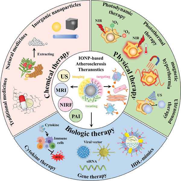
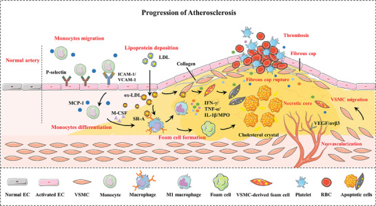
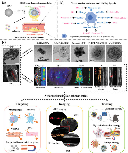
Atherosclerosis, a multifaceted chronic inflammatory disease, has a profound impact on cardiovascular health. However, the critical limitations of atherosclerosis management include the delayed detection of advanced stages, the intricate assessment of plaque stability, and the absence of efficacious therapeutic strategies. Nanotheranostic based on nanotechnology offers a novel paradigm for addressing these challenges by amalgamating advanced imaging capabilities with targeted therapeutic interventions. Meanwhile, iron oxide nanoparticles have emerged as compelling candidates for theranostic applications in atherosclerosis due to their magnetic resonance imaging capability and biosafety. This review delineates the current state and prospects of iron oxide nanoparticle-based nanotheranostics in the realm of atherosclerosis, including pivotal aspects of atherosclerosis development, the pertinent targeting strategies involved in disease pathogenesis, and the diagnostic and therapeutic roles of iron oxide nanoparticles. Furthermore, this review provides a comprehensive overview of theranostic nanomedicine approaches employing iron oxide nanoparticles, encompassing chemical therapy, physical stimulation therapy, and biological therapy. Finally, this review proposes and discusses the challenges and prospects associated with translating these innovative strategies into clinically viable anti-atherosclerosis interventions. In conclusion, this review offers new insights into the future of atherosclerosis theranostic, showcasing the remarkable potential of iron oxide-based nanoparticles as versatile tools in the battle against atherosclerosis.

Keywords: atherosclerosis; iron oxide nanoparticles; nanomedicines; theranostics.


Conflict of interest statement

The authors declare no conflict of interest.

Figures

Figure 1
Figure 1
Illustrative overview of therapeutic strategies in IONP‐based theranostics for atherosclerosis.
Figure 2
Figure 2
Schematic illustration of the development and pathological characteristics of atherosclerosis. ECs, endothelial cells; ox‐LDL, oxidized low‐density lipoprotein; ICAM‐1, intercellular adhesion molecule‐1; VCAM‐1, vascular cell adhesion molecule‐1; MCP‐1, monocyte chemotactic protein‐1; M‐CSF, macrophage colony‐stimulating factors; VSMCs, vascular smooth muscle cells; IFN‐γ, interferon‐γ; IL‐1β, interleukin‐1β; TNF‐α, tumor necrosis factor‐α; MPO, myeloperoxidase; VEGF, vascular endothelial growth factor.
Figure 3
Figure 3
a) Schematic representation of theranostic nanomedicine for atherosclerosis. b) Target marker molecules and binding ligands for atherosclerosis Theranostics. c) Diverse IONP‐based theranostic nanoplatforms for atherosclerosis targeting, imaging, and treatment. Representative image of IONPs. Reproduced with permission.[ 60 ] Copyright 2018, American Chemical Society. Image of MRI (up) and MM‐HDL NPs (TEM and figure in biologic therapy). Reproduced with permission.[ 61 ] Copyright 2020, American Chemical Society. Image of solid lipid nanoparticles (TEM and figure in chemical therapy). Reproduced with permission.[ 62 ] Copyright 2016, American Chemical Society. Image of CeO2‐ Fe3O4@LDH nanocomposites (TEM and figure in chemical therapy). Reproduced with permission.[ 63 ] Copyright 2019, American Chemical Society. Image of Au‐coated IONP. Reproduced with permission.[ 64 ] Copyright 2009, American Chemical Society. Image of Fe‐PFH‐PLGA/CS‐DS NPs. Reproduced with permission.[ 65 ] Copyright 2019, American Chemical Society. Image of SPECT‐CT. Reproduced with permission.[ 66 ] Copyright 2018, American Chemical Society. Image of PET. Reproduced under terms of the CC‐BY license.[ 67 ] Copyright 2021, The Authors, Published by American Chemical Society. Image of NIRF (up) and MRI (down). Reproduced under terms of the CC‐BY license.[ 68 ] Copyright 2022, The Authors, Published by MDPI, Basel, Switzerland. Image of NIRF (down) and nanoparticle figure in chemical therapy. Reproduced under terms of the CC‐BY license.[ 38 ] Copyright 2019, The Authors, Published by Informa UK Limited, trading as Taylor & Francis Group. Image of US and PAI. Reproduced under terms of the CC‐BY license.[ 69 ] Copyright 2021, Published by Wiley‐VCH. Image of physical stimulation therapy. Reproduced with permission.[ 70 ] Copyright 2022, American Chemical Society. Image of nanoparticles in biologic therapy. (middle) Reproduced with permission.[ 71 ] Copyright 2016, Elsevier B.V. (right) Reproduced with permission.[ 72 ] Copyright 2020, Elsevier Ltd.
Figure 4
Figure 4
a) Schematic illustration of RAP@Fe3O4‐PDA‐CD‐PEG‐PEI‐Profilin‐1‐Cy5.5 nanoparticles. b) Representative in vivo MR images of ApoE−/− mice with PFN1‐CD‐MNPs. c,d) NIRF of the aorta and general ORO staining of the carotid artery after 2 months of various treatments. Reproduced under terms of the CC‐BY license.[ 38 ] Copyright 2019, The Authors, Published by Informa UK Limited, trading as Taylor & Francis Group. e) Schematic illustration of the synthesis of theranostic liposomes Rap/ Fe3O4@VHP‐Lipo by self‐assembly. f) The fluorescence distribution in vivo for mice with three different treatments. g) The fluorescence enrichment of the aorta from three different mice groups. h) The treatment effectiveness testing of fluorescence imaging for mice from three different groups injected with Rap/ Fe3O4@VHP‐Lipo for 2 months. i) MRI T2 mapping sequence imaging to detect the treatment effect of mice in three different groups with different treatments for 2 months. Reproduced under terms of the CC‐BY license.[ 68 ] Copyright 2022, The Authors, Published by MDPI, Basel, Switzerland.
Figure 5
Figure 5
a) Schematic illustration of prostacyclin‐loaded SLN for image‐guided Therapy. Reproduced with permission.[ 62 ] Copyright 2016, American Chemical Society. b–d) Atheroma targeting and in vivo MRI ability of the nano‐emulsion platform modified with a fully human scFv‐Fc antibody. Reproduced under terms of the CC‐BY license.[ 74 ] Copyright 2021, The Authors, Published by MDPI, Basel, Switzerland. e) Schematic diagram of the structure and theranostic function of CeO2‐ Fe3O4@LDH nanocomposites. Reproduced with permission.[ 63 ] Copyright 2019, American Chemical Society. f) Mechanism of ROS Scavenging by CeO2 NPs. Reproduced with permission.[ 104 ] Copyright 2023, American Chemical Society.
Figure 6
Figure 6
a) Schematic illustration of phototherapy. Reproduced with permission.[ 106 ] Copyright 2020, Elsevier B.V. b) Schematic diagram of cell death by PDT. Reproduced with permission.[ 111 ] Copyright 2020, Elsevier B.V. c) Activatable photosensitization responds to H2S. Reproduced with permission.[ 123 ] Copyright 2018, American Chemical Society. d) Activatable photosensitization responds to pH change. Reproduced with permission.[ 124 ] Copyright 2016, American Chemical Society. e) Schematic illustration of PTT in the treatment of atherosclerosis. Reproduced with permission.[ 125 ] Copyright 2021, American Chemical Society. f) Schematic illustration of sequential photothermal/photodynamic ablation for activated macrophages. Reproduced with permission.[ 126 ] Copyright 2021, American Chemical Society. g) Near‐infrared thermal imaging images of PBS, ICG, Fe3O4, and Fe/ICG@HA after irradiation with an 808 nm laser. Reproduced with permission.[ 127 ] Copyright 2022, American Chemical Society. h) Small multifunctional nanoclusters and their capability of PTT. Reproduced with permission.[ 64 ] Copyright 2009, American Chemical Society.
Figure 7
Figure 7
a) Schematic illustration of MMSN@AT‐CS‐DS NPs for multi‐effective treatment of atherosclerosis. b,c) Characterization of MMSN‐CS‐DS in magnetocaloric effect, including heating curves (b) and in vitro temperature raising (c). d–f) The levels of TNF‐α, IL‐6, and IL‐1β in blood serum from different mouse groups. g) Therapeutic efficacy in atherosclerotic mouse. ORO staining of the aortic roots and aortic arches from different mouse groups. Reproduced with permission.[ 82 ] Copyright 2022, Elsevier B.V.
Figure 8
Figure 8
a) Schematic illustration of the synthesis process and measurement setup of Magneto‐Photothermal hybrids based on IONPs. b) Comparison of thermal properties of MHT, PTT, and DUAL. c) Schematic illustration of Magneto‐Photothermal therapy. Reproduced with permission.[ 133a ] Copyright 2023, American Chemical Society. d) Schematic diagram of MHT and PTT applying Iron‐based NPs in thrombolytic therapy. Reproduced with permission.[ 133c ] Copyright 2021, American Chemical Society. e) Schematic illustration of MHT and PTT based on Iron‐based nanoparticles in arterial inflammation therapy. Reproduced under terms of the CC‐BY license.[ 133b ] Copyright 2019, The Authors, Published by Elsevier Ltd.
Figure 9
Figure 9
a) Schematic illustration of the preparation and ADV effect of Fe‐PFH‐PLGA/CS‐DS NPs. b) US images of the B mode and contrast mode of Fe‐PFH‐PLGA/CS‐DS NPs in vitro. c) Effects of targeting and treatment in vivo shown by MRI. Reproduced with permission.[ 65 ] Copyright 2019, American Chemical Society. d) Schematic illustration of ultrasound‐triggered phase transition of MPmTN via the US. e) Schematic of the antithrombotic effect by ADV and the thrombolytic effect of MPmTN in vitro. f) Anti‐atherosclerosis effect of MPmTN in vivo. Reproduced with permission.[ 35 ] Copyright 2021, Royal Society of Chemistry.
Figure 10
Figure 10
a) Schematic illustration of the synthetic process and corresponding theranostic functionality for PFP–HMME@PLGA/MnFe2O4–Ram nanoplatform in multi‐effective treatment of atherosclerosis. b) In vivo MRI images of rabbit femoral plaque after injection of different nanoparticles. c) In vivo ultrasound imaging images of rabbit femoral plaque after injection of different nanoparticles. d) Representative histopathological staining of plaque sections presented the treatment outcomes of atherosclerotic rabbits. e) In vivo PA imaging of the rabbit femoral plaques after injection of indicated NPs at different time points. Reproduced under terms of the CC‐BY license.[ 69 ] Copyright 2021, Published by Wiley‐VCH.
Figure 11
Figure 11
a) Schematic representation of different components and actions of HDL‐mimicking NPs. b) MRI images of coronal slices from BALB/c mice injected with T1‐MM100‐HDL‐NPs. c) In vitro comparison of the cholesterol efflux property of indicated groups. d) In vitro comparison of the antioxidant properties of indicated groups. e) Lipid reduction profiles of BALB/c mice after being treated with MM‐loaded nanostructures for 24 h. Reproduced with permission.[ 61 ] Copyright 2020, American Chemical Society.
Figure 12
Figure 12
a) Schematic diagram of the radially symmetric transduction of LV/MNP complexes. b) Comparison between magnet configuration A (three images above) and magnet configuration B (three images below) in circumferential lentiviral transduction. c) Immunofluorescence staining of aortic comparing overexpression of VEGF in indicated groups. Reproduced with permission.[ 71 ] Copyright 2016, Elsevier B.V. d) Schematic diagram of the structure of IL10‐NC. e,f) Oil red O (ORO) staining of plaque and quantitative analysis showing the role in promoting plaque regression of IL10‐NC. g) Immunofluorescence staining of pro‐inflammatory cytokine IL‐1β showing the role in promoting inflammation resolution of IL10‐NC. Reproduced with permission.[ 72 ] Copyright 2020, Elsevier Ltd.
Figure 13
Figure 13
The future challenges and uncharted territories for the development of IONP‐based Atherosclerosis Theranostics.

Similar articles

Cited by

References

    1. a) Tsao C. W., Aday A. W., Almarzooq Z. I., Anderson C. A. M., Arora P., Avery C. L., Baker‐Smith C. M., Beaton A. Z., Boehme A. K., Buxton A. E., Commodore‐Mensah Y., Elkind M. S. V., Evenson K. R., Eze‐Nliam C., Fugar S., Generoso G., Heard D. G., Hiremath S., Ho J. E., Kalani R., Kazi D. S., Ko D., Levine D. A., Liu J., Ma J., Magnani J. W., Michos E. D., Mussolino M. E., Navaneethan S. D., Parikh N. I., et al., Circulation 2023, 147, e93; - PubMed
    2. b) Jebari‐Benslaiman S., Galicia‐Garcia U., Larrea‐Sebal A., Olaetxea J. R., Alloza I., Vandenbroeck K., Benito‐Vicente A., Martin C., Int. J. Mol. Sci. 2022, 23, 3346. - PMC - PubMed
    1. a) Mantovani A., Ballestri S., Lonardo A., Targher G., Dig. Dis. Sci. 2016, 61, 1246; - PubMed
    2. b) Libby P., Pasterkamp G., Crea F., Jang I. K., Circ. Res. 2019, 124, 150; - PMC - PubMed
    3. c) Falk E., Nakano M., Bentzon J. F., Finn A. V., Virmani R., Eur. Heart J. 2013, 34, 719; - PubMed
    4. d) Banerjee C., Chimowitz M. I., Circ. Res. 2017, 120, 502. - PMC - PubMed
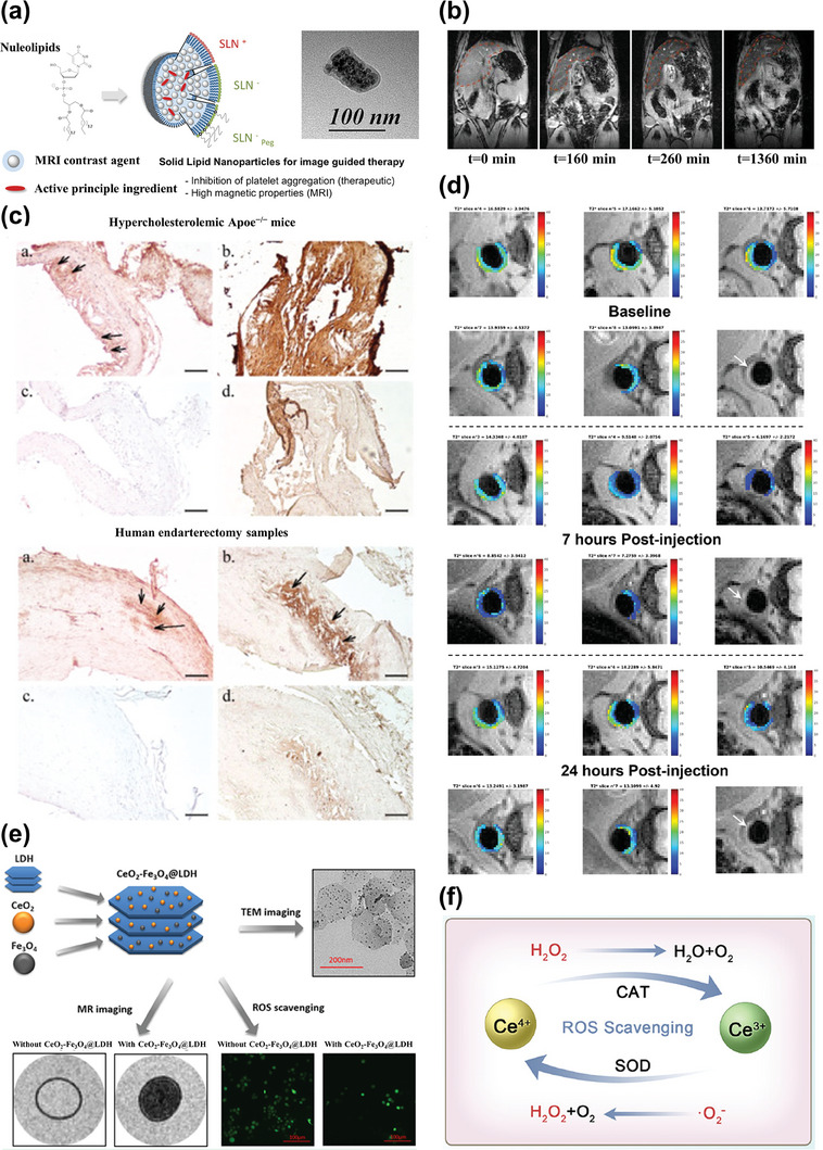
    1. Sugamata W., Nakamura T., Uematsu M., Kitta Y., Fujioka D., Saito Y., Kawabata K., Obata J. E., Watanabe Y., Watanabe K., Kugiyama K., J. Cardiol. 2014, 64, 179. - PubMed
    1. Chen J., Zhang X., Millican R., Sherwood J., Martin S., Jo H., Yoon Y. S., Brott B. C., Jun H. W., Adv. Drug Deliv. Rev. 2021, 170, 142. - PMC - PubMed
    1. Song Y., Jing H., Vong L. B., Wang J., Li N., Chin. Chem. Lett. 2022, 33, 1705.

Publication types

MeSH terms

LinkOut - more resources