. 2024 Feb 17;409(1):66.
doi: 10.1007/s00423-024-03246-7.

Diagnosis and treatment of patients with suspected mucinous cystic neoplasms of the liver: a retrospective cohort study

Affiliations

Diagnosis and treatment of patients with suspected mucinous cystic neoplasms of the liver: a retrospective cohort study

Alicia Furumaya et al. Langenbecks Arch Surg. .

Abstract

Purpose: Mucinous cystic neoplasms of the liver (MCN-L) are hepatic cysts with a low malignant potential. The recent European Association for the Study of the Liver (EASL) guidelines provide guidance on the imaging features and surgical management of MCN-L, yet are hampered by a lack of studies adhering to the revised World Health Organization (WHO) criteria. This study attempted to validate the new 2022 EASL-guidelines in a retrospective cohort study of patients who underwent surgery for suspected MCN-L.

Methods: Patients undergoing surgery for suspected MCN-L in a single center between 2010 and 2020 were included. Imaging features were assessed according to the EASL guidelines and were compared to final pathological diagnoses, according to the WHO criteria.

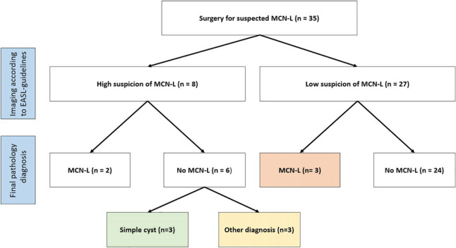
Results: In total, 35 patients were included. In three patients, there were no worrisome imaging features, yet final pathological diagnosis showed MCN-L. Contrarily, six patients with worrisome imaging features did not have MCN-L. Five patients were diagnosed with MCN-L on final pathology. The sensitivity of the EASL-guidelines for the diagnosis of MCN-L was 40% (95%CI: 5.3-85%) and the specificity was 80% (95% CI: 61-92%).

Conclusion: Although the new EASL-guidelines provide some guidance, they could not reliably distinguish MCN-L from other cysts in our series. Thus, preoperative diagnosis of MCN-L remains challenging and we should be careful in selecting surgical strategies based on these criteria.

Keywords: Cyst; Liver; Pathology; Radiology; Surgery.


Conflict of interest statement

None.

Figures

Fig. 1
Fig. 1
Representative slides of a simple hepatic cyst and mucinous cystic neoplasm of the liver A. Simple hepatic cyst, B-D. Mucinous cystic neoplasm of the liver with ovarian-like stroma and positive estrogen receptor (C) and progesterone receptor (D) staining
Fig. 2
Fig. 2
Application of the EASL criteria in our (high risk) population Orange (diagonal pattern filling) indicates patients that would be undertreated according to EASL-guidelines. Characteristics of these patients are shown in Table 2. Green (horizontal pattern filling) and yellow (vertical pattern filling) indicates patients that would be overtreated according to EASL-guidelines. Characteristics of these patients are shown in Table 3 and Supplementary Table 1, respectively
Fig. 3
Fig. 3
Representative CT and MRI images of patients in whom imaging assessment according to the EASL-guidelines did not correspond to the final pathological diagnosis A. CT-scan of a 52-year-old female patient with a liver cyst with low suspicion of MCN-L based on imaging assessment according to the EASL-guidelines. Final pathological diagnosis was MCN-L. B and C. CT-scan and MRI images of a 59-year-old patient with a liver cyst with high suspicion of MCN-L based on imaging assessment according to the EASL-guidelines. Final pathological diagnosis was simple hepatic cyst. A. The cyst measuring 8.7 cm, located in segment 4b, showed no nodularity or thick septations (no major worrisome features). Thin septations and less than three co-existent cysts were seen (two minor worrisome features). No upstream biliary dilatation, haemorrhage, or perilesional perfusional change were seen. B and C. The cyst measuring 10.6 cm in diameter, located in the right hemiliver, showed thick septations (one major worrisome feature), indicated by the yellow arrow. The cysts showed upstream biliary dilation (figure C) and less than three co-existent hepatic cysts (two minor worrisome features). No perilesional perfusional change or haemorrhage was observed

References

    1. Smith-Bindman R, Miglioretti DL, Johnson E, et al. Use of diagnostic imaging studies and associated radiation exposure for patients enrolled in large integrated health care systems, 1996–2010. JAMA. 2012;307(22):2400–2409. doi: 10.1001/jama.2012.5960. - DOI - PMC - PubMed
    1. Berland LL, Silverman SG, Gore RM, et al. Managing Incidental Findings on Abdominal CT: White Paper of the ACR Incidental Findings Committee. J Am Coll Radiol. 2010;7(10):754–773. doi: 10.1016/j.jacr.2010.06.013. - DOI - PubMed
    1. Marrero JA, Ahn J, Reddy RK. on behalf of the Practice Parameters Committee of the American College of G ACG Clinical Guideline: The Diagnosis and Management of Focal Liver Lesions. Am J Gastroenterol. 2014;109(9):1328–1347. doi: 10.1038/ajg.2014.213. - DOI - PubMed
    1. Choi HK, Lee JK, Lee KH, et al. Differential diagnosis for intrahepatic biliary cystadenoma and hepatic simple cyst: significance of cystic fluid analysis and radiologic findings. J Clin Gastroenterol. 2010;44(4):289–293. doi: 10.1097/MCG.0b013e3181b5c789. - DOI - PubMed
    1. Fuks D, Voitot H, Paradis V, Belghiti J, Vilgrain V, Farges O. Intracystic concentrations of tumour markers for the diagnosis of cystic liver lesions. Br J Surg. 2014;101(4):408–416. doi: 10.1002/bjs.9414. - DOI - PubMed