. 2024 Feb 17;24(1):64.
doi: 10.1186/s12893-024-02354-x.

Functional outcomes of different surgical treatments for common peroneal nerve injuries: a retrospective comparative study

Affiliations

Functional outcomes of different surgical treatments for common peroneal nerve injuries: a retrospective comparative study

Zhen Pang et al. BMC Surg. .

Abstract

Background: This study aims to assess the recovery patterns and factors influencing outcomes in patients with common peroneal nerve (CPN) injury.

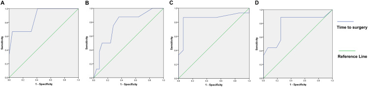
Methods: This retrospective study included 45 patients with CPN injuries treated between 2009 and 2019 in Jing'an District Central Hospital. The surgical interventions were categorized into three groups: neurolysis (group A; n = 34 patients), nerve repair (group B; n = 5 patients) and tendon transfer (group C; n = 6 patients). Preoperative and postoperative sensorimotor functions were evaluated using the British Medical Research Council grading system. The outcome of measures included the numeric rating scale, walking ability, numbness and satisfaction. Receiver operating characteristic (ROC) curve analysis was utilized to determine the optimal time interval between injury and surgery for predicting postoperative foot dorsiflexion function, toe dorsiflexion function, and sensory function.

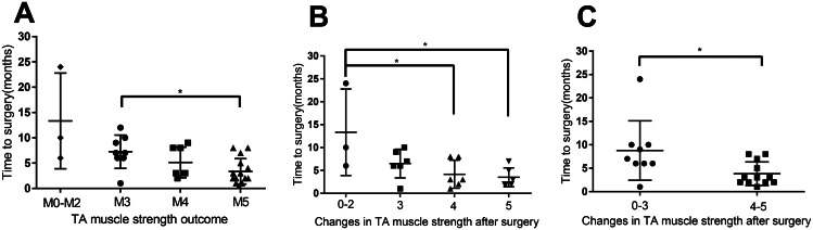
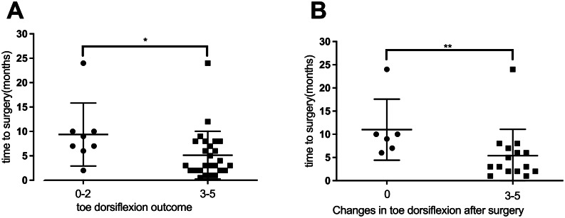
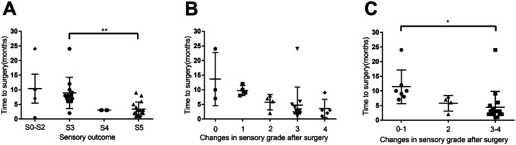
Results: Surgical interventions led to improvements in foot dorsiflexion strength in all patient groups, enabling most to regain independent walking ability. Group A (underwent neurolysis) had significant sensory function restoration (P < 0.001), and three patients in Group B (underwent nerve repair) had sensory improvements. ROC analysis revealed that the optimal time interval for achieving M3 foot dorsiflexion recovery was 9.5 months, with an area under the curve (AUC) of 0.871 (95% CI = 0.661-1.000, P = 0.040). For M4 foot dorsiflexion recovery, the optimal cut-off was 5.5 months, with an AUC of 0.785 (95% CI = 0.575-0.995, P = 0.020). When using M3 toe dorsiflexion recovery or S4 sensory function recovery as the gold standard, the optimal cut-off remained at 5.5 months, with AUCs of 0.768 (95% CI = 0.582-0.953, P = 0.025) and 0.853 (95% CI = 0.693-1.000, P = 0.001), respectively.

Conclusions: Our study highlights the importance of early surgical intervention in CPN injury recovery, with optimal outcomes achieved when surgery is performed within 5.5 to 9.5 months post-injury. These findings provide guidance for clinicians in tailoring treatment plans to the specific characteristics and requirements of CPN injury patients.

Keywords: Common peroneal nerve injury (CPN injury); Nerve repair; Neurolysis; Tendon transfer.


Conflict of interest statement

The authors declare no competing interests.

Figures

Fig. 1
Fig. 1
The release of the nerve distally. The white arrow indicates the common peroneal nerve after release. This is a lateral incision at the fibular head on the right leg, with the popliteal fossa on the right and the calf on the left
Fig. 2
Fig. 2
Suturing of the sural nerves between the common peroneal nerve stumps. The white arrow indicates the distal end of the common peroneal nerve, the gray arrow indicates the proximal end of the common peroneal nerve, and the white asterisk indicates the transplanted nerve
Fig. 3
Fig. 3
The posterior tibialis tendon was delivered from the wound on the medial calf. The gray arrow indicates the posterior tibialis tendon. The incision outlined by the white dotted line is used to find and resect the insertion of the posterior tibialis tendon. See Fig. 4 for the specific surgical operation
Fig. 4
Fig. 4
Fixing the tendon by sutures. The posterior tibialis tendon indicated by the white arrow is sutured to the third cuneiform bone. This is a front-and-rear view with the toe in front and the heel behind
Fig. 5
Fig. 5
The tibialis anterior muscle force among the three groups. A. Relationship between the time from symptom onset to neurolysis and post-surgery muscle strength (M3 vs. M5). Patients with M5 muscle strength after surgery (3.37 ± 2.43) had a significantly shorter period from symptom onset to surgical treatment compared to those with M3 muscle strength (7.25 ± 3.07). P=0.011. Data from one patient, whose muscle strength had recovered to M5, was excluded from the analysis due to an unusually long time interval (24 months) between symptom onset and neurolysis. When including this patient’s data, the time interval for patients with M5 was 4.66 ± 5.52, and the P value was 0.0549. B-C. Duration from symptom onset to neurolysis in relation to changes in tibialis anterior muscle force. B. Patients with 4 (4.16 ± 2.79) or 5 (3.5 ± 1.89) grade muscle strength improvements exhibited shorter time intervals than those with 0–2 improvements (13.33 ± 7.72). P=0.03 (compared to 4 grade improvements), P=0.02 (compared to 5 grade improvements). C. Patients with 4-5 grade muscle strength improvements (3.83 ± 2.41) had shorter time intervals than those with 0–3 improvements (8.78 ± 5.98). P=0.03. *, P<0.05; error bars represent the standard deviation (SD). n=33 for panel A, n=21 for panels B-C. TA, tibialis anterior
Fig. 6
Fig. 6
The toe dorsiflexion muscle strength among the three groups. A. Relationship between the time from symptom onset to neurolysis and post-surgery toe dorsiflexion muscle strength (M0-M2 vs. M3-M5). Patients with M3 to M5 muscle strength (5.14 ± 3.06) had significantly shorter time intervals between symptom onset and surgical treatment compared to patients with M0 to M2 muscle strength (9.38 ± 5.74). P=0.04. B. Graph illustrating that patients with 3 to 5 grade muscle strength improvements had shorter time intervals (5.4 ± 5.5) than those without improvements (11 ± 6). P=0.0074. *, P<0.05; **, P<0.01; error bars represent the standard deviation (SD). n=34 for panel A, n=21 for panel B
Fig. 7
Fig. 7
The sensory grade analysis among the three groups. A. Relationship between the time from symptom onset to neurolysis and sensory grade (Grade 3 vs. Grade 5). Patients with Grade 5 sensory function (3.38 ± 2.37) had a significantly shorter period from symptom onset to surgical treatment compared to those with Grade 3 (8.92 ± 2.43). P=0.002. B-C. Time from symptom onset to treatment for patients with different changes in sensory grade. C. Patients with 3 to 4 grade sensory function improvements exhibited shorter intervals (4.41 ± 5.26) than those with 0–1 improvement (11.43 ± 5.34). P=0.02. *, P<0.05; **, P<0.01; error bars represent the standard deviation (SD). n=34 for panel A, n=28 for panels B-C
Fig. 8
Fig. 8
ROC curves for time to surgery. A. ROC analysis using M3 foot dorsiflexion recovery as the gold standard, with a best cut-off value of 9.5 months (AUC=0.871, 95% CI=0.661-1.000, P=0.04). B. ROC analysis using M3 toe dorsiflexion recovery as the gold standard, with a best cut-off value of 5.5 months (AUC=0.768, 95% CI=0.582-0.953, P=0.025). C. ROC analysis using S4 sensory function recovery as the gold standard, with a best cut-off value of 5.5 months (AUC=0.853, 95% CI=0.693-1.000, P=0.001). D. ROC analysis using M4 foot dorsiflexion recovery as the gold standard, with a best cut-off value of 5.5 months (AUC=0.785, 95% CI=0.575-0.995, P=0.02). n=25 for panels A and D; n=33 for panel B; n=30 for panel C. ROC, Receiver Operating Characteristic; AUC, Area Under the ROC Curve

References

    1. Horteur C, Forli A, Corcella D, Pailhe R, Lateur G, Saragaglia D. Short- and long-term results of common peroneal nerve injuries treated by neurolysis, direct suture or nerve graft. Eur J Orthop Surg Traumatol. 2019;29(4):893–8. doi: 10.1007/s00590-018-2354-0. - DOI - PubMed
    1. George SC, Boyce DE. An evidence-based structured review to assess the results of common peroneal nerve repair. Plast Reconstr Surg. 2014;134(2):302e–11e. doi: 10.1097/PRS.0000000000000318. - DOI - PubMed
    1. Daniels SP, Ross AB, Sneag DB, Gardon SN, Li G, Hanna AS, Tuite MJ. Can MR neurography of the common peroneal nerve predict a residual motor deficit in patients with foot drop? Skeletal Radiol. 2023;52(4):751–61. doi: 10.1007/s00256-022-04214-2. - DOI - PubMed
    1. Ho B, Khan Z, Switaj PJ, Ochenjele G, Fuchs D, Dahl W, Cederna P, Kung TA, Kadakia AR. Treatment of peroneal nerve injuries with simultaneous tendon transfer and nerve exploration. J Orthop Surg Res. 2014;9:67. doi: 10.1186/s13018-014-0067-6. - DOI - PMC - PubMed
    1. Rasulic L, Savic A, Vitosevic F, Samardzic M, Zivkovic B, Micovic M, Bascarevic V, Puzovic V, Joksimovic B, Novakovic N, et al. Iatrogenic peripheral nerve injuries-Surgical treatment and outcome: 10 years’ experience. World Neurosurg. 2017;103:841–51. doi: 10.1016/j.wneu.2017.04.099. - DOI - PubMed

LinkOut - more resources