. 2024 Feb 19;22(1):69.
doi: 10.1186/s12951-024-02335-5.

The oncolytic bacteria-mediated delivery system of CCDC25 nucleic acid drug inhibits neutrophil extracellular traps induced tumor metastasis

Affiliations

The oncolytic bacteria-mediated delivery system of CCDC25 nucleic acid drug inhibits neutrophil extracellular traps induced tumor metastasis

Li-Na Liu et al. J Nanobiotechnology. .

Abstract

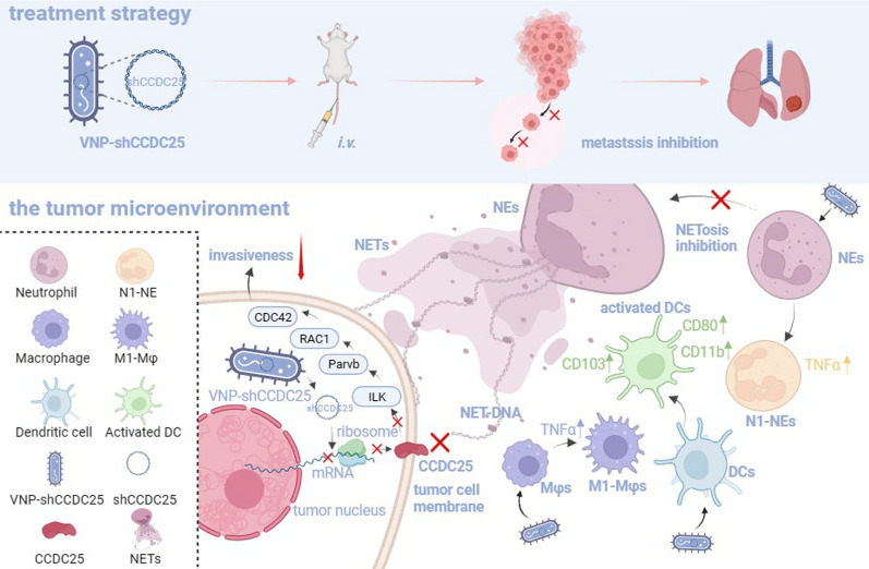
Background: Neutrophil extracellular traps (NETs), antibacterial weapons of neutrophils (NEs), have been found to play a crucial role in cancer metastasis in recent years. More and more cancer research is focusing on anti-NETs. However, almost all anti-NETs treatments have limitations such as large side effects and limited efficacy. Therefore, exploring new anti-NETs therapeutic strategies is a long-term goal.

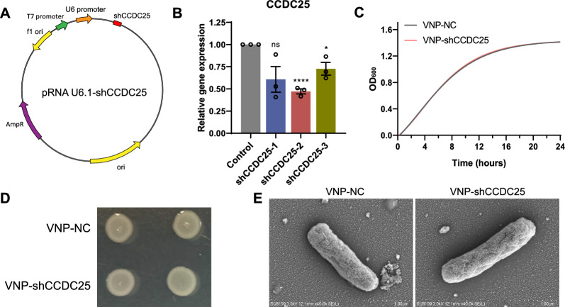
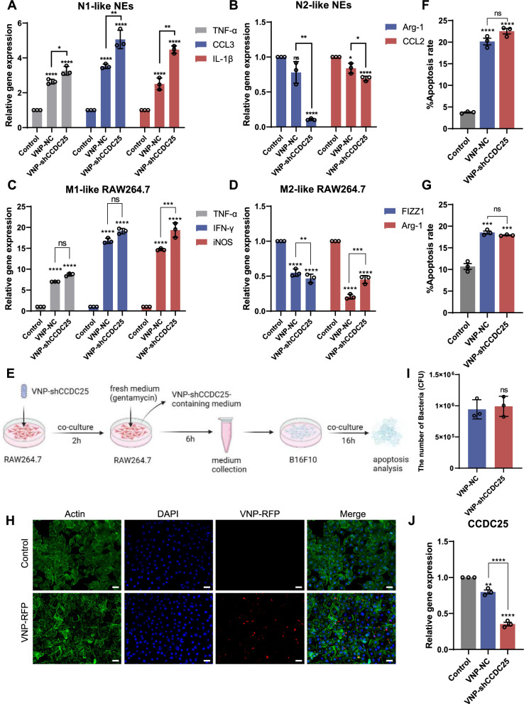
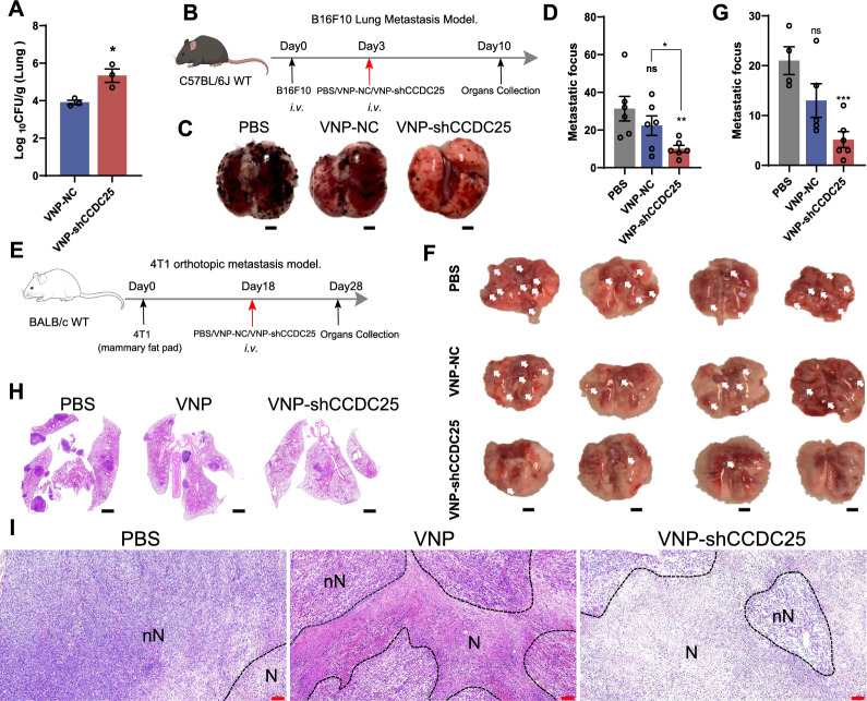
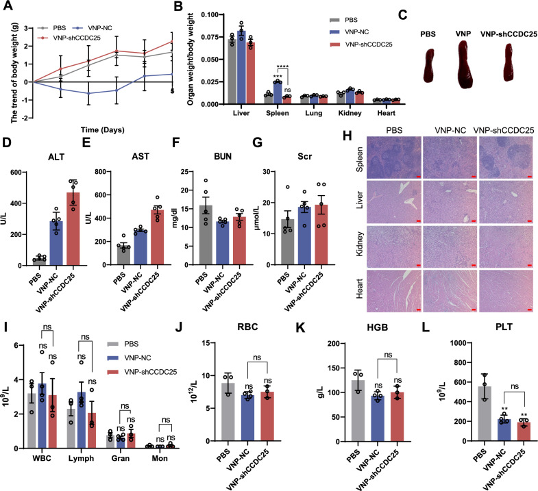
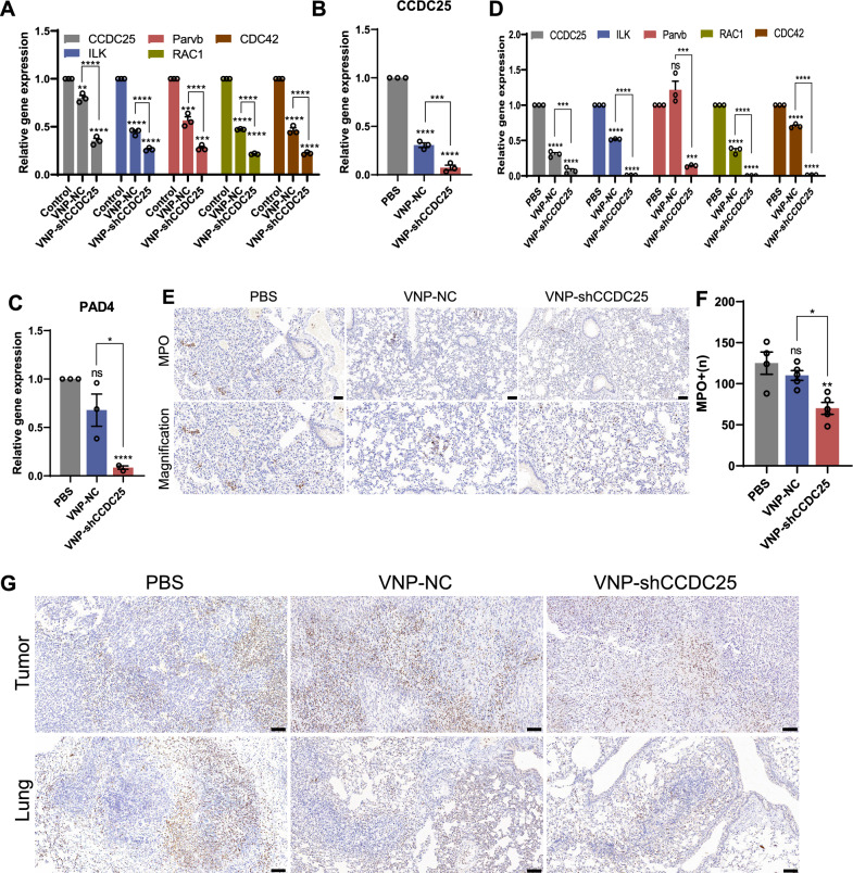
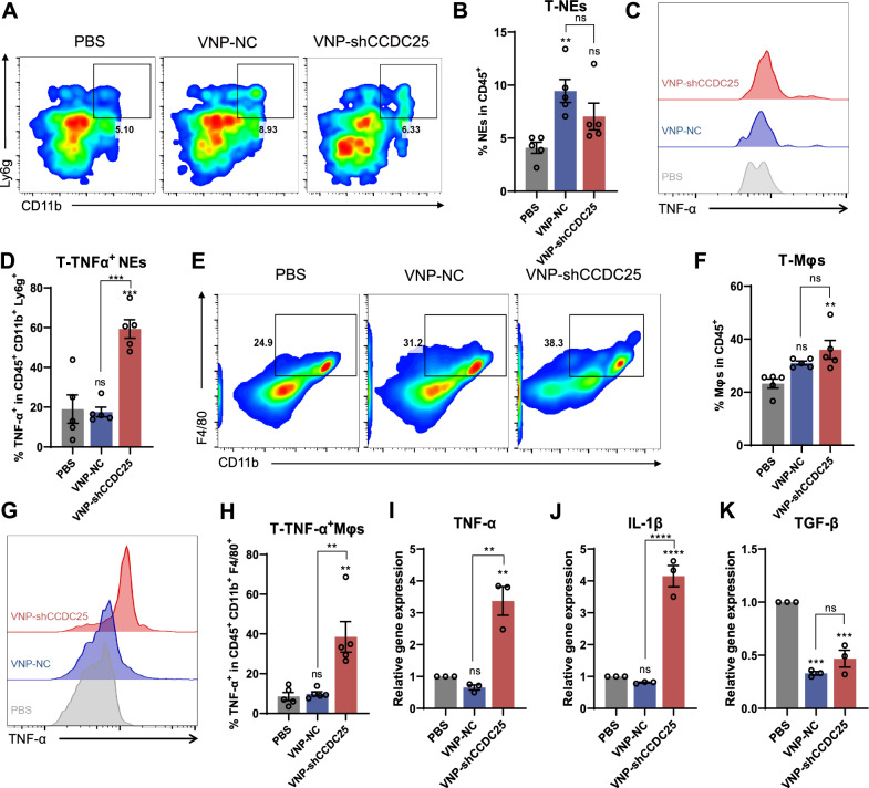
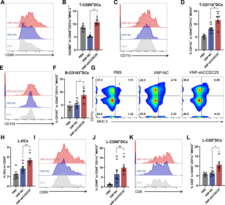
Results: The transmembrane protein coiled-coil domain containing 25 (CCDC25) on tumor cell membranes can bind NETs-DNA with high specificity and affinity, enabling tumor cells to sense NETs and thus promote distant metastasis. We transformed shCCDC25 into VNP20009 (VNP), an oncolytic bacterium, to generate VNP-shCCDC25 and performed preclinical evaluation of the inhibitory effect of shCCDC25 on cancer metastasis in B16F10 lung metastasis and 4T1 orthotopic lung metastasis models. VNP-shCCDC25 effectively blocked the downstream prometastatic signaling pathway of CCDC25 at tumor sites and reduced the formation of NETs while recruiting more neutrophils and macrophages to the tumor core, ultimately leading to excellent metastasis inhibition in the two lung metastasis models.

Conclusion: This study is a pioneer in focusing on the effect of anti-NET treatment on CCDC25. shCCDC25 is effectively delivered to tumor sites via the help of oncolytic bacteria and has broad application in the inhibition of cancer metastasis via anti-NETs.

Keywords: CCDC25; Metastasis; NETs; Neutrophils; Nucleic acid delivery; VNP20009.


Conflict of interest statement

The authors declare that they have no competing interests.

Figures

Fig. 1
Fig. 1
Construction and characterization of VNP-shCCDC25. A Schematic diagram of pRNA U6.1- shCCDC25. B The expression of CCDC25 in B16F10 cells after transfection with pRNA U6.1-shCCDC25 was analyzed via qPCR. C Bacteria growth curves of VNP-NC and VNP-shCCDC25 (n = 6). D Colonial morphology of VNP-NC and VNP-shCCDC25. E Scanning electron microscopes (SEM) of VNP-NC and VNP-shCCDC25. Data are shown as the mean ± SD. **** p < 0.0001, *** p < 0.001, ** p < 0.01, * p < 0.05, ns: no significance
Fig. 2
Fig. 2
VNP-shCCDC25 stimulates NEs and macrophage activation and infect cells in vitro. A The expression of N1 polarization markers of NEs after VNP-shCCDC25 treatment was analyzed via qPCR. B The expression of N2 polarization markers of NEs after VNP-shCCDC25 treatment was analyzed via qPCR. C The expression of M1 polarization markers of RAW264.7 cells after VNP-shCCDC25 treatment was analyzed via qPCR. D The expression of M2 polarization markers of RAW264.7 cells after VNP-shCCDC25 treatment was analyzed via qPCR. E Schematic diagram of RAW264.7 cell medium stimulated by VNP-shCCDC25 promotes apoptosis of B16F10 cells. After VNP-shCCDC25 was co-cultured with RAW264.7 cells for 2 h, the medium was changed to fresh medium containing gentamycin, and the incubation was continued for 6 h. After 6 h, the RAW264.7 cell medium was collected and co-incubated with B16F10 cells for 16 h. Finally, B16F10 cells were collected for the apoptosis assay. F The apoptosis levels of B16F10 cells after incubated for 16 h with RAW264.7 cell medium, which was stimulated with VNP-NC, VNP-shCCDC25, or not (n = 3). G The apoptosis levels of B16F10 cells after incubated with VNP-NC, VNP-shCCDC25, or not for 16 h (n = 3). H The fluorescent pictures of B16F10 cells incubated with VNP-RFP (MOI = 100:1) or not; actin (green), DAPI (blue), VNP-RFP (red). Scale bars: 25 μm. I The titer of bacterium colonized in the B16F10 cells after co-incubated with VNP-NC or VNP-shCCDC25 (n = 3) for 16 h. J The expression of CCDC25 in B16F10 cells after incubated with VNP-shCCDC25 was analyzed via qPCR. Data are shown as the mean ± SD. **** p < 0.0001, *** p < 0.001, ** p < 0.01, * p < 0.05, ns: no significance
Fig. 3
Fig. 3
VNP-shCCDC25 inhibits tumor metastasis. A The titer of bacterium colonized in the lung tumor at 4 h post i.v. with 1 × 106 CFU VNP-NC or VNP-shCCDC25 (n = 3). B The treatments schedule for VNP-shCCDC25 inhibition of tumor metastasis in B16F10 lung metastasis model: 3 days after B16F10 cells injection, PBS, VNP-NC, or VNP-shCCDC25 was administrated by i.v. and the tissues were collected at 10 days. C, D The pictures and numbers of the lung metastasis foci in B16F10 lung metastasis model (n = 6). Scale bars: 2000 μm. E The treatment schedule for VNP-shCCDC25 inhibition of tumor metastasis in 4T1 orthotopic lung metastasis model: 18 days after 4T1 cells injection, PBS, VNP-NC, or VNP-shCCDC25 was administrated by i.v. and the tissues were collected at 28 days. F, G The pictures and numbers of the lung metastasis foci in 4T1 orthotopic lung metastasis model. Scale bars: 2000 μm. H H&E staining of the lung metastasis foci after treatments in 4T1 orthotopic lung metastasis model. Scale bars: 1000 μm. I H&E staining of situ tumors after treatments in 4T1 orthotopic lung metastasis model. Scale bar: 200 µm. N: necrotic region, nN: nonnecrotic region. Data are shown as the mean ± SD. **** p < 0.0001, *** p < 0.001, ** p < 0.01, * p < 0.05, ns: no significance
Fig. 4
Fig. 4
Safety assessment of VNP-shCCDC25 in vivo. A The changing trend of body weight of mouse was monitored daily after treatments (n = 6). B The ratio of organ weight to body weight of mouse in different administration groups (n = 3). C The picture of spleen after administrations. DG Serological analysis of ALT (D), AST (E), BUN (F), and Scr (G) in serum 5 days after treatments (n = 5). H H&E staining of organs in different administration groups. Scale bars: 200 μm. IL Routine blood test after 5 days of treatment (n = 5). Data are shown as the mean ± SD. **** p < 0.0001, *** p < 0.001, ** p < 0.01, * p < 0.05, ns: no significance
Fig. 5
Fig. 5
VNP-shCCDC25 blocks the downstream pro-metastasis signaling pathway of CCDC25 and reduces the formation of NETs. A 16 h after co-incubation with VNP-shCCDC25 and B16F10 cells (MOI = 10:1), the expression level of genes in the downstream prometastasis signaling pathway of CCDC25 in B16F10 cells was analyzed via qPCR. B After VNP-shCCDC25 treatment, the expression level of CCDC25 in the metastatic foci of B16F10 lung metastasis model was analyzed via qPCR. C After VNP-shCCDC25 treatment, the expression level of PAD4 in the metastatic foci of B16F10 lung metastasis model was analyzed via qPCR. D In 4T1 orthotopic lung metastasis model, the expression level of genes in the downstream prometastasis signaling pathway of CCDC25 in the lung tissue after administrations was analyzed via qPCR. E, F The number of MPO in metastatic foci of B16F10 lung metastasis model was analyzed via immunohistochemistry (IHC). G IHC analysis of MPO expression in metastatic foci of 4T1 orthotopic lung metastasis model. Data are shown as the mean ± SD. **** p < 0.0001, *** p < 0.001, ** p < 0.01, * p < 0.05, ns: no significance
Fig. 6
Fig. 6
The TME were remodeled after VNP-shCCDC25 administrations. A, B The percentage of tumor-infiltrating NEs was analyzed via FACS, 5 days after treatments. C, D The percentage of TNF-α positive cells in tumor-infiltrating NEs was analyzed via FACS. E Similarly, the percentage of tumor-infiltrating Mφs was analyzed via FACS, and the statistic diagrams were shown in (F). G The FACS histogram plot of TNF-α positive cells in tumor-infiltrating Mφs. H The statistic diagrams of TNF-α+ Mφs in tumor-infiltrating Mφs. I-K The mRNA expression level of TNF-αIL-1βTGF-β in the metastatic foci of B16F10 lung metastasis model was analyzed via qPCR. Data are shown as the mean ± SD. **** p < 0.0001, *** p < 0.001, ** p < 0.01, * p < 0.05, ns: no significance
Fig. 7
Fig. 7
VNP-shCCDC25 stimulated DCs maturation. A, B The percentage of CD80 positive cells in tumor-infiltrating DCs was analyzed via FACS. C, D The percentage of CD11b positive cells in tumor-infiltrating DCs was analyzed via FACS. E, F 5 days after administrations, the level of CD103 in DCs in peripheral blood was analyzed via FACS. G The percentage of DCs in TdLNs. And the statistic diagrams were shown in (H). I, J In TdLNs, the percentage of CD80+DCs was analyzed via FACS. K, L Similarly, CD8+ DCs in TdLNs was evaluated. Data are shown as the mean ± SD. **** p < 0.0001, *** p < 0.001, ** p < 0.01, * p < 0.05, ns: no significance
Fig. 8
Fig. 8
Schematic diagram of VNP-shCCDC25 blocking CCDC25-NET-DNA and remodeling immunity in B16F10-bearing mice and 4T1-bearing mice. Overall, VNP-shCCDC25 inhibits tumor lung metastasis after intravenous injection in mice. Specifically, VNP-shCCDC25 mediates tumor lung metastasis inhibition through the following 3 mechanisms. (1) VNP-shCCDC25 knocked down CCDC25 and its downstream prometastasis signaling pathway ILK-Parvb-RAC1-CDC42 after successful invasion of tumor cells, which in turn reduced the invasiveness of tumor cells. (2) The NETosis at tumor site was somewhat inhibited after knocking down CCDC25. (3) In TME, elevated infiltration rates of M1-Mφs and N1-NEs with antitumor phenotypes as well as effective activation of DCs were observed, suggesting that VNP-shCCDC25 can remodel TME and ultimately achieve tumor metastasis suppression

Similar articles

Cited by

References

    1. De Meo ML, Spicer JD. The role of neutrophil extracellular traps in cancer progression and metastasis. Semin Immunol. 2021;57:101595. doi: 10.1016/j.smim.2022.101595. - DOI - PubMed
    1. Papayannopoulos V. Neutrophil extracellular traps in immunity and disease. Nat Rev Immunol. 2018;18:134–147. doi: 10.1038/nri.2017.105. - DOI - PubMed
    1. Cristinziano L, Modestino L, Antonelli A, Marone G, Simon HU, Varricchi G, Galdiero MR. Neutrophil extracellular traps in cancer. Semin Cancer Biol. 2022;79:91–104. doi: 10.1016/j.semcancer.2021.07.011. - DOI - PubMed
    1. Masucci MT, Minopoli M, Del Vecchio S, Carriero MV. The emerging role of neutrophil extracellular traps (NETs) in tumor progression and metastasis. Front Immunol. 2020;11:1749. doi: 10.3389/fimmu.2020.01749. - DOI - PMC - PubMed
    1. Saffarzadeh M. Neutrophil extracellular traps as a drug target to counteract chronic and acute inflammation. Curr Pharm Biotechnol. 2018;19:1196–1202. doi: 10.2174/1389201020666190111164145. - DOI - PubMed

MeSH terms