. 2024 Feb 18;14(1):4008.
doi: 10.1038/s41598-024-53843-1.

Toxoflavin analog D43 exerts antiproliferative effects on breast cancer by inducing ROS-mediated apoptosis and DNA damage

Affiliations

Toxoflavin analog D43 exerts antiproliferative effects on breast cancer by inducing ROS-mediated apoptosis and DNA damage

Tingyue Wu et al. Sci Rep. .

Abstract

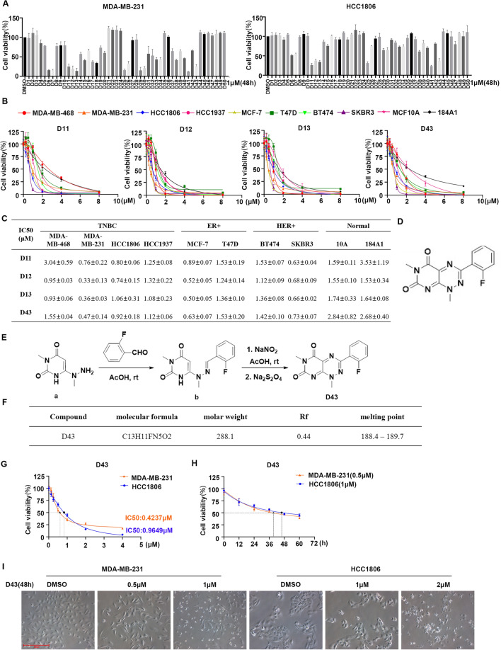
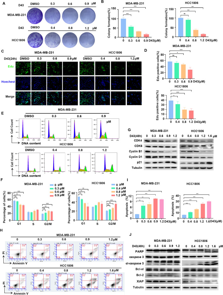
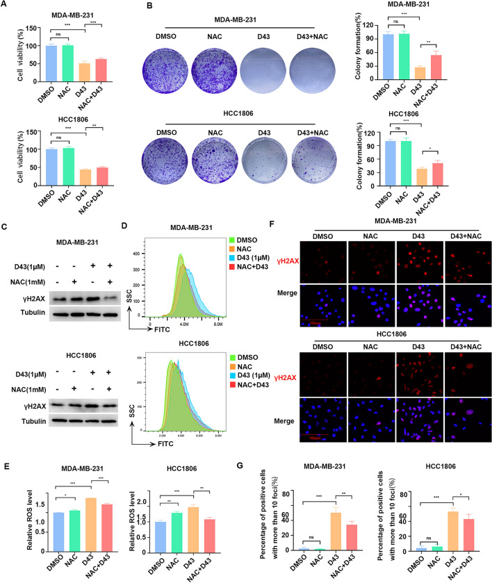
Triple-negative breast cancer (TNBC) is regarded as the deadliest subtype of breast cancer because of its high heterogeneity, aggressiveness, and limited treatment options. Toxoflavin has been reported to possess antitumor activity. In this study, a series of toxoflavin analogs were synthesized, among which D43 displayed a significant dose-dependent inhibitory effect on the proliferation of TNBC cells (MDA-MB-231 and HCC1806). Additionally, D43 inhibited DNA synthesis in TNBC cells, leading to cell cycle arrest at the G2/M phase. Furthermore, D43 consistently promoted intracellular ROS generation, induced DNA damage, and resulted in apoptosis in TNBC cells. These effects could be reversed by N-acetylcysteine. Moreover, D43 significantly inhibited the growth of breast cancer patient-derived organoids and xenografts with a favorable biosafety profile. In conclusion, D43 is a potent anticancer agent that elicits significant antiproliferation, oxidative stress, apoptosis, and DNA damage effects in TNBC cells, and D43 holds promise as a potential candidate for the treatment of TNBC.

Keywords: DNA damage; N-acetylcysteine (NAC); Patient-derived breast cancer organoids (PDO); ROS; Toxoflavin.


Conflict of interest statement

The authors declare no competing interests.

Figures

Figure 1
Figure 1
Compound D43 inhibits the growth of TNBC cells. (A) SRB experiments were performed to detect the cell viability of HCC1806 and MDA-MB-231 cells after treatment with 50 compounds (48 h, 1 μM). (B) IC50 of D11, D12, D13 and D43 in the breast cancer cell lines MDA-MB-468, MDA-MB-231, HCC1806, HCC1937, MCF-7, T47D, BT474, and SKBR3 and the immortalized breast epithelial cell lines MCF10A and 184A1. (C) Statistical values of IC50 data. (D) The chemical structure of D43. (E) The synthesis process of D43. (F) The physicochemical properties of D43. (G) IC50 of D43 in MDA-MB-231 and HCC1806 breast cancer cell lines. (H) The antitumor effect of D43 in MDA-MB-231 (0.5 μM) and HCC1806 (1 μM) cells was detected over time. (I) Microscopic morphology of MDA-MB-231 and HCC1806 cells treated with D43.
Figure 2
Figure 2
D43 inhibits DNA synthesis and survival of TNBC cells. (A) Representative picture of colony formation in MAD-MB-231 and HCC1806 cells incubated with a D43 concentration gradient for 14 days. (B) D43 inhibited the colony formation of TNBC cells. Individual colonies were counted under a microscope. (C) D43 inhibits the DNA synthesis ability of cells. An EdU probe was used to detect the DNA synthesis ability of MDA-MB-231 and HCC1806 cells treated with different concentrations of D43 for 24 h. Blue represents Hoechst staining, and green represents EdU staining. (D) The proportion of EdU-positive cells was counted. (E) D43 increased the number of cells in the G2/M phase. Cells were dyed with PI and analyzed with flow cytometry after incubation with D43 (48 h). (F) The proportion of each stage of the cell cycle changes is shown. (G) D43 regulated cell cycle-related proteins. Western blot images showing the expression levels of cyclin B1, cyclin D1, CDK4, CDK6, and p21 proteins in MDA-MB-231 and HCC1806 cells treated with D43 at different concentrations for 48 h with tubulin as a loading control. (H) The proportion of apoptotic subsets was analyzed by flow cytometry after Annexin V-PI double staining with D43 in a concentration gradient treatment for 48 h. (I) Statistics showing the proportion of Annexin V-positive cells. (J) D43 regulates apoptosis-related proteins. Western blotting was used to detect the expression of apoptosis-related proteins, including PARP, Caspase3 and their splicing forms, and the anti-apoptotic proteins Bcl-xl, Bcl-2 and XIAP were also detected. The samples derive from the same experiment and that blots were processed in parallel. Original blots are presented in Supplementary Material. Data are representative of three independent experiments. *p < 0.05, **p < 0.01 and ***p < 0.001; ns, not significant.
Figure 3
Figure 3
D43 induces DNA damage by increasing the level of ROS in TNBC cells. (A) RNA sequencing of HCC1806 cells treated with D43 (1 μM) for 24 h revealed a KEGG enrichment scatterplot. (B) D43 caused DNA damage in a concentration-dependent manner. Western blot assays were performed to detect the expression of ATR, p-CHK1, CHK1, and γH2AX in MDA-MB-231 and HCC1806 cells treated with D43 at different concentrations for 48 h with tubulin as a loading control. (C) D43 caused DNA damage in a time-dependent manner. After treatment of D43 with a time gradient (0, 6, 12, 24, 36, 48 h), the above proteins were also detected by Western blotting. The samples derive from the same experiment and that blots were processed in parallel. Original blots are presented in Supplementary Material. (D) D43 stably increased the ROS level in TNBC cells. MDA-MB-231 and HCC1806 cells treated with a D43 concentration gradient for 48 h were labeled with the fluorescent probe DCFH-DA, and ROS levels were detected by flow cytometry. (E) Statistics of relative ROS levels. (F) D43 was able to activate the NRF2-HO-1 signaling axis in response to oxidative stress in a dose-dependent manner. Western blot assays were performed to detect the expression of NRF2 and HO-1 after treatment with D43 at different concentrations for 48 h. (G) D43 treatment resulted in a higher number of γ-H2AX foci. MDA-MB-231 and HCC1806 cells were treated with a D43 concentration gradient for 24 h, and the expression of γ-H2AX was detected by immunofluorescence staining and recorded by high-resolution fluorescence microscopy. (H) Four samples were taken from each treatment group to count the proportion of positive cells with more than 10 foci in the cells. Data are representative of three independent experiments. *p < 0.05, **p < 0.01 and ***p < 0.001; ns, not significant.
Figure 4
Figure 4
NAC rescued D43-induced DNA damage and ROS upregulation. (A) NAC partially alleviated the cell death induced by D43 in HCC1806 and MDA-MB-231 cells. After pretreatment with NAC (1 mM) for 6 h, MDA-MB-231 (0.5 μM) and HCC1806 (1 μM) cells were treated with D43 for 48 h, and cell viability was verified by SRB. (B) Cell proliferation was verified by colony formation assay after treatment with NAC and D43, respectively, or in combination. (C) Western blot analysis showed that pretreatment with NAC (1 mM, 6 h) antagonized D43-induced increase of γH2AX protein levels. The samples derive from the same experiment and that blots were processed in parallel. Original blots are presented in Supplementary Material. (D) NAC effectively antagonized the D43-induced upregulation of ROS. MDA-MB-231 and HCC1806 cells were treated with D43 or NAC for 48 h and labeled with the fluorescent probe DCFH-DA, and ROS levels were measured by flow cytometry. (E) Statistics of relative ROS levels. (F) NAC antagonized the upregulation of γ-H2AX foci formation induced by D43. After NAC pretreatment for 6 h, MDA-MB-231 (0.5 μM) and HCC1806 (1 μM) cells were treated with D43 for 24 h, and the expression of γ-H2AX was detected by immunofluorescence staining and recorded by high-resolution fluorescence microscopy. (G) Four samples were taken from each treatment group to count the proportion of positive cells with more than 10 foci in the cells. Data are representative of three independent experiments. *p < 0.05, **p < 0.01 and ***p < 0.001; ns, not significant.
Figure 5
Figure 5
D43 inhibits the growth of patient-derived TNBC organoids and xenograft tumors. (A) Tissue information of TNBC patients sampled from Yunnan Cancer Hospital. (B) Treatment with D43 compromised the structural integrity of TNBC PDOs. Morphological records of breast tumor organoids at different D43 treatment concentrations for different days. The red arrow points to the disintegrating or completely disintegrating TNBC PDOs. (C) D43 inhibited the ATP activity of TNBC PDOs. An ATPase activity kit was used to detect the changes in the viability of TNBC PDO cells treated with different D43 concentrations for 5 days. (D) D43 had a significant inhibitory effect on the growth of TNBC PDOs. The area of PDOs was calculated by ImageJ, and the growth curve was drawn. (E) Schematic diagram of subcutaneous tumor model construction in nude mice and administration time and frequency. (F) D43 significantly suppressed tumor growth in nude mice. After 15 days of continuous administration, the mice were sacrificed by cervical dislocation, and tumors were removed and photographed. (G) The tumor weights of control (DMSO) and experimental (5 mg/kg) mice are shown. (H) The tumor volume was weighed every 3 days after the beginning of the drug administration treatment and is shown as a line chart. (I) Changes in body weight of mice before and after D43 administration. (JL) Immunohistochemistry was used to detect the expression of Ki-67 and γH2AX in tumor tissues, and γH2AX immunoreactivity was scored semiquantitatively. *p < 0.05, **p < 0.01 and ***p < 0.001; ns, not significant.

Similar articles

Cited by

References

    1. Sung H, Ferlay J, Siegel RL, et al. Global cancer statistics 2020: GLOBOCAN estimates of incidence and mortality worldwide for 36 cancers in 185 countries. CA Cancer J. Clin. 2021;71(3):209–249. doi: 10.3322/caac.21660. - DOI - PubMed
    1. Tan PH, Ellis I, Allison K, et al. The 2019 World Health Organization classification of tumours of the breast. Histopathology. 2020;77(2):181–185. doi: 10.1111/his.14091. - DOI - PubMed
    1. Barzaman K, Karami J, Zarei Z, et al. Breast cancer: Biology, biomarkers, and treatments. Int. Immunopharmacol. 2020;84:106535. doi: 10.1016/j.intimp.2020.106535. - DOI - PubMed
    1. Liang Y, Zhang H, Song X, et al. Metastatic heterogeneity of breast cancer: Molecular mechanism and potential therapeutic targets. Semin. Cancer Biol. 2020;60:14–27. doi: 10.1016/j.semcancer.2019.08.012. - DOI - PubMed
    1. Derakhshan F, Reis-Filho JS. Pathogenesis of triple-negative breast cancer. Annu. Rev. Pathol. 2022;17:181–204. doi: 10.1146/annurev-pathol-042420-093238. - DOI - PMC - PubMed